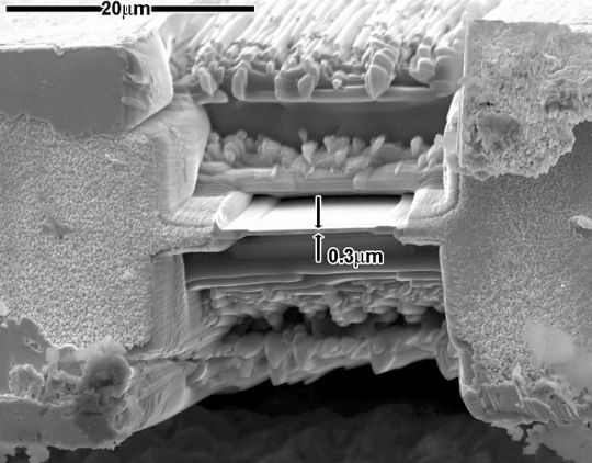
#scanning transmission electron microscopy
Photo
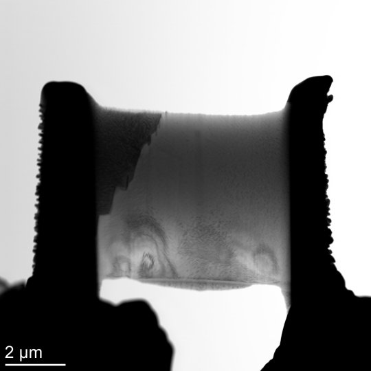
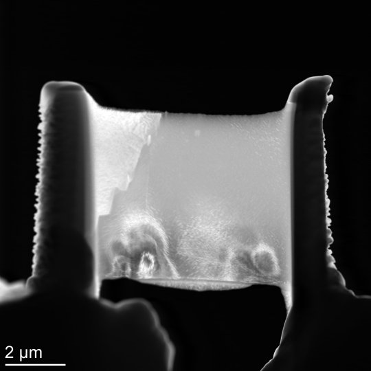
#transmission electron microscopy#bright field#dark field#HRTEM#Fourier transform#nickel alloy#scanning transmission electron microscopy#original content
5 notes
·
View notes
Text

A Journey into the World of Microscopy: From Humble Beginnings to High-Tech Magnification
The science of looking into the hidden invisible Microscopy has transformed our understanding of the world around us. It can explore the universe beyond the reach of our naked eyes, with complex cellular structures, red blood cells, viruses and other viruses and microorganisms taking on amazing perspectives
The history of the microscope is a fascinating story of human curiosity, scientific genius, and relentless exploration. From the humble beginnings of simple magnifying glasses to the sophistication of modern electronic microscopes, the invention of microscopes has shaped our understanding of the microscopic world
In the 1600s, Dutch opticians such as Hans and Zachary Janssen are credited with inventing the first microscope. Known for this hybrid microscope, many lenses were used to magnify objects up to 30 times.At the end of the 17th century, Antony van Leeuwenhoek, Dutch draper some changed our perception of thumbnails. Armed with a well-made single-lens microscope, and explored the hidden reaches of nature. In 1674, Leeuwenhoek discovered microorganisms in lake water, which he aptly named “animalcules”. His discovery laid the foundations of biology and inspired generations of scientists. This incredible feat allowed him to uncover a hidden universe – the first sightings of bacteria, red blood cells, and other microorganisms.
Formation of the scientific environment (17th-19th centuries): Leeuwenhoek’s discoveries boosted scientific research. Robert Hooke, an English scientist, established these developments. In 1665, his book "Micrographia" recorded his observations with a compound microscope. Notably, the term "cell" was coined by Hooke when he examined cork tissue, laying the foundation for cell biology.Microscope systems flourished throughout the 18th and 19th centuries Joseph Lister and other scientists addressed the limitations of the early lenses, introducing improvements that reduced image distortion.
Beyond the Limits of Light: The Beginning of the New Age (19th-20th century): As the 19th century progressed, the limitations of optical microscopy became apparent and scientists yearned for a tool which can go deeper into cells. This research culminated in the development of the electron microscope in the 1930s. The 20th century was revolutionary with the invention of the electron microscope. Unlike light microscopes, which use visible light, electron microscopes use electron beams to achieve much higher magnification.Formation of the scientific environment (17th-19th centuries): Leeuwenhoek’s discoveries boosted scientific research. Robert Hooke, an English scientist, established these developments. In 1665, his book "Micrographia" recorded his observations with a compound microscope. Notably, the term "cell" was coined by Hooke when he examined cork tissue, laying the foundation for cell biology.Microscope systems flourished throughout the 18th and 19th centuries Joseph Lister and other scientists addressed the limitations of the early lenses, introducing improvements that reduced image distortion.
Beyond the Limits of Light: The Beginning of the New Age (19th-20th century): As the 19th century progressed, the limitations of optical microscopy became apparent and scientists yearned for a tool which can go deeper into cells. This research culminated in the development of the electron microscope in the 1930s. The 20th century was revolutionary with the invention of the electron microscope. Unlike light microscopes, which use visible light, electron microscopes use electron beams to achieve much higher magnification.
In the 1930s, German experts Max Knoll and Ernst Ruska made the first electron microscope. This tool let us see tiny things like cells and even atoms by using electron beams, not light, getting images many times bigger. This cool invention showed us the tiny parts inside cells, viruses, and stuff too small to see before. The 1900s brought even more cool microscopes. New kinds like phase-contrast and confocal microscopy let scientists look at live cells without using stuff that could hurt them. Now, the world of looking at tiny things is getting even better. Today, we have high-tech microscopes that use computers and lasers. These let us see and even change tiny things in ways we never could before.
Modern Microscopy's Diverse Arsenal - Today, the field of microscopy boasts a diverse range of specialized instruments, each tailored to address specific scientific needs. Here's a glimpse into some remarkable examples:
Scanning Electron Microscope (SEM): Imagine a high-tech camera that captures images using a beam of electrons instead of light. That's the essence of a SEM. By scanning the surface of a sample with a focused electron beam, SEMs generate detailed information about its topography and composition. This makes them ideal for studying the intricate structures of materials like insect wings, microchips, and even pollen grains.
Transmission Electron Microscope (TEM): While SEMs provide exceptional surface detail, TEMs take us a step further. They function by transmitting a beam of electrons through a very thin sample, allowing us to observe its internal structure. TEMs are the go-to instruments for visualizing the intricate world of viruses, organelles within cells, and macromolecules like proteins.
Confocal Microscopy: Ever wished to focus on a specific layer within a thick biological sample and blur out the rest? Confocal microscopy makes this possible. It utilizes a laser beam to precisely illuminate a chosen plane within the sample, effectively eliminating information from out-of-focus regions. This allows researchers to create sharp, three-dimensional images of cells, tissues, and even small organisms.
Atomic Force Microscopy (AFM): This technique takes a completely different approach, venturing into the realm of physical interaction. AFM employs a tiny cantilever, akin to a microscopic feeler, to physically scan the surface of a sample. By measuring the minute forces between the cantilever and the sample's surface, AFM can map its topography at an atomic level. This provides invaluable insights into the properties of materials at an unimaginable scale, making it crucial for research in fields like nanotechnology and surface science.
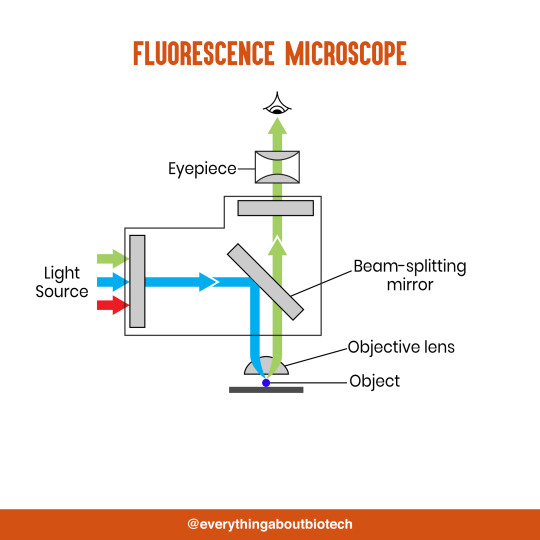
Fluorescence Microscopy: Imagine illuminating a sample with specific wavelengths of light and observing it glowing in response. That's the essence of fluorescence microscopy. This technique utilizes fluorescent molecules or tags that bind to specific structures within a cell or tissue. When excited by light, these tags emit their own light, highlighting the target structures with remarkable clarity. This allows researchers to visualize specific proteins, DNA, or even pathogens within biological samples.
Super-resolution Microscopy (SRM): Overcoming the limitations imposed by the wavelength of light, SRM techniques like STED (Stimulated Emission Depletion) and PALM (Photoactivated Localization Microscopy) achieve resolutions surpassing the diffraction limit. This allows researchers to visualize structures as small as 20 nanometers, enabling the observation of intricate cellular machinery and the dynamics of individual molecules within living cells.
Cryo-Electron Microscopy (Cryo-EM): This powerful technique takes a snapshot of biological samples in their near-life state. Samples are rapidly frozen at ultra-low temperatures, preserving their native structure and minimizing damage caused by traditional fixation methods. Cryo-EM has been instrumental in determining the three-dimensional structures of complex molecules like proteins and viruses, providing crucial insights into their function and potential drug targets.
Correlative Microscopy: Combining the strengths of multiple microscopy techniques, correlative microscopy offers a comprehensive view of biological samples. For instance, researchers can utilize fluorescence microscopy to identify specific structures within a cell and then switch to electron microscopy to examine those structures in high detail. This integrated approach provides a deeper understanding of cellular processes and their underlying mechanisms.
Light Sheet Microscopy (LSM): Imagine illuminating a thin slice of a sample within a living organism. LSM achieves this feat by focusing a laser beam into a thin sheet of light, minimizing photobleaching and phototoxicity – damaging effects caused by prolonged exposure to light. This allows researchers to observe dynamic processes within living organisms over extended periods, providing valuable insights into cellular behavior and development.
Expansion Microscopy (ExM): This innovative technique physically expands biological samples by several folds while preserving their structural integrity. This expansion allows for better resolution and visualization of intricate cellular structures that would otherwise be difficult to distinguish using traditional microscopy methods. ExM holds immense potential for studying the organization and function of organelles within cells.
Scanning Near-Field Optical Microscopy (SNOM): This innovative technique pushes the boundaries of resolution by utilizing a tiny probe that interacts with the sample at an extremely close range. SNOM can not only image the surface features of a sample with exceptional detail but also probe its optical properties at the nanoscale. This opens doors for research in areas like material science and photonics, allowing scientists to study the behavior of light at the interface between materials.
X-ray Microscopy: Stepping outside the realm of light and electrons, X-ray microscopy offers unique capabilities. By utilizing high-energy X-rays, this technique can penetrate deep into samples, making it ideal for studying the internal structure of dense materials like bones and minerals. Additionally, it allows for the visualization of elements within a sample, providing valuable information about their distribution and composition.
From revealing the building blocks of life to aiding in the development of new medicines, the microscope has played an undeniable role in shaping our scientific understanding. As technology continues to evolve, one can only imagine the future breakthroughs this remarkable invention holds in unveiling the secrets of our universe, both seen and unseen. These advancements hold the potential to revolutionize our understanding of biological processes, develop new materials with extraordinary properties, and ultimately pave the way for breakthroughs in medicine, nanotechnology, and countless other fields. As we continue to refine and develop novel microscopy techniques and the future holds immense promise for further groundbreaking discoveries that will undoubtedly revolutionize our perception of the world around us.
#science sculpt#life science#science#molecular biology#biology#biotechnology#artists on tumblr#microscopy#microscope#Scanning Electron Microscope#Transmission Electron Microscope#Confocal Microscopy#Atomic Force Microscopy#Fluorescence Microscopy#Expansion Microscopy#X-ray Microscopy#Super-resolution Microscopy#Light Sheet Microscopy#illustration#illustrator#illustrative art#education#educate yourself#techniques in biotechnology#scientific research#the glass scientists#scientific illustration#scientific advancements
6 notes
·
View notes
Text
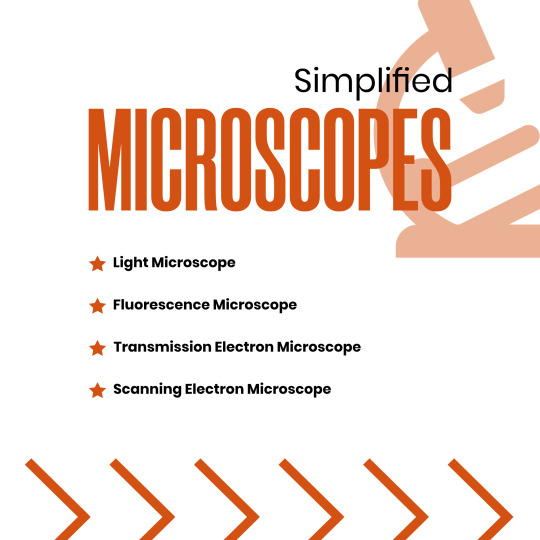


Microscopes Simplified
Light Microscope
Fluorescence Microscope
Transmission Electron Microscope
Scanning Electron Microscope
#microscope#light microscope#Fluorescence Microscope#Transmission Electron Microscope#Scanning Electron Microscope#microscopic#science#microscopy#microscopic life#biotechnology#biotech#biology#biochemistry#studyblr#molecularbiology#notes#class notes#students#science illustration#illustration#illustragram#scientific illustration#graphic design#graphicwork#study notes#study#study motivation#study tips#studygram
82 notes
·
View notes
Text
Uses of Different Types of Microscopes in Forensics
Uses of Different Types of Microscopes in Forensics
The forensic and microbiological labs include a variety of microscopes that may be used. The value of microscopes is increased by how widely they may be used and modified.
#microscope #forensicscience
(more…)

View On WordPress
#comparison microscope#comparison microscope in Forensic Science#dark-field microscope in forensic science#Fluorescent microscope#Forensic science#forensic science notes#microscopy notes#polarising microscope#Scanning Electron Microscope#stereoscopic microscope#stereoscopic microscope uses in forensic science#Transmission Electron Microscope#use of compound microscope in forensic science#use of digital microscope in forensic science#use of fluorescent microscope in forensic science#use of inverted microscope in forensic science#use of phase contrast microscopy in forensic science#use of polarising microscope in forensic science#use of scanning electron microscope#use of scanning electron microscope in forensic science#Uses of Different types of Microscope In Forensics
7 notes
·
View notes
Text
In an effort to understand how and why 2D interfaces take on the structures they do, researchers at the University of Illinois Urbana-Champaign have developed a method to visualize the thermally-induced rearrangement of 2D materials, atom-by-atom, from twisted to aligned structures using transmission electron microscopy (TEM). They observed a new and unexpected mechanism for this process where a new grain was seeded within one monolayer, whose structure was templated by the adjacent layer. Being able to control the macroscopic twist between layers allows for more control over the properties of the entire system.
17 notes
·
View notes
Text
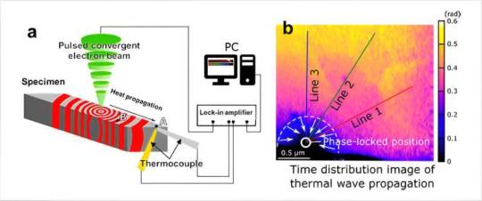
New electron microscopy technique for thermal diffusion measurements
A NIMS research team has developed a technique that enables the nanoscale observation of heat propagation paths and behavior within material specimens. This was achieved using a scanning transmission electron microscope (STEM) capable of emitting a pulsed electron beam and a nanosized thermocouple—a high-precision temperature measurement device developed by NIMS. The research is published in Science Advances.
Public interest in energy conservation and recycling has grown considerably in recent years. This change has inspired scientists to develop next-generation materials/devices capable of controlling and utilizing heat with a high degree of precision, including thermoelectric devices able to convert waste heat into electricity and heat dissipation composites that can cool electronic components exposed to high temperatures.
Read more.
#Materials Science#Science#Electron microscopy#Diffusion#Temperature#Heat flow#Thermoelectric#Waste heat
19 notes
·
View notes
Text
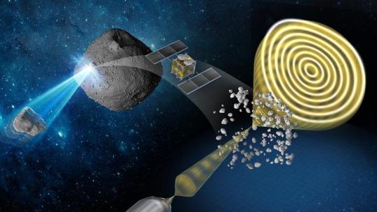
The effects of interplanetary space on asteroid Ryugu
Samples reveal evidence of changes experienced by the surface of asteroid Ryugu, some probably due to micrometeoroid bombardment.
Analyzing samples retrieved from the asteroid Ryugu by the Japanese Space Agency’s Hayabusa2 spacecraft has revealed new insights into the magnetic and physical bombardment environment of interplanetary space. The results of the study, carried out by Professor Yuki Kimura at Hokkaido University and co-workers at 13 other institutions in Japan, are published in the journal Nature Communications.
The investigations used electron waves penetrating the samples to reveal details of their structure and magnetic and electric properties, a technique called electron holography.
Hayabusa2 reached asteroid Ryugu on 27 June 2018, collected samples during two delicate touchdowns, and then returned the jettisoned samples to Earth in December 2020. The spacecraft is now continuing its journey through space, with plans for it to observe two other asteroids in 2029 and 2031.
One advantage of collecting samples directly from an asteroid is that it allows researchers to examine long-term effects of its exposure to the environment of space. The ‘solar wind’ of high energy particles from the sun and bombardment by micrometeoroids cause changes known as space-weathering. It is impossible to study these changes precisely using most of the meteorite samples that land naturally on Earth, partly due to their origin from the internal parts of an asteroid, and also due to the effects of their fiery descent through the atmosphere.
“The signatures of space weathering we have detected directly will give us a better understanding of some of the phenomena occurring in the Solar System,” says Kimura. He explains that the strength of the magnetic field in the early solar system decreased as planets formed, and measuring the remnant magnetization on asteroids can reveal information about the magnetic field in the very early stages of the solar system.
Kimura adds, “In future work, our results could also help to reveal the relative ages of surfaces on airless bodies and assist in the accurate interpretation of remote sensing data obtained from these bodies.”
One particularly interesting finding was that small mineral grains called framboids, composed of magnetite, a form of iron oxide, had completely lost their normal magnetic properties. The researchers suggest this was due to collision with high velocity micrometeoroids between 2 and 20 micrometers in diameter. The framboids were surrounded by thousands of metallic iron nanoparticles. Future studies of these nanoparticles will hopefully reveal insights into the magnetic field that the asteroid has experienced over long periods of time.
“Although our study is primarily for fundamental scientific interest and understanding, it could also help estimate the degree of degradation likely to be caused by space dust impacting robotic or manned spacecraft at high velocity,” Kimura concludes.
TOP IMAGE....Conceptual illustration of the study Credit Yuki Kimura
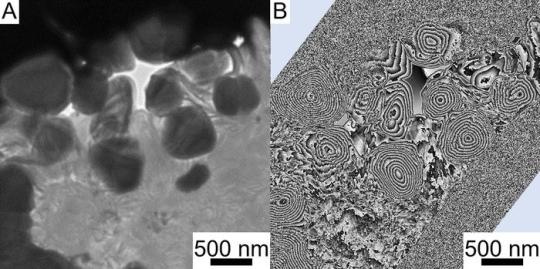
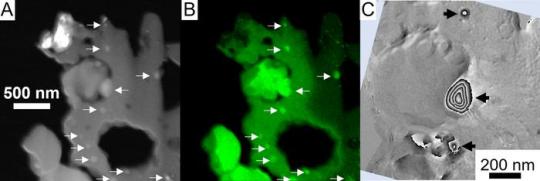
CENTRE IMAGE....Magnetite (round particles) particles cut from a Ryugu sample. (A) Bright field transmission electron microscopy image. (B) Magnetic flux distribution image obtained by electron holography. The concentric circular stripes seen inside the particles correspond to magnetic lines of force. They are called vortex magnetic domain structures and are more stable than ordinary hard disks, which can record magnetic fields for more than 4.6 billion years. (Yuki Kimura, et al. Nature Communications. April 29, 2024) Credit Yuki Kimura, et al. Nature Communications. April 29, 2024
LOWER IMAGE....Iron nanoparticles distributed around pseudo-magnetite. (A) Dark-field image taken with a scanning transmission electron microscope. (B) Corresponding iron distribution image. White arrows indicate iron nanoparticles. (C) Magnetic flux distribution image of the central region of A and B. No magnetic field lines can be seen in the pseudo-magnetite, whereas concentric vortex-like magnetic domain structures can be seen inside the iron particles as shown by black arrows. (Yuki Kimura, et al. Nature Communications. April 29, 2024) Credit Yuki Kimura, et al. Nature Communications. April 29, 2024
6 notes
·
View notes
Text
women in scanning and transmission electron microscopy
9 notes
·
View notes
Text
The data collected at Diamond contributed to a wider study of the space weathering signatures on the asteroid.
The Scientists did a chemical analysis of the astroid to see the elements and the element isotype it is made of with ZANES X-ray device. Then looked through an electron microscope ePSIC to see the grain of rock on the asteroid.
The building blocks of Ryugu are remnants of interactions between water, minerals, and organics in the early Solar System prior to the formation of Earth.
X-ray Absorption Near Edge Spectroscopy (XANES).
Diamond’s electron Physical Science Imaging Centre (ePSIC)
Without a protective atmosphere, space-exposed surfaces of airless Solar System bodies gradually experience an alteration in composition, structure and optical properties through Space Weathering
Solar wind irradiation and high-velocity micrometeoroid bombardment dominate space weathering for all airless bodies.
The mineralogy of most Ryugu grains investigated by (scanning) transmission electron microscopy is similar to that of CI chondrites, which are the most chemically primitive materials in the Solar System
#astronomy#asteroid#space weathering#science#science news#scientific research#space#outer space#solar system
4 notes
·
View notes
Photo
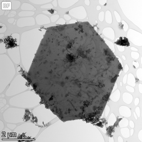
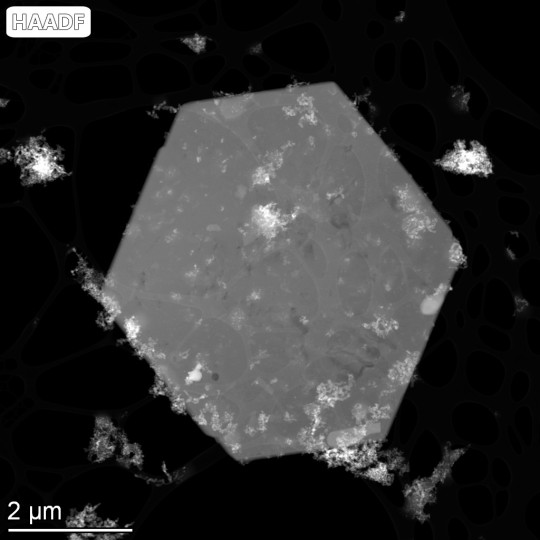
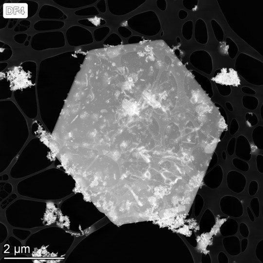

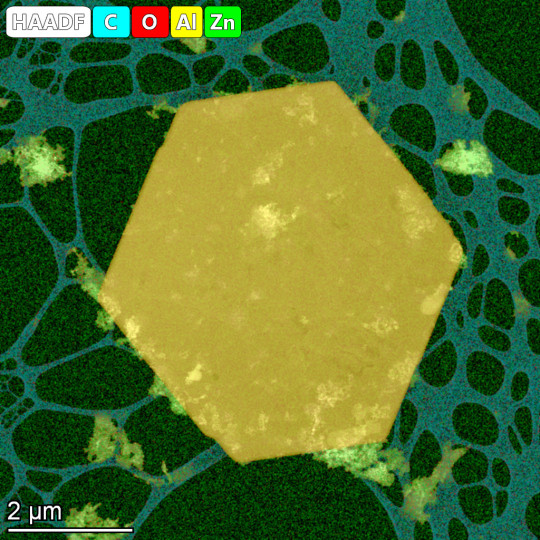


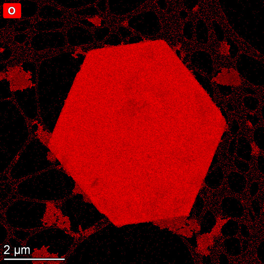
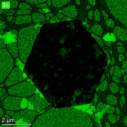
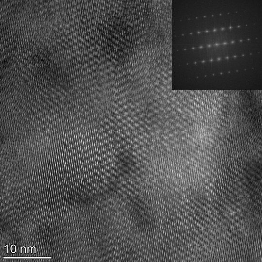
#electron microscopy#transmission electron microscopy#scanning transmission electron microscopy#lattice imaging#Fourier transform#energy dispersive x-ray spectroscopy#bright field image#dark field image#high angle annular dark field#original content
9 notes
·
View notes
Text
Scanning & Transmission Electron Microscopy Reveals Graphene & Parasites in CoV-19 Vaccines[88]
Scanning & Transmission Electron Microscopy Reveals Graphene & Parasites in CoV-19 Vaccines[88]
— Read on www.drrobertyoung.com/post/transmission-electron-microscopy-reveals-graphene-oxide-in-cov-19-vaccines
View On WordPress
0 notes
Text
Types of Microscopes
1. Simple Microscope
2. Compound Microscope
3. Phase Contrast Microscope
4. Fluorescence Microscope
5. Electron Microscope
6. Scanning Electron Microscope (SEM)
7. Transmission Electron Microscope (TEM)
8. Dark Field Microscope
9. Dissecting Microscope (Stereo Microscope)
10. Digital Microscope
11. Scanning Probe Microscope (SPM)
12. Atomic Force Microscope (ATM)
13. Inverted Microscope
14. Acoustic Microscope
15. X-Ray Microscope
16. Polarizing Microscope
17. Metallurgical Microscope
18. Pocket Microscope
19. USB Microscope
20. Confocal Microscope
21. Laser Scanning Microscope
22. Differential Interference Contrast Microscope (DIC)
23. Near-field Scanning Optical Microscope (NSOM)
24. Raman Microscope
25. Super-resolution Microscope
26. Cryo-electron Microscope
27. Time-lapse Microscope
There is a wide range of microscopy techniques and instruments used in various fields of science and research.
#forensic#forensics#criminology#forensic science#evidence#criminalistic#forensic field#crime#forensic science notes#crime scene investigation#electron microscope#microscope
6 notes
·
View notes
Text


Materials Characterization: Transmission electron microscopy
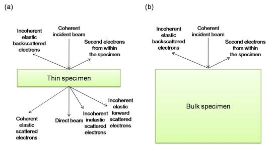
As with other forms of electron microscopy, transmission electron microscopy (TEM) uses a beam of electrons to 'illuminate' the specimen of interest and provide information and images. As the name would suggest, the electrons used in this technique are transmitted through the sample, requiring ultrathin specimens (typically less than 100nm in thickness). The thinner the specimen, the clearer the resulting images. The resolution of TEM can be less than a nanometer, though the specific machine resolution can vary based on a number of factors.
There are several techniques to produce TEM samples. For solid, bulk inorganic specimens, historically, mechanical polishing, chemical etching, and electropolishing have been used, though these techniques are time consuming and cannot easily be used to extract TEM foils from specific locations. These days, focused ion beam milling is often used to extract foils from targeted areas, as shown in image 2 above.
In addition to images such as the one shown in image 4 above, TEM can also produce diffraction patterns (as in image 3) which can be used to determine the site-specific crystal structure, as well as its orientation. TEM can also provide chemical information, including, if the resolution is high enough, on an atom-by-atom basis.
Many variations of the technique exist, including scanning transmission electron microscopy (STEM), cryo-TEM, aberration corrected TEM (more common in modern instruments), and the ability to conduct various in-situ/environmental experiments during imaging. While TEM can be used to collect extremely detailed small scale information about a sample, it should be noted that the size of the specimens mean that, particularly for bulk specimens, results may not be representative of the sample overall.
Sources/Further reading: ( 1 - images 1, 4, 5 ) ( 2 - images 2 and 3 ) ( 3 ) ( 4 ) ( 5 )
#Materials Science#Science#Transmission electron microscopy#Electron microscopy#Materials Characterization#MyMSEPost
34 notes
·
View notes
Text
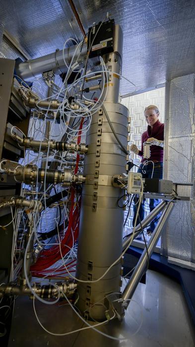
Hydrogen detected in lunar samples, points to resource availability for space exploration
U.S. Naval Research Laboratory (NRL) researchers have discovered solar-wind hydrogen in lunar samples, which indicates that water on the surface of the Moon may provide a vital resource for future lunar bases and longer-range space exploration. Space-based resource identification is a key factor in planning for civilian- and government-led space exploration.
“Hydrogen has the potential to be a resource that can be used directly on the lunar surface when there are more regular or permanent installations there,” said Dr. Katherine D. Burgess, geologist in NRL’s Materials Science and Technology Division. “Locating resources and understanding how to collect them prior to getting to the Moon is going to be incredibly valuable for space exploration.”
The Apollo lunar soil samples were provided by a NASA-funded research mission to NRL scientists for investigation and testing. The research team, led by scientists in NRL’s Materials Science and Technology Division, continues to study lunar surface and asteroidal samples to gain understanding of how surfaces interact with the space environment, which is known as space weathering. Previous testing from additional Apollo samples confirmed location of solar wind helium in lunar soil grains.
“This is the first time scientists have demonstrated detection of hydrogen-bearing species within vesicles in lunar samples,” said Dr. Burgess. “Previously, the same team at NRL used state-of-the-art techniques such as scanning transmission electron microscopy and electron energy loss spectroscopy to detect helium in lunar samples, and other researchers have found water in other planetary samples, but this is the first publication to show hydrogen in-situ in lunar samples.”
The research article was published to the Communications Earth & Environment journal on Wednesday, Nov. 15, 2023.
About the U.S. Naval Research Laboratory
NRL is a scientific and engineering command dedicated to research that drives innovative advances for the U.S. Navy and the U.S. Marine Corps from the seafloor to space and in the information domain. NRL is located in Washington, D.C., with major field sites in Stennis Space Center, Mississippi, Key West, Florida, and Monterey, California, and employs approximately 3,000 civilian scientists, engineers and support personnel.
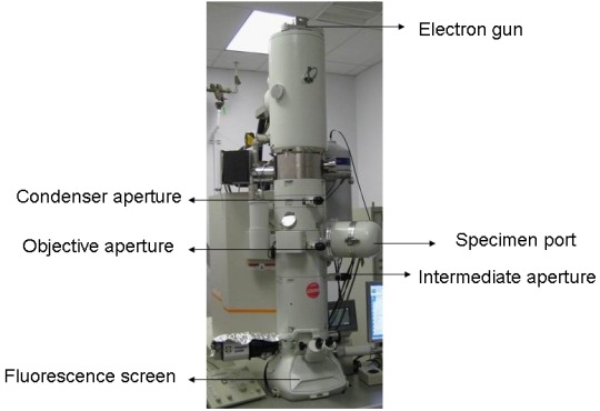
IMAGE....Katherine Burgess, U.S. Naval Research Laboratory geologist, places a magazine holding several transmission electron microscopy samples into a Nion UltraSTEM-200x microscope in Washington, D.C., November 16, 2023. Burgess studies lunar grains that were collected during Apollo missions to the moon. (U.S. Navy photo by Jonathan Steffen) Credit Jonathan Steffen/U.S. Naval Research Laboratory
2 notes
·
View notes
Text

What Are The Applications Of Vacuum Pumps In Experimental Research?
(1)Sample preparation: Vacuum pumps are commonly used to remove air and other gases from samples, such as in vacuum drying or freeze drying, to improve sample quality and stability.
(2)Thin film deposition: Vacuum pumps are used to create and maintain a vacuum environment for thin film deposition techniques, such as physical vapor deposition (PVD) and chemical vapor deposition (CVD).
(3)Mass spectrometry: Vacuum pumps are used to create and maintain the high vacuum conditions necessary for mass spectrometry analysis.
(4)Electron microscopy: Vacuum pumps are used to create and maintain the high vacuum conditions necessary for electron microscopy techniques, such as scanning electron microscopy (SEM) and transmission electron microscopy (TEM).
(5)Nuclear magnetic resonance (NMR) spectroscopy: Vacuum pumps are used to create and maintain the high vacuum conditions necessary for NMR spectroscopy analysis.
(6)Accelerator science: Vacuum pumps are used in particle accelerators to create and maintain the high vacuum conditions necessary for the operation of the accelerator.
(7)Space simulation: Vacuum pumps are used in space simulation chambers to create and maintain the vacuum environment necessary for testing spacecraft components and systems.
0 notes
Quote
The sky is the limit with regards to the societal impact nanomaterials can have on the lives. However, in this study, it is shown that their potential is out of this world. The planet Mars has an abundant source of calcium sulfate minerals and in this work, it is shown that these deposits can be the basis of transformative nanomaterials to potentially support future space endeavors. Vitally, the methods applied are low cost and require no specialized instruments of great expertise, strengthening the potential involvement of nanotechnology in sustaining Martian inhabitation. Through a scalable eco-friendly liquid processing technique performed on two common terrestrial gypsum, this simple method presented a cost-efficient procedure to yield suspensions of large aspect ratio anhydrite nanobelts with long-term stability that are characterized through scanning electron microscopy and Raman spectroscopy. Transmission electron microscopy shows nanobelts to have a mesocrystal structure, with distinct nanoparticle constituents making up the lattice. Unexpectedly, anhydrite nanobelts have remarkable electronic properties, namely a bandgap that is easily tuned between semiconducting (≈2.2 eV) and insulating (≈4 eV) behaviors through dimensional control measured via atomic force microscopy. To demonstrate the application potential of the nanobelts; optoelectronic, electrochemical, and nanocomposite measurements are made.
Quasi–1D Anhydrite Nanobelts from the Sustainable Liquid Exfoliation of Terrestrial Gypsum for Future Martian‐Based Electronics - Wei - Advanced Functional Materials - Wiley Online Library
0 notes
Last Seen Blogs

ptilou
BOYS BOYS BOYS

p-aradichlorobenzene
Your Adventure Log Has Vanished!!!
ptilou
BOYS BOYS BOYS

minka1
Untitled
minka1
Untitled