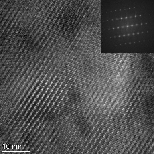
#scanning electron microscopy
Text
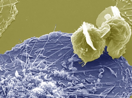
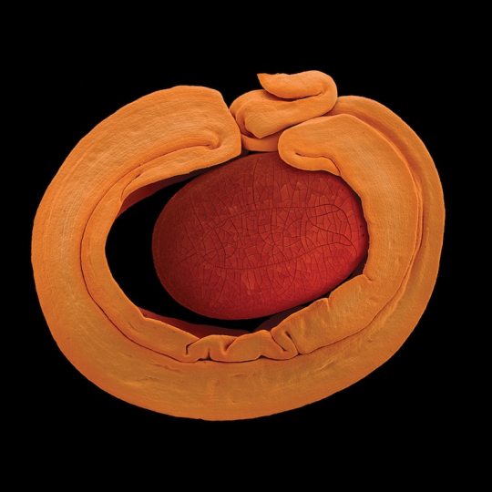
Infection by Fusion
Fusing HIV-infected CD4 T cells with macrophages – cells of the immune system that are a reservoir for HIV in tissue – infects them effectively, providing insight into the mechanisms of persistent infection
Read the published research paper here
Image from work by Rémi Mascarau and colleagues
Institut de Pharmacologie et Biologie Structurale (IPBS), Université de Toulouse, Centre National de la Recherche Scientifique, Université Toulouse III - Paul Sabatier (UPS), Toulouse, France
Image originally published with a Creative Commons Attribution 4.0 International (CC BY 4.0)
Published in Journal of Cell Biology, March 2023
You can also follow BPoD on Instagram, Twitter and Facebook
#science#biomedicine#biology#hiv#immune system#macrophages#t cells#cd4#electron microscopy#scanning electron microscopy#viral infections
7 notes
·
View notes
Photo
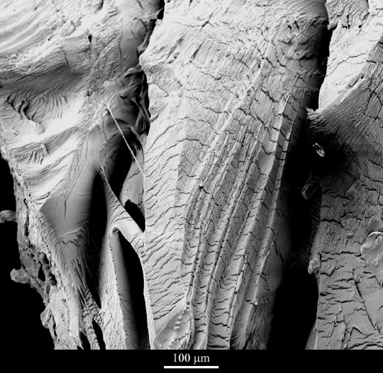
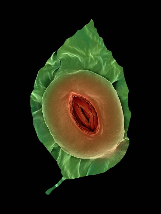
Ductile tearing near a fracture surface in polycarbonate
Applications
Polycarbonate is a clear and relatively tough plastic used to make shatterproof windows, lenses and even helmets. It is also used to make compact discs.
Sample preparation
To induce crazing and fracture in polycarbonate, acetone is used. This results in the otherwise tough material becoming very brittle
Technique
Scanning electron microscopy (SEM)
Length bar
100 μm
Further information
The fracture surface is dominated by ductile tearing, and also shows linear voids with fine transverse filaments. These are the remnants of crazes which act both as a precursor to cracking and as a toughening mechanism. In some areas, more angular brittle fracture surfaces are also apparent. The fine-scale ridging of the surface, parallel to the principal stress direction (which is approximately vertical) morphology is the result of fibrous crystals being induced by cold-drawing.
Contributor
J A Curran
Organisation
Department of Materials Science and Metallurgy, University of Cambridge
Source.
#Materials Science#Science#Polycarbonate#Materials failure#Polymers#Scanning electron microscopy#Magnified view#University of Cambridge#DoITPoMS
23 notes
·
View notes
Text
A Journey into the World of Microscopy: From Humble Beginnings to High-Tech Magnification
The science of looking into the hidden invisible Microscopy has transformed our understanding of the world around us. It can explore the universe beyond the reach of our naked eyes, with complex cellular structures, red blood cells, viruses and other viruses and microorganisms taking on amazing perspectives
The history of the microscope is a fascinating story of human curiosity, scientific genius, and relentless exploration. From the humble beginnings of simple magnifying glasses to the sophistication of modern electronic microscopes, the invention of microscopes has shaped our understanding of the microscopic world
In the 1600s, Dutch opticians such as Hans and Zachary Janssen are credited with inventing the first microscope. Known for this hybrid microscope, many lenses were used to magnify objects up to 30 times.At the end of the 17th century, Antony van Leeuwenhoek, Dutch draper some changed our perception of thumbnails. Armed with a well-made single-lens microscope, and explored the hidden reaches of nature. In 1674, Leeuwenhoek discovered microorganisms in lake water, which he aptly named “animalcules”. His discovery laid the foundations of biology and inspired generations of scientists. This incredible feat allowed him to uncover a hidden universe – the first sightings of bacteria, red blood cells, and other microorganisms.
Formation of the scientific environment (17th-19th centuries): Leeuwenhoek’s discoveries boosted scientific research. Robert Hooke, an English scientist, established these developments. In 1665, his book "Micrographia" recorded his observations with a compound microscope. Notably, the term "cell" was coined by Hooke when he examined cork tissue, laying the foundation for cell biology.Microscope systems flourished throughout the 18th and 19th centuries Joseph Lister and other scientists addressed the limitations of the early lenses, introducing improvements that reduced image distortion.
Beyond the Limits of Light: The Beginning of the New Age (19th-20th century): As the 19th century progressed, the limitations of optical microscopy became apparent and scientists yearned for a tool which can go deeper into cells. This research culminated in the development of the electron microscope in the 1930s. The 20th century was revolutionary with the invention of the electron microscope. Unlike light microscopes, which use visible light, electron microscopes use electron beams to achieve much higher magnification.Formation of the scientific environment (17th-19th centuries): Leeuwenhoek’s discoveries boosted scientific research. Robert Hooke, an English scientist, established these developments. In 1665, his book "Micrographia" recorded his observations with a compound microscope. Notably, the term "cell" was coined by Hooke when he examined cork tissue, laying the foundation for cell biology.Microscope systems flourished throughout the 18th and 19th centuries Joseph Lister and other scientists addressed the limitations of the early lenses, introducing improvements that reduced image distortion.
Beyond the Limits of Light: The Beginning of the New Age (19th-20th century): As the 19th century progressed, the limitations of optical microscopy became apparent and scientists yearned for a tool which can go deeper into cells. This research culminated in the development of the electron microscope in the 1930s. The 20th century was revolutionary with the invention of the electron microscope. Unlike light microscopes, which use visible light, electron microscopes use electron beams to achieve much higher magnification.
In the 1930s, German experts Max Knoll and Ernst Ruska made the first electron microscope. This tool let us see tiny things like cells and even atoms by using electron beams, not light, getting images many times bigger. This cool invention showed us the tiny parts inside cells, viruses, and stuff too small to see before. The 1900s brought even more cool microscopes. New kinds like phase-contrast and confocal microscopy let scientists look at live cells without using stuff that could hurt them. Now, the world of looking at tiny things is getting even better. Today, we have high-tech microscopes that use computers and lasers. These let us see and even change tiny things in ways we never could before.
Modern Microscopy's Diverse Arsenal - Today, the field of microscopy boasts a diverse range of specialized instruments, each tailored to address specific scientific needs. Here's a glimpse into some remarkable examples:
Scanning Electron Microscope (SEM): Imagine a high-tech camera that captures images using a beam of electrons instead of light. That's the essence of a SEM. By scanning the surface of a sample with a focused electron beam, SEMs generate detailed information about its topography and composition. This makes them ideal for studying the intricate structures of materials like insect wings, microchips, and even pollen grains.
Transmission Electron Microscope (TEM): While SEMs provide exceptional surface detail, TEMs take us a step further. They function by transmitting a beam of electrons through a very thin sample, allowing us to observe its internal structure. TEMs are the go-to instruments for visualizing the intricate world of viruses, organelles within cells, and macromolecules like proteins.
Confocal Microscopy: Ever wished to focus on a specific layer within a thick biological sample and blur out the rest? Confocal microscopy makes this possible. It utilizes a laser beam to precisely illuminate a chosen plane within the sample, effectively eliminating information from out-of-focus regions. This allows researchers to create sharp, three-dimensional images of cells, tissues, and even small organisms.
Atomic Force Microscopy (AFM): This technique takes a completely different approach, venturing into the realm of physical interaction. AFM employs a tiny cantilever, akin to a microscopic feeler, to physically scan the surface of a sample. By measuring the minute forces between the cantilever and the sample's surface, AFM can map its topography at an atomic level. This provides invaluable insights into the properties of materials at an unimaginable scale, making it crucial for research in fields like nanotechnology and surface science.
Fluorescence Microscopy: Imagine illuminating a sample with specific wavelengths of light and observing it glowing in response. That's the essence of fluorescence microscopy. This technique utilizes fluorescent molecules or tags that bind to specific structures within a cell or tissue. When excited by light, these tags emit their own light, highlighting the target structures with remarkable clarity. This allows researchers to visualize specific proteins, DNA, or even pathogens within biological samples.
Super-resolution Microscopy (SRM): Overcoming the limitations imposed by the wavelength of light, SRM techniques like STED (Stimulated Emission Depletion) and PALM (Photoactivated Localization Microscopy) achieve resolutions surpassing the diffraction limit. This allows researchers to visualize structures as small as 20 nanometers, enabling the observation of intricate cellular machinery and the dynamics of individual molecules within living cells.
Cryo-Electron Microscopy (Cryo-EM): This powerful technique takes a snapshot of biological samples in their near-life state. Samples are rapidly frozen at ultra-low temperatures, preserving their native structure and minimizing damage caused by traditional fixation methods. Cryo-EM has been instrumental in determining the three-dimensional structures of complex molecules like proteins and viruses, providing crucial insights into their function and potential drug targets.
Correlative Microscopy: Combining the strengths of multiple microscopy techniques, correlative microscopy offers a comprehensive view of biological samples. For instance, researchers can utilize fluorescence microscopy to identify specific structures within a cell and then switch to electron microscopy to examine those structures in high detail. This integrated approach provides a deeper understanding of cellular processes and their underlying mechanisms.
Light Sheet Microscopy (LSM): Imagine illuminating a thin slice of a sample within a living organism. LSM achieves this feat by focusing a laser beam into a thin sheet of light, minimizing photobleaching and phototoxicity – damaging effects caused by prolonged exposure to light. This allows researchers to observe dynamic processes within living organisms over extended periods, providing valuable insights into cellular behavior and development.
Expansion Microscopy (ExM): This innovative technique physically expands biological samples by several folds while preserving their structural integrity. This expansion allows for better resolution and visualization of intricate cellular structures that would otherwise be difficult to distinguish using traditional microscopy methods. ExM holds immense potential for studying the organization and function of organelles within cells.
Scanning Near-Field Optical Microscopy (SNOM): This innovative technique pushes the boundaries of resolution by utilizing a tiny probe that interacts with the sample at an extremely close range. SNOM can not only image the surface features of a sample with exceptional detail but also probe its optical properties at the nanoscale. This opens doors for research in areas like material science and photonics, allowing scientists to study the behavior of light at the interface between materials.
X-ray Microscopy: Stepping outside the realm of light and electrons, X-ray microscopy offers unique capabilities. By utilizing high-energy X-rays, this technique can penetrate deep into samples, making it ideal for studying the internal structure of dense materials like bones and minerals. Additionally, it allows for the visualization of elements within a sample, providing valuable information about their distribution and composition.
From revealing the building blocks of life to aiding in the development of new medicines, the microscope has played an undeniable role in shaping our scientific understanding. As technology continues to evolve, one can only imagine the future breakthroughs this remarkable invention holds in unveiling the secrets of our universe, both seen and unseen. These advancements hold the potential to revolutionize our understanding of biological processes, develop new materials with extraordinary properties, and ultimately pave the way for breakthroughs in medicine, nanotechnology, and countless other fields. As we continue to refine and develop novel microscopy techniques and the future holds immense promise for further groundbreaking discoveries that will undoubtedly revolutionize our perception of the world around us.
#science sculpt#life science#science#molecular biology#biology#biotechnology#artists on tumblr#microscopy#microscope#Scanning Electron Microscope#Transmission Electron Microscope#Confocal Microscopy#Atomic Force Microscopy#Fluorescence Microscopy#Expansion Microscopy#X-ray Microscopy#Super-resolution Microscopy#Light Sheet Microscopy#illustration#illustrator#illustrative art#education#educate yourself#techniques in biotechnology#scientific research#the glass scientists#scientific illustration#scientific advancements
6 notes
·
View notes
Photo

Scanning electron micrograph of a greenfly eye, by Kevin Mackenzie, University of Aberdeen by ZEISS Microscopy https://flic.kr/p/rv9NR2
12 notes
·
View notes
Text
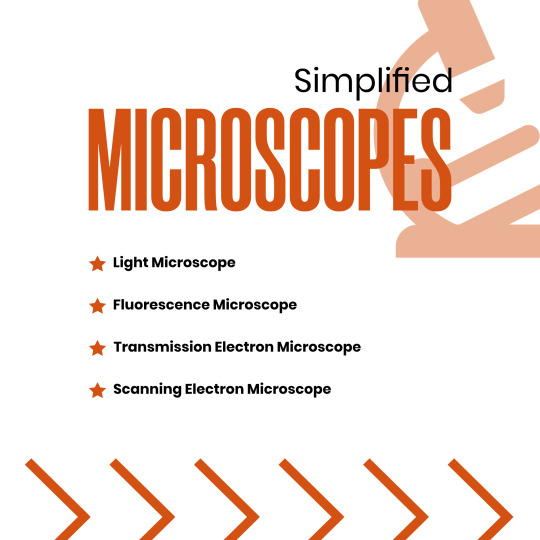

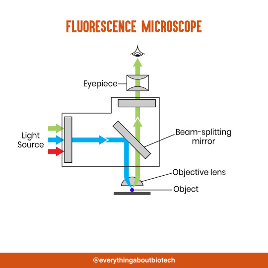


Microscopes Simplified
Light Microscope
Fluorescence Microscope
Transmission Electron Microscope
Scanning Electron Microscope
#microscope#light microscope#Fluorescence Microscope#Transmission Electron Microscope#Scanning Electron Microscope#microscopic#science#microscopy#microscopic life#biotechnology#biotech#biology#biochemistry#studyblr#molecularbiology#notes#class notes#students#science illustration#illustration#illustragram#scientific illustration#graphic design#graphicwork#study notes#study#study motivation#study tips#studygram
82 notes
·
View notes
Text
An example of induced aerenchyma occurs in maize (corn; Zea mays) (Figure 24.19).
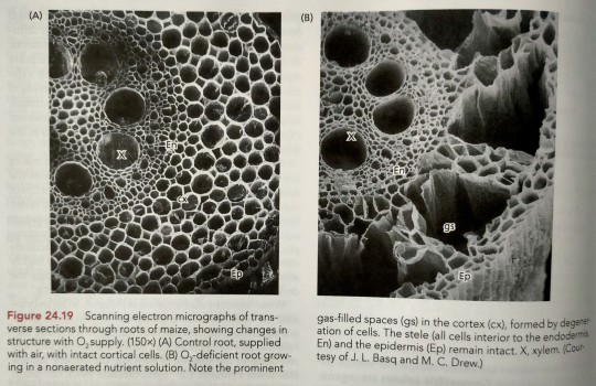
"Plant Physiology and Development" int'l 6e - Taiz, L., Zeiger, E., Møller, I.M., Murphy, A.
#book quotes#plant physiology and development#nonfiction#textbook#corn#maize#zea mays#trypophobia#scanning electron micrograph#microscopy#aerenchyma#plant cells
8 notes
·
View notes
Photo
#transmission electron microscopy#bright field#dark field#HRTEM#Fourier transform#nickel alloy#scanning transmission electron microscopy#original content
5 notes
·
View notes
Text
Uses of Different Types of Microscopes in Forensics
Uses of Different Types of Microscopes in Forensics
The forensic and microbiological labs include a variety of microscopes that may be used. The value of microscopes is increased by how widely they may be used and modified.
#microscope #forensicscience
(more…)

View On WordPress
#comparison microscope#comparison microscope in Forensic Science#dark-field microscope in forensic science#Fluorescent microscope#Forensic science#forensic science notes#microscopy notes#polarising microscope#Scanning Electron Microscope#stereoscopic microscope#stereoscopic microscope uses in forensic science#Transmission Electron Microscope#use of compound microscope in forensic science#use of digital microscope in forensic science#use of fluorescent microscope in forensic science#use of inverted microscope in forensic science#use of phase contrast microscopy in forensic science#use of polarising microscope in forensic science#use of scanning electron microscope#use of scanning electron microscope in forensic science#Uses of Different types of Microscope In Forensics
7 notes
·
View notes
Text
youtube
This video shows how a Scanning Electron Microscope works? And how to use a virtual scanning electron microscope to acquire a good image. It is a great learning and teaching tool. You can access the virtual TEM at https://myscope-explore.org/virtualSE... developed by Microscopy Australia and Thermofisher Scientific.
#scanning#electronmicroscope#electronmicroscopy#electronbeam#image#images#nano#lotus#butterfly#lens#electrons#education#digitalliteracy#onlinelearning#microscope#microscopy#teaching#fiber#forensics#hair#arthropods#wool#superhydrophobic#waterrepellent#e-learning#virtuallearning#digitaltools#digitaleducation#openeducationalresources#openeducation
1 note
·
View note
Photo
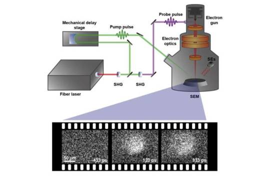
With scanning ultrafast electron microscopy, researchers unveil hot photocarrier transport properties of cubic boron
In a study that confirms its promise as the next-generation semiconductor material, UC Santa Barbara researchers have directly visualized the photocarrier transport properties of cubic boron arsenide single crystals.
"We were able to visualize how the charge moves in our sample," said Bolin Liao, an assistant professor of mechanical engineering in the College of Engineering. Using the only scanning ultrafast electron microscopy (SUEM) setup in operation at a U.S. university, he and his team were able to make "movies" of the generation and transport processes of a photoexcited charge in this relatively little-studied III-V semiconductor material, which has recently been recognized as having extraordinary electrical and thermal properties. In the process, they found another beneficial property that adds to the material's potential as the next great semiconductor.
Their research, conducted in collaboration with physics professor Zhifeng Ren's group at the University of Houston, who specialize in fabricating high-quality single crystals of cubic boron arsenide, appears in the journal Matter.
Read more.
#Materials Science#Science#Electron microscopy#Boron arsenide#Boron#Arsenic#Scanning electron microscopy#Semiconductors#UC Santa Barbara
9 notes
·
View notes
Text
Met a mad scientist today.
#my post#he was wearing a hawaiian shirt under his lab coat and spoke about scanning electron microscopy#with the most passion i have witnessed in my lfe#it was amazing#also i looked at some unspeakably small things today and i don't know what i'm feeling but it surely is Something
0 notes
Text

Archaeologists Uncover a Bronze Belt Fitting From an Unknown Pagan Cult
team of archaeologists from Masaryk University have uncovered a bronze belt fitting from an unknown pagan cult in the village of Lány, located in the Central Bohemian Region of the Czech Republic.
The belt fitting dates from the 8th century AD and depicts a snake devouring a frog-like creature that appears in Germanic, Avar, and Slavic mythology.
Such representations are related to the cosmogonic myth of the world’s creation which are found at various sites across Central Europe, while the interaction between the frog and the snake can be linked to fertility cult practices.
According to the researchers, the belt fitting provides evidence of a previously unknown pagan cult that connected diverse populations of varying origins during the early Middle Ages before the advent of Christianity which began in the 9th century AD.

The discovery in Lány belongs to the group of so-called Avar belt fittings, which were mainly produced in Central Europe in the 7th and 8th centuries AD. It was likely worn by an Avar, a Northeast Caucasian ethnic group who settled in the Carpathian Basin, however, it could also be from one of the cultures influenced by Avar cultural practices.
Using an X-ray fluorescence analysis (EDXRF), scanning electron microscopy (SEM), a lead isotope analysis, and 3D digital morphometry, an analysis of the belt fitting revealed that the greater part of the bronze was heavily gilded and was cast by using a wax model.
A chemical analysis of the lead isotopes in the bronze alloy indicates that the copper used in the production was mined in the Slovak Red Mountains, while the morphometric analysis suggests that some of the fittings originate from the same workshop.
The results of the study have been published in the Journal of Archaeological Science.
By Mark Milligan.

#Archaeologists Uncover a Bronze Belt Fitting From an Unknown Pagan Cult#Czech Republic#village of Lány#bronze#Avar belt fittings#ancient artifacts#archeology#archeolgst#history#history news#ancient history#ancient culture#ancient civilizations#avar culture
41 notes
·
View notes
Text
In an effort to understand how and why 2D interfaces take on the structures they do, researchers at the University of Illinois Urbana-Champaign have developed a method to visualize the thermally-induced rearrangement of 2D materials, atom-by-atom, from twisted to aligned structures using transmission electron microscopy (TEM). They observed a new and unexpected mechanism for this process where a new grain was seeded within one monolayer, whose structure was templated by the adjacent layer. Being able to control the macroscopic twist between layers allows for more control over the properties of the entire system.
17 notes
·
View notes
Note
What do you have the male/male cables in the lab for?
a method of electron microscopy we do in the lab is called STM, or scanning tunneling microscope. STMs involve electrons quantum tunneling between an atomically sharp tip and a sample of interest. The flow of hopping electrons, ie the magnitude of the tunneling current, is later interpreted as an atomic image. This is a useful technique for understanding the surface structure and electrical properties of materials.
thats all fine and dandy but to get true atomic resolution, you need an insanely sharp tip since you want the electrons to tunnel around as small of a point as possible. Theres a number of ways to do this, however one of the methods we used for a long time was well........We'd have some very fine tungsten wire. we hook the wire up to one end of the widow maker (ie the male/male cable) that's plugged into an AC power supply, and hook the other end around a little gold hoop...and then they both get submerged into KOH, which is highly corrosive and has a ph anywhere from 10-13. we let them sit in the corrosive base for a small period of time until the base and the AC current cause the tungsten wire to etch away and eventually break off at the surface, which generates a very fine, precise tip point that can be used for imaging.
so dont worry folks, not only am i working with male/male cables that are open and supplied with an active AC current, we're also putting that current through a liquid corrosive enough to immediately burn your skin on contact.
#in case youre wondering there are a million safer ways to do this#our PI just loves hunting us for sport#between making us work on a live 20k powerbox and running 35 amps of current through open wires
36 notes
·
View notes
Text

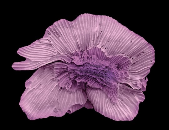
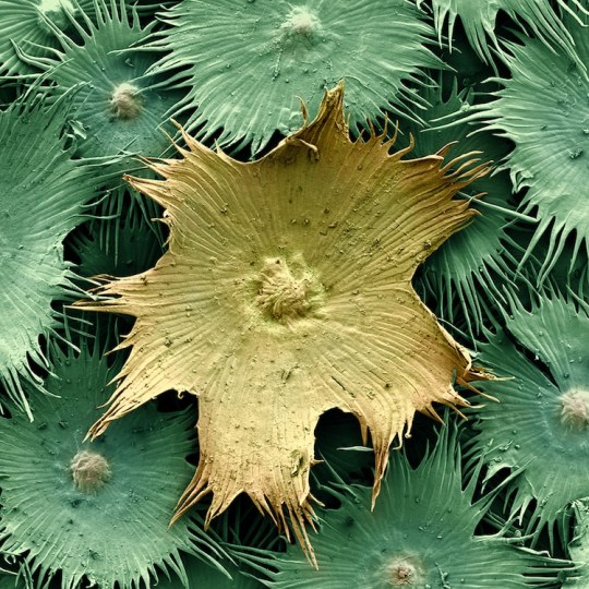
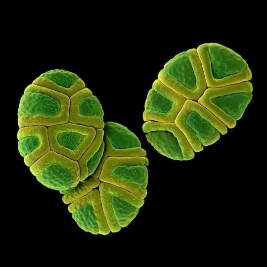
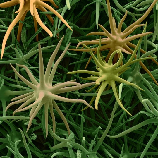
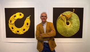
Rob Kesseler is a visual artist and Emeritus Professor of Arts, Design & Science at Central Saint Martins, London. For the past twenty years he has worked with botanical scientists and molecular biologists around the world to explore the living world at a microscopic level. Using a range of complex microscopy processes he creates multi-frame composite images of plant organs.
Using scanning electron microscopy and a mix of microscopic, scientific, digital, and manual processes, artist Rob Kesseler develops coloured micrographs of the intricate patterns within pollen and seed grains, plant cells, and leaf structures. The photographs feature specifics of cellular composition that are undetectable without magnification.
Kesseler tells that as a child, his father gifted him a microscope, marking a pivotal moment in his creative career. “What the microscope gave me was an unprecedented view of nature, a second vision,” he writes, “and awareness that there existed another world of forms, colours and patterns beyond what I could normally see.” The artist says his use of color is inspired by the time he spends researching and observing, and that just like nature, he employs it to attract attention.
32 notes
·
View notes
Last Seen Blogs

inlovewithrose
nessy

airheaven
Air Heaven

perfunctory-satisfaction
Beautiful Oblivion

rclaude2123-blog
Chrishelles Towing LLC - (410) 900-4512

magna-art
Magna's Art