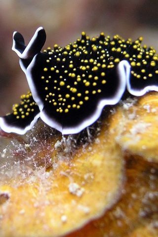
#planktonic
Video

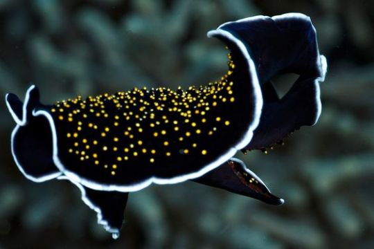
flickr
Tomopteris sp. by Alexander Semenov
#tomopteris#polychaeta#worm#plankton#planktonic#Aquatilis#Aquatilis Expedition#blackwater#Maldives#night#underwatermacro#macro#under water#flickr#marine life
4 notes
·
View notes
Text
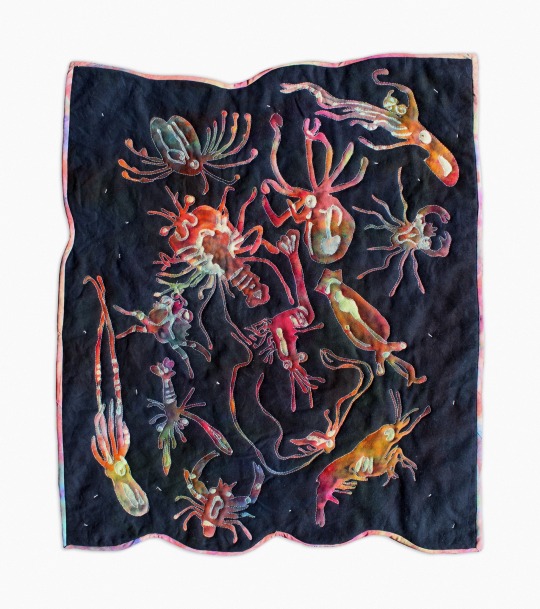
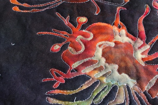
Blackwater Dive
2024, hand bleached and dyed denim, cotton batting and thread
inspired by blackwater photography of plankton! this was my first time layering bleach painting. All the silhouettes were painted with bleaching gel, loosely tie dyed, and then bleached again to make the highlights. I quilted the piece using my free motion foot to outline each individual animal and tacked down the rest of the quilt with small satin stitches that remind me of marine snow. I dyed bias tape to match. super happy with this one and excited to show it in a gallery setting soon!
#my art#queer artist#fiber art#quilt#plankton#marine biology#art#hand dyed#bleach painting#quilting#sewing#sewing machine#shrimp#crustacean#cephalopod#squid#crab#lobster
8K notes
·
View notes
Text

#a e s t h e t i c#retro aesthetic#vhs aesthetic#nostalgiacore#glow#lofi and chill#retrowave#neonvibes#synthwave#spongecore#chum bucket#spongebob#video games#aesthetic gif#plankton#abandoned
8K notes
·
View notes
Text
Chil becoming a union organizer for half foots makes so much sense because literally any time a half foot was in chil’s vicinity he was looking out for them
Like when marcille & senshi transformed
And with mickbell
#dungeon meshi#dunmeshi#chilchuck#dunmeshi spoilers#dungeon meshi spoilers#mfs acting like chil already being a union man was a huge plot point instead of smth mickbell mentioned once#damn#chill out…get it? no ok#but I’m about to pull out the plankton meme fr
8K notes
·
View notes
Text

Full Plankton Moon over Maldives l Petr Horálek via NASA APOD
#nasa#apod#space#astrophotography#astronomy#maldives#plankton#moon#full moon#bioluminecent#sea#night#sky#stars#galaxy#universe#planets#solar system#earth
7K notes
·
View notes
Text

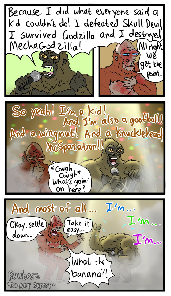


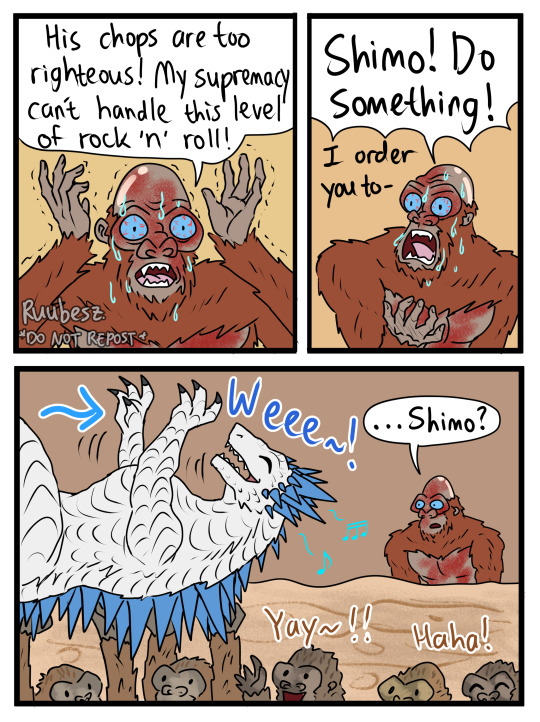

Godzilla x Kong New Empire but it's the Spongebob Movie
I had this idea BEFORE the movie even came out lol
This took longer than I thought! Please appreciate it!
youtube
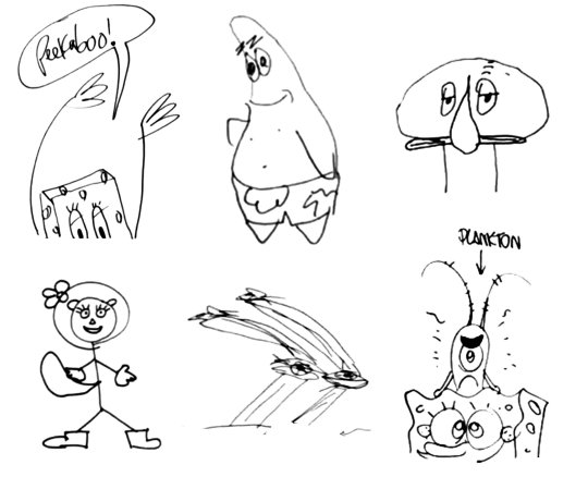
#godzilla#godzilla x kong: the new empire#kong#mothra#scar king#shimo#spongebob squarepants#patrick star#plankton#it's Godzilla x Kong but Spongebob lol#I'M A GOOFY GOOBER#YEAH!!!!#I actually love the song#i watched this movie like godzillion times#im serious#anyway hope yall like it#pls don't hate me#also enjoy goji with heels#lmao#my boi rockin' those heels lmao#do not repost#my art#Youtube
1K notes
·
View notes
Text
Yes, I know we’ve got the Ides of March coming on March 15, but you know what other important day we’re remembering this week??
March 14.

THE DAY THAT KRABS FRIES
9K notes
·
View notes
Text
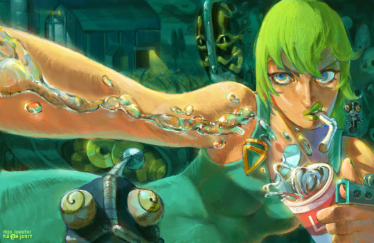
F.F.
The Intergalactic Icon
#art#digital art#artists on tumblr#fanart#jjba#artwork#illustration#jjba fanart#painting#jojo#stone ocean#foo fighters#f.f.#atroe#plankton#jjba part 6#stand#digital painting#jojo’s bizarre adventure
1K notes
·
View notes
Text
Brain herniation death in pediatric cerebral lyme borreliosis followed by kidney organ donation by Alan B MacDonald in Journal of Clinical and Medical Images, Case Reports (JCMICR)

Abstract
Fatal pediatric patient outcomes in chronic Lyme borreliosis are rarely identified. One sudden cardiac Lyme death in a seventeen-year-old black adolescent has been autopsy confirmed and reported in 2017 from Westchester, New York four years after his death in 2013. Herein is a case report of a pediatric Lyme disease brain death in a nineteen-month-old South Asian infant whose duration of disease was twelve months after sustaining three ixodid tick bites, to his sudden death from cerebellar tonsil herniation. This infant’s kidneys were transplanted into an unknown recipient.
Keywords: Lyme; borrelia; biofilm; spirochete; planktonic; pediatric; fatal; autopsy; seronegative; transplantation; FISH; IHC; transplantation.
Introduction
Lyme disease focused autopsies can establish that borrelia burgdorferi spirochetes resided in human tissues at the moment of death and thereby establish Lyme borreliosis as a contributing circumstance to patient death. One hundred and twenty-three adult and child borrelia burgdorferi positive autopsy cases are found in the peer reviewed international medical literature. These publications establish the postmortem diagnosis of active Lyme disease by microscopic documentation of borrelia burgdorferi in autopsy tissues. (https:/vimeo.com/117418013 Compendium of Peer Reviewed Autopsies in Lyme Borreliosis).
All the peer reviewed autopsy evidence for active Lyme borreliosis at moment of death accrues from the deployment of borrelia focused autopsy techniques which utilize special staining techniques to visualize the borrelia pathogens, which are otherwise invisible in standard H&E staining methods. None of the autopsy positive Lyme borreliosis case studies relied on conventional H&E stains for detection of borrelia infection in tissue sites.
The special category of Lyme disease in pregnancy fetal autopsies offers examples of gestational Lyme borreliosis with fetal death at term pregnancy [1]. This circumstance is an intra-uterine equivalent of chronic adult (“long haul” at Johns Hopkins Lyme clinic) Lyme borreliosis. Perinatal Lyme borreliosis infant deaths occurring hours [2] to days after term delivery [1] represent a biological equivalent of the notorious history congenital syphilis with excess stillbirths. In congenital syphilis, an additional scenario of late “tardive “manifestations of intrauterine infection with treponema pallidum where adolescent survivors of congenital Syphilis demonstrated tabes dorsalis [3]. The Centers for Disease Control have prevented human organ transplantation of Lyme infected hearts harvested from three asymptomatic adults who died suddenly [4]. Special silver stains performed on the candidate human hearts from deceased adults identified borrelia burgdorferi spirochetes in the Warthin Starry method. Aldred Scott Warthin M.D. at the University of Michigan perfected this method because, like borrelia spirochetes, the spirochetes of treponema pallidum are invisible in routine H&E-stained tissue.
This case report describes a sentinel case of human-to-human organ transplantation of infant kidneys from a patient with twelve months of active (post tick bite transmitted but undiagnosed) Lyme borreliosis infection. The infected kidneys were not examined in silver stains prior to release to a waiting recipient patient. Clinical proof of Lyme borreliosis during the lifetime of this infant donor was established by retrospective diagnosis from photographs of an erythema migrans skin lesion which appeared during the administration of amoxicillin age seven months in this infant. The mother of this infant took the photograph and was convinced that her son had erythema migrans (EM.) but was unable to persuade her pediatricians to make an EM diagnosis of active Lyme borreliosis.
A complete forensic autopsy examination failed to identify borrelia burgdorferi infection because mandatory Warthin Starry stains [MMWR] were not deployed. After the kidney transplantation and after the issuance of a final forensic autopsy report, additional advanced borrelia focused Fluorescence in situ Hybridization (FISH) studies identified “high infectious burden” borrelia burgdorferi infection in the infant donor’s autopsy brain and heart autopsy tissues. By any metric the active blood borne disseminated borrelia infections of brain and heart discovered at autopsy were also present in the unexamined harvested kidneys too.
Case Report
A nineteen-month-old South Asian male whose family skin color was brown, was identified as a potential kidney donor after he suffered in hospital brain death due to cerebellar tonsil herniation two days after initiation of mechanical ventilation in a pediatric intensive care. He had immediately before hospitalization, suffered an out of hospital spontaneous and unexpected cardiac arrest, from which he was immediately and successfully resuscitated by family members at his home. He resided in a United States Atlantic corridor state where Lyme disease is endemic. Past obstetrical history revealed that he was the product of a spontaneous uncomplicated vaginal home delivery at term by a midwife. His prenatal and perinatal two-month medical history was unremarkable. He was breastfed. Between six months and seven months of age, the infant experienced three ixodid tick bites in skin of scalp, skin of external ear helix, and skin of external ear canal). No erythema migrans lesions were identified in skin adjacent to the bite sites. He later, at age seven months, developed noteworthy skin redness of the entire ear skin helices and fever. A clinical diagnosis of “external otitis” was rendered by the pediatrician. The child was treated with oral amoxicillin for seven days. At age seven months, a targetoid eight centimeters in diameter red ring flat lesion developed on the thigh skin with central clearing and with central small vesiculations appeared coincident with oral amoxicillin therapy (Figure 1).
The red ring lesion was photographed by the mother and presented to her five pediatrician consultants for comment. All pediatricians declined a medical diagnosis of the targetoid erythematous patch. The next twelve months were marked by many separate febrile events with associated red patches on head, neck, trunk, abdomen, and extremities skin and which were diagnosed as “pediatric exanthems” of uncertain etiology. Failure to thrive, irritability, poor feeding, weight loss, and one episode of Seventh Nerve Palsy announced by the child’s facial asymmetry were recorded by his mother. Because both of his parents had been concurrently diagnosed and received antibiotic treatment for seropositive Lyme disease while their child was ill, the parents requested that Lyme serologies be obtained on their child. Quest Laboratories Inc., and LabCorp Inc and Igenex Laboratories Inc. Lyme ELISA numerical measurement results were inconclusive. Antibodies to three borrelia burgdorferi bands (41kd (IgG and IGM)) and “indefinite” band 31 kd.(IgM) were reported as “Negative” in western immunoblots for Lyme disease at Igenex laboratories. The triple band immunoblot results were not followed up with follow-up testing at infant age 12-19 months by five pediatricians (Dermatology, Immunology, Infectious disease, Rheumatology board certified consultants), This infant subsequently “failed” in Prevnar immunization because he failed to produce Prevnar related antibodies post vaccination and a second Prevnar immunization was administered.
A forensic autopsy, mandated by organ donor pre-transplantation protocols, utilizing only hematoxylin and eosin stains (H&E), authorized the release of the child’s kidneys to a recipient patient who was a California resident. CDC requisite silver stains to rule out presence of borrelia spirochetes in tissue were omitted. Noteworthy autopsy findings were areas of deep brain gray matter sites with “lymphocytic cuffing in blood vessels” which was attributed sequelae of the patient’s fulminant cerebellar tonsillar herniation with destruction of the underlying brain stem respiratory centers. Numerous axonal spheroids of the thalamus were reported. The axonal spheroids pathology significance was not discussed. The lack of deployment of Warthin Starry [5]. silver stains for detection of possible borrelia spirochetes and other bacterial infectomes was explained as the result of “budgetary cutbacks” at the office of the medical examiner. The family and the medical examiner, in subsequent negotiations about of the final report at ninety days post autopsy, reached agreement that additional DNA based diagnostic research studies for detection of borrelia burgdorferi infection might be undertaken by an outside pathologist with a research interest in DNA based detection of residual borrelia deposits in the autopsy tissue blocks, without any obligation by the medical examiner to amend the finalized and certified original forensic autopsy report. The medical examiner released only five recut glass slides to this author from tissue paraffin blocks (brain and heart) examined in the original forensic autopsy. A fluorescence in situ Hybridization (FISH) protocol using previously published and validated Borrelia burgdorferi specific Molecular Beacon DNA probes (Flagellin B gene Bbo0147) Probe Sequence: (nucleotide) TGG GAG TTT CTG GTA AGA TTAA; --- fluorescein isethionate label 5’ position [7] and additional protein Immunohistochemistry studies [8]. chromogen deposition at sites of bound anti-borrelia rabbit antibody (Diaminobenzidine) resulting in a brown chromogen product deposition on specific tissue sites. Ventana iView DAB kit [IHC Kit for detection of Rabbit antibodies] (product #: Roche Tissue Diagnostics; Catalogue760-091– according to the manufacturer’s instructions with final chromogen color development; ABCAM Inc. www.abcam.com (Catalogue ab20950) Borrelia polyclonal antibodies: harvested from rabbit immunized with total borrelia proteins in a pure culture digestion product.
Figure 1: The patient’s brown skin color renders the exact outermost borders of the flat erythematous ring to be indistinct but the “central clearing” and inner border zone of erythema in the EM lesion is distinct.
Figure 2: Biofilm community borrelia burgdorferi in specialized colony type morphology showed discrete bright signal fluorescent spirochetes surrounded by a veil like lower signal intensity fluorescent gel-like extracellular matrix. FISH method with DNA probe for Flagellin B gene of borrelia burgdorferi. (BBO 0147) Probe Sequence: (nucleotide) TGG GAG TTT CTG GTA AGA TTAA; --- fluorescein isethionate label 5’ position.
Figure 3: The biofilms were confirmed in separate Immunohistochemistry IHC studies for detection of borrelia specific protein antigens. Chromogen deposition at sites of bound anti Borrelia rabbit antibody (Diaminobenzidine) resulting in a brown chromogen product deposition on specific tissue sites. Ventana iView DAB kit [IHC Kit for detection of Rabbit antibodies] (product #: Roche Tissue Diagnostics; Catalogue760-091– according to the manufacturer’s instructions with final chromogen color development; ABCAM Inc. www.abcam.com (Catalogue ab20950) Borrelia polyclonal antibodies: harvested from rabbit immunized with total borrelia proteins in a pure culture digestion product.
Note: Immune reactive borrelia infectomes in biofilms produced brown color products which mirrored the fluorescent signal biofilms in FISH method studies.
Figure 4: Singleton (planktonic form) borrelia spirochetes were observed in FISH studies as multiple separate borrelia burgdorferi microbes in a single microscopic field of view. FISH method with borrelia burgdorferi DNA probe for gene BBO 0147 -Flagellin B. (Probe Sequence: (nucleotide) TGG GAG TTT CTG GTA AGA TTAA; --- fluorescein isethionate label 5’ position).
Figure 5: Warthin Starry Stain of Autopsy Brain: Red arrow points to an elongated borrelia burgdorferi spirochete. Abundant Round body spirochetal forms (Black color) are present.
Figure 6: Warthin Starry stain of Autopsy Brain: Blue color arrows designate a grouping of borrelia burgdorferi Spirochetes in brain tissue. Round body spirochetal forms (black color).
Figure 7: Murine Monoclonal Antibodies CD 10 bind to Outer Surface Protein A (OSP A) - Red fluorescent label) A single borrelia burgdorferi spirochete is present at the 9 o’clock position.
Figure 8: A single biofilm community of Borrelia burgdorferi spirochetes is present. (gift from J.L Benach PhD).
Figure 9: Murine Monoclonal Antibodies CD 10 bind to Outer Surface Protein A (OSP A) - Red fluorescent label) A single biofilm community of Borrelia burgdorferi spirochetes is present. (gift from J.L Benach PhD).
The actual infectious “burden” is visually manifest in microscopic images of single borrelia microbes and of biofilm colonies of borrelia microbes in autopsy tissues based on the numerical counts of borrelia infectomes in tissue sites. Multiple separate borrelia infectomes in a single 400x magnification field of view of this infant’s tissues far exceed the observed numerical counts of borrelia spirochetes in microscopy in any published previous borrelia focused Lyme disease autopsy case [1]. Past published Lyme disease autopsies (gestational) have displayed only rare single borrelia spirochetes [2] (i.e., one spirochete per microscopic field of view). It is noteworthy that the “one spirochete per microscopic field of view” observation also applies to borrelia burgdorferi spirochetes in the erythema migrans lesion [10].
The Westchester adolescent Lyme disease cardiac death autopsy [6], by comparison, disclosed only a single borrelia spirochete in the autopsy heart adolescent decedent. Biofilm colonies of borrelia infectomes were not seen in the adolescent patient autopsy
Commentary
1. A diagnosis of pediatric Lyme borreliosis in this unfortunate infant was justified while the infant was alive based solely on the photograph of the pathognomonic erythema migrans lesion (EM)
2. This represents a secondary type of EM lesion as opposed to a tick bite site EM lesion [10], because it appeared during empirical amoxicillin therapy prescribed for presumed external ear region otitis. Secondary and recrudescent EM lesions have been previously published [11].
3. Five pediatric subspecialists failed to recognize the erythema migrans pattern in the photograph provided by the child’s mother. The lesion was remarkable for central clearing and central vesiculation. Nearly identical vesiculated erythema migrans skin lesions are displayed on the Johns Hopkins Lyme Disease website. https://www.HopkinsLyme.org.
4.The pediatrician diagnosis of “external otitis” might, in retrospect, was likely a “red ear helix “skin sign of North American borrelia chondritis [12]. which can announce pediatric Lyme borreliosis
5. European borrelia lymphocytoma in childhood may present with either cartilage site bright red erythema of the skin overlying the ear cartilage tissues [12] or alternately, red skin inflammation of the non-cartilaginous soft earlobes without extension of the erythema over the remaining external ear region, It is pertinent to recall that the child’s father’s Lyme disease symptoms included the “red ear” clinical sign of borrelia chondritis as well as facial nerve palsy and facial asymmetry which were mirrored in his infant son and discussed with the attending pediatricians
son and discussed with the attending pediatricians.
3. Negative serology results in thrice measured Lyme antibody tests (ELISA and Immunoblot) in the last fifteen months of life in this infant were “false Flags of wellness” in this chronically ill child. The child’s unambiguous erythema migrans lesion, merited a mandatory diagnosis of Lyme disease. Sero-positive results in ELISA and Western blot Lyme testing are never required for diagnosis of Lyme disease in erythema migrans positive patients because the CDC provides guidance that the EM lesion alone is quantum suffit for presumptive diagnosis.
Immaturity of the immune system of pediatric patients of ages less than 24 months [14] is well described. This developmental circumstance in perinatal infants renders negative antibody detection test results such as Lyme serologies at this stage of life less than definitive. Negative serology test results always include the possibility of “false negative” serologies in a biologically positive patient in all serologically defined human medical illnesses.
6. The forensic autopsy product in this patient failed the “Standard of AUTOPSY care “. Failure to deploy diagnostic Warthin Starry silver stains to cerebral and cardiac tissues [MMWR} from this infant denied the patient’s family and the kidney recipient of the opportunity to prevent the release of infected kidneys to an innocent recipient infant. This can be objectively and severely critiqued as the medical examiner’s failure to deploy the well-publicized CDC free and pro bono Warthin starry stains.
Biofilm colonies in autopsy disseminated sites demonstrate that a “chronic” and persistent Lyme borreliosis infection was present.
A recent adult Lyme disease autopsy describes high density borrelia burgdorferi biofilms in kidney, liver, brain, and heart in a fifty-three-year-old woman with a 16-year Lyme illness. This borreliosis was not cured by multiple rounds intravenous multiple antibiotic treatments over decade prior to death. The autopsy, performed at the College of Physicians and Surgeons of Columbia University, and follow-up special molecular borrelia detection studies at the University of New Haven showed multiorgan site biofilm borrelia colonies confirmed by FISH and IHC methods [15].
A new paradigm of autopsy established chronic biofilm positive Lyme borreliosis is now defined. The molecular basis for the adult and pediatric “post-Lyme Disease Syndrome” may now viewed through the lens of persistent DNA and protein detections in autopsy. Analogous autopsy findings from tertiary long term syphilis infections with Warthin starry stains have also established chronic syphilis infections with visible treponema pallidum spirochetes at death in autopsy tissues [3].
6. There is no clinical information available concerning the health status of the unknown recipient patient post kidney transplantation in this case. Noteworthy is the observation that extensive chronic disseminated borrelia infection of autopsy kidneys in one patient’s 16 year of illness was manifest as numerous biofilms of borrelia infection in her kidneys ( as well as in brain, heart, and liver sites) [15].
7. Two pediatric “Failure to Diagnose” Lyme fatalities in patients whose skin color was brown or black remind all practitioners that the diagnosis of erythema migrans lesions may be difficult or impossible in persons of color [6].
Litigation is currently underway in Westchester by the family of the 17-year-old patient. The family of this infant has decided against legal action against their child’s pediatric providers but hopes that the mistakes in their child’s pediatric antemortem and postmortem care will not be repeated.
Conflict of Interest declaration
The author declares that he has no conflict of interest in connection with this manuscript.
Acknowledgments
This research was supported by the Lindorf Family Foundation, in Orem Utah. Neil Spector M.D. kindly referred the family of this child to this author for the purpose of obtaining pro bono advanced molecular diagnostic FISH method testing to detect Borrelia burgdorferi genomic signatures in autopsy tissues. Dr. Spector survived end stage Lyme Borreliosis induced cardiac failure requiring heart transplantation with full cardiac recovery. (“Gone in a Heartbeat”, N.S. Spector, MD, Triton Books, 2015) This manuscript would not have been possible without Dr. Neil Spector’s referral.
Dedication
This manuscript is dedicated to the parents (T.K. and B.K.) and to the brothers and sisters of this infant
For more details : https://jcmimagescasereports.org/author-guidelines/
#Lyme#borrelia#biofilm#spirochete#planktonic#pediatric#fatal#autopsy#seronegative#transplantation#FISH#IHC#DNA#Alan B MacDonald#JCMICR
0 notes
Text
I was at a hospital and a old fat Italian man to my right had an entire canvas and was painting tits, and to my left was a depressed 1950s detective. Plankton from SpongeBob was the nurse.
#dream#text#hospital#italian#painting#art#depression#detective#plankton#spongebob#spongebob squarepants#nurse#queueueueueueueueueueueueueue
1K notes
·
View notes
Text


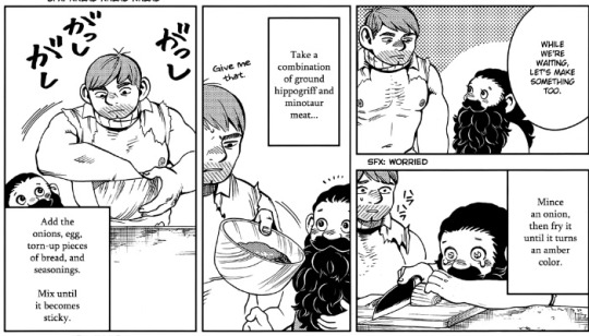
Ya ain’t supposed to mate
(commissions open)
#boys art#comic#comics#doodles#spongebob squarepants#patrick Star#squidward tentacles#mr. krabs#mrs. puff#plankton#squidbob#plankbob#patbob#flats the flounder#spot#Karen#squilliam fancyson
1K notes
·
View notes
Text

slipper lobster (larvae) painting from today as I’m out enjoying the weather! message me if interested in this piece and I’ll give you details!
2K notes
·
View notes
Photo
The main cast of “SpongeBob SquarePants” as drawn by their voice actors.
First row: SpongeBob SquarePants by Tom Kenny, Patrick Star by Bill Fagerbakke, Squidward Tentacles by Rodger Bumpass
Second row: Sandy Cheeks by Carolyn Lawrence, Eugene Krabs by Clancy Brown, Sheldon Plankton (perched atop SpongeBob’s head) by Mr. Lawrence
BONUS VIDEO
#🌟#SBSP#Official Art#🏆Hall of Fame🏆#I guess? I mean it’s by the cast members and Mr. Lawrence is the head writer as well as a storyboard artist on the show soooo...🤷♀️#SpongeBob SquarePants#Tom Kenny#Patrick Star#Bill Fagerbakke#Squidward Tentacles#Rodger Bumpass#Sandy Cheeks#Carolyn Lawrence#Eugene Krabs#Clancy Brown#Sheldon Plankton#Mr. Lawrence
9K notes
·
View notes
Text

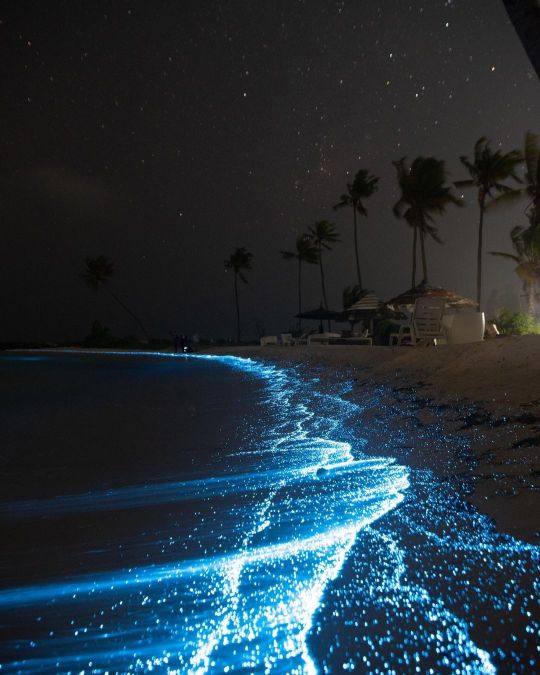
by nihthu
#bioluminecent#bioluminescence#aesthetic#nature#naturecore#icean#oceancore#beach#plankton#maldives#travel#travelcore#photography#landscape#curators on tumblr#up
7K notes
·
View notes
Text


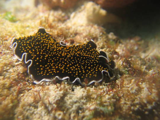


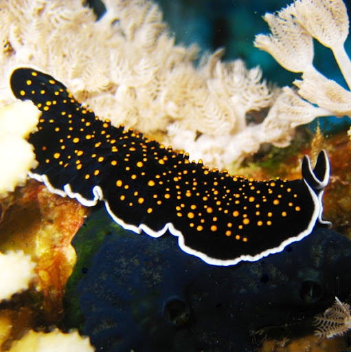


Червь плоский желтопятнистый (Thysanozoon havomaculatum) . Морской червь из класса ресничные черви, или турбеллярий. Принадлежит отряду Поликлад. Имеет уплощённое, овальное тело, покрытое ресничным эпителием. Длина этого симпатичного создания около 4 см, а толщина всего пару миллиметров. Передний конец его тела несёт пару щупалец. Мускулатура этого плоского червя многослойная, хорошо развитая, благодаря ей он способен подниматься в толщу воды за счёт ундулирующих движений краёв тела, а не только обитать на дне. Этот свободноживущий плоский червь питается преимущественно как хищник, водными беспозвоночными. Встречается в морях вокруг Австралии и Индонезии.
Yellow-spotted flatworm (Thysanozoon havomaculatum). A marine worm from the class of ciliated worms, or turbellarians. Belongs to the Polyclad squad. It has a flattened, oval body covered with ciliated epithelium. The length of this cute creature is about 4 cm, and the thickness is only a couple of millimeters. The front end of its body bears a pair of tentacles. The musculature of this flatworm is multi-layered, well developed, thanks to it it is able to rise into the water column due to undulating movements of the edges of the body, and not just live on the bottom. This free-living flatworm feeds primarily as a predator on aquatic invertebrates. It is found in the seas around Australia and Indonesia.
Источник:https://t.me/+t0G9OYaBjn9kNTBi, //www.webdive.ru/fotocat.php?t=11&id=20542&mode=view, http://mylongdongbay.blogspot.com/2007/07/save-ocean-cherish-marine-lives.html, /ru.pinterest.com/pin/630644754053053958/, http://www.akkiira.com/hiramushi/yoimiyaminohiramusi-swimming.html, //foxford.ru/wiki/biologiya/tip-ploskie-chervi?utm_referrer=https%3A%2F%2Fwww.google.com%2F.
#fauna#video#animal video#marine life#marine biology#nature#aquatic animals#Polyclad#Yellow-spotted flatworm#ocean#sea#corals#plankton#animal photography#nature aesthetic#видео#фауна#природа#Поликлады#Червь плоский желтопятнистый#океан#кораллы#планктон#море
850 notes
·
View notes
Last Seen Blogs

moonkid-art
bts fan art n things

lonelylost-and-stranded
lonely lost & stranded.

codymania
Sin título

corpsesurfing
Always Another Secret

ta-nizzle
Tahani