#desmosome
Text
Blonde boy shoots creamy cum on his carpet
Loirinha Amadora na Webcam
Tamil boy hand work gay sex video From Jail to Jizz
Antique dealer anal fucked in her shop
Thick Latina Jolla is handcuffed and punished
Fitness Rooms Interracial lesbian shower threesome with shy Latina
Novinha tira a roupa mete de quatro e leva gozada na bunda
dick flash en bus y ella mira
Small Waist Fatt Ass Gemini gets Fucked for 10 mins and a multiple times
Looner trans TRAILER
#prinos#winned#insp#cyclecar#nabobesses#vodka#piquette#tabuing#furole#nondeistical#gumball#diastyle#desmosome#bedrugging#amorism#Koblick#reproportion#reabstracted#grafiti#self-tuition
0 notes
Text
Cell Junctions
-- there are three types of junctions:
-- tight
-- desmosome
-- gap
-- tight junctions seal epithelial cells to one another
-- desmosomes have “spotted” seals, where keratin filaments anchor the two cells
-- gap junctions have channels to allow the passage of ions and molecules
#medblr#studyblr#notes#my notes#medical notes#medblr notes#med notes#anatomy and physiology#anatomy#physiology#biology#microbiology#histology#cells#cell notes#anatomy notes#physiology notes#biology notes#bio#bio notes#microbiology notes
52 notes
·
View notes
Text
Tree of Life 6: Metazoa (animals)
Brace yourselves, this one’s long.
[Disclaimer: taxonomy is a complex, ever-changing field, and this overview is certainly not going to be exhaustive, especially concerning extinct groups]
← Part 5 (Unikonta: amoebae, fungi, and such)
Part 6.5 (Porifera, Cnidaria, Plathyhelminthes, Annelida) →
We finally come to Animalia, or Metazoa “animals beyond” (as opposed to Protozoa, which are no longer considered animals at all). Perhaps you won’t find it easy to think of what sponges, chickens, oysters, starfish, and millipedes have in common. Here are the distinctive features that zoologists have identified for Metazoa:
First of all, all animals are heterotrophs, i.e. they cannot produce their own organic molecules, though very few species have become partially autotrophic thanks to symbiosis.
All animals have a diplont life cycle (see part 3), which goes as follows. It starts with two gametes, a female egg cell and a male sperm cell, which has a single flagellum (or, rarely, none), and fuses into the egg cell thanks to an anterior organ called acrosome. Each gamete is haploid, meaning it carries one copy of each gene; the fusion of the gametes produces a diploid zygote, which has two copies of each gene. The zygote multiplies by mitosis to form a multicellular body in which each cell is also diploid (unlike fungi and plants). Animals are always multicellular; while some fungi, namely yeasts, have reverted to unicellularity (see part 5), no animal has done so. Finally, each individual produces new haploid gametes, male and/or female, by meiosis, which discards half the genome in those cells.
(All multicellular organisms -- not just animals, but plants and fungi too -- go through a unicellular stage at some point in their life cycle. Why is it so, given that many species can regenerate their body from small fragments and reproduce by division? Probably to keep cancerous mutations in check: if you start as a single cell, either that cell will be healthy, and mutant lineages will have to start all over again; or it will be mutant, and so will be all its descendants, with no healthy cells to exploit, so that the mutation will destroy itself.)
During the development from zygote to multicellular body, the embryo folds to form an inner cavity, and the cells organize themselves in two or three germ layers: an internal endoderm, an external ectoderm, and in most cases a mesoderm which lies in between. Generally speaking, the endoderm forms the lining of the digestive tube with annex glands (e.g. the liver), as well as our lungs; the ectoderm forms the outer coating of the body, with skin, armor, and sense organs, as well as the nervous system; the mesoderm forms other organs (e.g. kidneys, sex glands), bones, and muscles.
Animals have at least two types of tissues: epithelial, which cover surfaces, and connective, which fill up volumes. Epithelial tissues, such as skin and mucous membranes, are made out of one or more layers of tightly packed cells growing over a basal lamina which supplies food. The cells are linked to each other by junctions such as desmosomes, which sew cell membranes to each other making the whole tissue more resistant. In connective tissues, cells are spaced wide apart, sunk in an extracellular matrix. Usually the matrix is a mix of gelatin and elastic fibers made out of proteins, most importantly collagen; but sometimes it may be liquid, as in blood, or built out of hard mineral, as in bone. To these two basic kinds, most animals add two more: muscles and nerves.
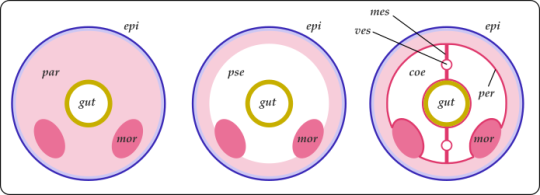
Cross-sections of three generic animals. Left to right, acoelomate, pseudocoelomate, eucoelomate. The endoderm (in yellow) forms the gut; the ectoderm (in blue) forms the outer surface; the mesoderm (in pink) fills the space in between.
The body of an acoelomate, e.g. a tapeworm, is filled with a mass of mesodermal cells called parenchym (par), in which organs (such as kidneys or gonads, mor) and the gut are immersed. The body is surrounded by an ectodermal epidermis (epi). Simple enough, but the organs are vulnerable to squishing.
In a pseudocoelomate, e.g. a hookworm, the other organs are immersed in a fluid-filled cavity called pseudocoelom (pse). Since water is not compressible, it gives the body a fixed volume and some mechanical resistance, as well as distributing substances, and storing products. Unfortunately a puncture wound is easily fatal.
In an eucoelomate, e.g. an earthworm, the mesoderm forms a lining or peritoneum (per) that wraps around the coelom (coe) and divides it in sections, as well as mesenteries (mes) that keep organs in place. The separate chambers of the coelom help against liquid loss and allow new kinds of movement, such as burrowing in sand. However, blood vessels (ves) are now required to distribute materials.
This is not a strict evolutionary sequence: animal phyla change from one to the other depending on their niche and conditions.
The animal kingdom is -- no wonder -- vast and complex. The most important high-level categories are known as phyla, commonly used in studies of animal diversity and large-scale evolution; here, I’ve written the names of phyla in all caps. The phyla Mollusca, Arthropoda, and the above-phylum group Deuterostomia will all get their own posts; the phyla Porifera, Cnidaria, Platyhelminthes, and Annelida will be described together in part 6.5.
?? 0. † Vendobionta: Let’s start with the strangest, shan’t we? Vendobionta is almost certainly not a clade, but a convenience term for all the weird macroscopic organisms that flourished in the Ediacaran period (once known as Vendian), between 580 and 540 Ma. It’s not clear even that they were all animals, let alone where they might fit in the tree: some have been described as algae, fungi, colonies of protists, or even representatives of a completely lost kingdom of life. In general, they didn’t have visible mouths or sense organs, nor any way to move; they probably (?) absorbed nutrients from water, grazed on mats of bacterial slime, and/or were photosynthetic. They suddenly disappeared at the beginning of the Cambrian, presumably because new organisms with eyes, jaws, and muscles predated them into extinction and/or consumed all their food.
† Trilobozoa “three-lobed animals”: These Vendobionts are radially symmetrical: their body, circular as a whole, is composed of three segments rotated around the central axis. This is quite unique: no living animal has a rotational or a three-fold symmetry (although some Cnidarians have a non-rotational, six-fold one). Branching grooves (a ciliated organ to collect food, maybe?) converge at the center of the upper surface. The most famous representative is † Tribrachidium, found in Russia and Australia.
† Medusoidea “jellyfish-like”: These too are flattened and radially symmetrical, often with concentric rings. The largest, like † Aspidella and † Cyclomedusa, could grow several cm wide. † Arkarua adami has a peculiar five-fold symmetry, like Echinoderms, though it’s unilkely to be related. Sometimes included among the † Medusoidea is † Eoandromeda octobrachiata, a strange galaxy-like being with eight spiral arm coiling out from the center.
† Petalonamae “petals from Namibia”: A set of two groups with plant-like shapes. † Rangeomorpha, most common in Newfoundland, had frond-like fractal structures -- possibly an adaptation to absorb as much food and oxygen as possible from the water. (Maybe sunlight too, but it seems some lived too deep for photosythesis.) Two, three, or six rows of branching fronds emerged from a central axis. Some, like † Charnia, stood upright in the water; others, like † Fractofusus, lay on the seabed. † Erniettomorpha such as † Pteridinium, found in Namibia, had lateral "vanes” formed by rows of sacs.
† Proarticulata “before articulated ones”: The most familiar, maybe. They had flattened bodies and bilateral (which is to say, left-right) symmetry. They also seemed to have internal branching canals that might be remains of a digestive system. † Dickinsonia grew as a thin segmented disk up to a meter long; it might actually be a Ctenophoran (see below), showing both righ-left and front-back symmetry. † Spriggina, from the very end of the Ediacaran, head a sort of “head” shield and rows of segments that might not have been exactly symmetrical, but somewhat offset.

A selection of Vendobionta. Far left: † Charnia masoni († Rangeomorpha, England). (Museo delle Scienze di Trento, via Wikipedia) Upper row, from left: † Spriggina floundersi († Proarticulata, Australia) (Wikipedia); † Ernietta plateauensis († Erniettomorpha, Namibia) (Ivantsov & Zagrevskaya 2021). Lower row, from left: †Tribrachidium heraldicum († Trilobozoa, Australia) (I. & Z. 2021); † Dickinsonia costata († Proarticulata, Australia) (Verisimilus, Wikipedia); † Mawsonites spriggi († Medusoidea) (Nordelch, Wikipedia)
1. PORIFERA “hole-carriers”. The (possibly paraphyletic) phylum of sponges. In general, they have a system of internal ciliated canals collecting food from the water that streams in through lateral pores, and out through a channel at the top. The whole structure is kept up by a basket of various composition. See part 6.5 for more.
? 2. PLACOZOA “plate animals” Honestly, it’s a bit embarassing to call this thing an animal. Until a few years ago, phylum Placozoa counted a grand total of one species, Trichoplax adhaerens from tropical seas (now, the count is three). Its body is simply a single layer of epithelial tissue surrounding a thin syncitium (i.e. a network of fused cells that retain separate nuclei). On the lower side, the epithelium has cilia and digestive glands; on the upper, it contains little crystal spheres that might serve as defense. Trichoplax reproduces by splitting, and feeds by crawling over bacterial mats; it secretes digestive enzymes to break food down, but each epithelial cell takes it up on its own. Recent studies (e.g. Laumer & al 2019) suggest that Placozoa might belong next to Cnidaria.

Trichoplax adharens, top view and cross-section. The whole thing is about 0.5 mm across and 0.025 mm thick. (Oliver Voigt, Wikipedia; Hickman & al 2008)
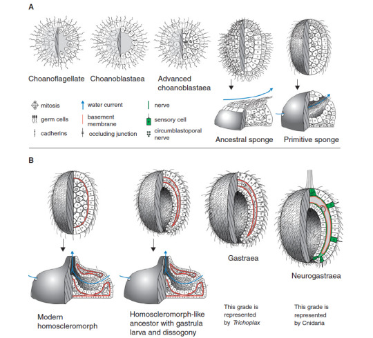
A model of the origin of animals, starting from a colony of Choanoflagellates (see part 5), passing through the ancestors of sponges, and ending with the likely common ancestor of Eumetazoans, which gave rise to both us and jellyfish. (Nielsen 2012)
3. Eumetazoa “animals well beyond”: All animals more complex than that pass through a process called gastrulation: when the embryo is still just a little ball of cells, it folds inwards producing a hollow or archenteron, the future gut. It communicates with the exterior through an opening called blastopore, the first prototype of a mouth. The two layers of cells produced by the folding, inner and outer, become the endoderm and ectoderm. Thanks to this hollow, Eumetazoans practice extracellular digestion: the uptake of food is not a responsibility of individual cells, like in sponges and Placozoans, but rather food is degraded in the internal cavity by digestive enzymes, and the resulting simple molecules are distributed to all tissues. In Eumetazoans we also see the first appearance of the nervous system, which allows coordinated action of the whole body.
In recent classifications, it’s being phased out in favor of Parahoxozoa, the animals defined by the presence of Hox genes, which specify different body regions and ensure that each anatomical structure forms in the correct part of the body; this would include Cnidaria and perhaps Placozoa too, but would exclude Ctenophora.
3a. CNIDARIA “nettles”: The phylum of corals, sea anemones, jellyfish, man-o’-wars, and curious parasites that have long been believed to be protists. They have radial symmetry (4-, 6-, or 8-fold), a single opening where the radia meet in the center, and specialized stinging cells for defense and hunting. See part 6.5 for more detail.
?? 3b. CTENOPHORA “comb-carriers”: What a strange situation! Ctenophorans have distinct tissues, appendages, a nervous system, and even have all three germ layers, unlike Porifera, Placozoa, and Cnidaria which lack the mesoderm. This would put them next to Bilateria (see below); except that recent molecular studies (e.g. Laumer &al 2019) insist on placing them in a basal position, further away from us than even sponges! An artifact of unusually fast evolution rates, perhaps?
Most Ctenophorans (e.g. Mertensia or Pleurobrachia) have bag-shaped bodies with the mouth at one pole (serving both to eat and to expel waste) and a branching, blind-end gut. Eight rows of combs, each of which is a band of cilia, run along the sides: hence the common name of “comb jellies”. In fact, they are the largest organisms on Earth that swim with cilia instead of muscles. Two tentacles may be retracted into pouches or extended to catch preys thanks to colloblasts, specialized glue-producing cells. Others, like Beroë, lost their tentacles; Coeloplana flattened its body to crawl on the seabed; the “Venus’ girdle” (Cestum veneris) extended it sideways to swim like a lopsided worm. Class † Scleroctenophora, endowed with armor plates, is known from the Cambrian but now extinct.
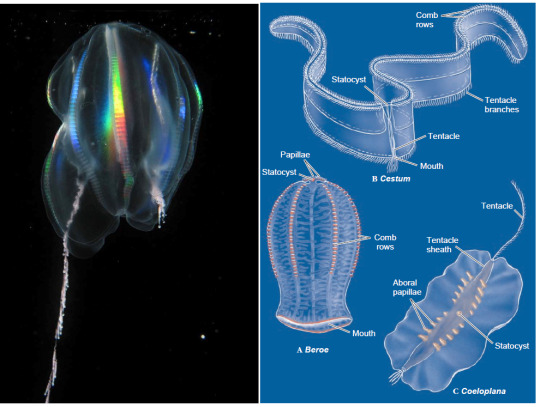
A variety of comb jellies. Left: Mertensia ovum, with one tentacle retracted. The rainbow shimmer of the comb rows is a distinctive feature of Ctenophora. Right: Beroë, Cestum, and Coeloplana. The mouth of Coeloplana is on the bottom, its lining forming the whole lower surface. Note that the body of Cestum is not long and narrow -- it’s short and very broad! (Kevin Raskoff, Wikipedia; Hickman & al 2008)
3c. Bilateria “two-sided”: The name says it all: animals in this clade have bilateral symmetry, with a left and a right side that are (in general) mirror images of each other, while keeping a distinction (thanks to signalling gradients during embryo development) between a front and back end. This allows them to concentrate their feeding and sensorial apparatus in the front, when the body first encounters new stuff, while the expulsion of waste and gametes moves to the back: a good plan if you’re an organism that moves consistently in one direction. In general, Bilaterians have a linear gut with an anterior mouth and a posterior anus, where food always flows from front to back.
3c1. Acoelomorpha: A group of marine worms, with thin bodies a few mm long. The simplest sort of Bilaterians: unlike almost all others, they don’t have a distinct mouth and anus, but a single opening at the center of their ventral surface, like jellyfish. That opening, which serves both as mouth and anus, does not lead into a gut (not even a jellyfish-style cavity), but onto a simple mass of endodermal cells that break down food. The body is covered by a thin ciliated epidermis; between these two layers, there are mesodemal muscles, nerves, ovaries, and testicles (they’re all hermaphroditic). That’s... about it. It’s not clear whether this simplicity is primitive, or a secondary loss of traits. They comprise three phyla, ACOELA, NEMERTODERMATIDA, and also XENOTURBELLIDA, whose 2 species were until recently classified next to Echinoderms (e.g. starfish).
3c2. Protostomia “mouth first”: This group is defined by a number of features, but few of its members possess them all. In theory, when the embryo folds to create a proto-gut, the resulting opening, or blastopore, should give rise to the mouth, with the anus appearing later; in Deuterostomes the opposite is true. In practice, this is only true in some cases. The nervous system is formed by a number of ganglia strewn along paired ventral nerve cords; these cords form a loop around the mouth, which in some cases (e.g. in spiders and octopodes) becomes a brain. Many groups of Protostomes develop from planktonic larvae that swim and collect food with bands of cilia.
3c2a. ? Mesozoa “middle animals” So called because they were once thought to lie between the simpler sponges and the more complex Eumetazoa. All there is to their body is a single layer of ciliated cells surrounding a mass of maturing eggs. In fact, they are descendants of relatively complex Protostomes that shed most of their body structures as a consequence of living as parasites: ORTHONECTIDA live in the body cavities of various marine invertebrates, RHOMBOZOA only in the kidneys of squids and octopodes.
Left: a generic Acoela, possibly the model from which all Bilaterian animals from snails to pigeons and from wasps to starfish developed. Right: the Mesozoan Rhopalura (Nemertodermatida; female above, male below).
3c2b. Gnathifera “jaw-carriers”: A diverse clade of invertebrates. Most are microscopic, and form a pretty good part of marine animal plankton. They have some variety of jaws made out of chitin, a nitrogen-rich sugar that we already saw in the cell wall of Fungi (see part 5).
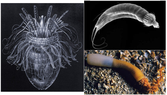
? 3c2b1. CHAETOGNATHA “hairy jaws”: They go by the common name of “arrow worms”. Unlike most worms, they swim in open water, with undulations of their flattened body which extends sideways into fish-like fins. They even have, like vertebrates, a post-anal tail that doesn’t contain the intestine. The head has a large hood that conceals rows of chitinous grasping spines.
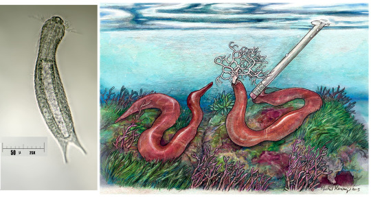
3c2b2. GNATHOSTOMULIDA “little jawed mouths”: Delicate mm-sized worms that live between grains of sand on beaches and seafloors. They have rasping jaws to scrape bacteria and fungi off sand grains and into a blind-end gut.
3c2b3. MICROGNATHOZOA “animals with tiny jaws”: A phylum with only one species, Limnognathia maerski on Greenland. A tiny wrinkled thing only 0.15 mm long, but with bizarrely complex retractile jaws, made out of 15 different elements. All other structures are very simple; it uses cilia to move and feed. Only females have ever been found.
3c2b4. ROTIFERA “wheel-carriers” or Syndermata “fused skins”: Another of the big ones. They are named after the wheel-like ciliate crown (corona) which, beating, creates the water current they use to swim and feed. Almost always microscopic, they have a great variety of body shapes, sac-like, worm-like, or spiny, sometimes with a thick segmented armor. They have little jaws and usually a foot with adhesive glands to stick to a substrate, which can fold onto itself like a spyglass. Under the cuticle, their skin is a syncytium, that is, fused cells that share their cytoplasm. Rotiferan class Bdelloidea is famous for having only females, which reproduce by parthenogenesis, their males having gone extinct some 25 million years ago. Until recently considered a separate phylum, Acanthocephala (“spiny heads”) is a group of Rotifers that specialized for life as parasites, losing jaws and gut, and developing an eversible spiny proboscis to attach to the intestine of vertebrates.
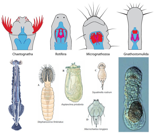
Variety of Gnathifera. On top: the chitinous jaws of Gnathiferan phyla, in red (Nicolas Bekkouche, Wikipedia). Below, from left: the arrowworm Pterosagitta draco (Chaetognatha), with its grasping spines extruded (Animal Diversity Web); some examples of Rotifera (Hickman & al 2008); Limnognathia maerski, only species of Micrognathozoa, carrying an enormous, highly decorated egg (Brusca 2016).
3c2c. Rouphozoa “sucking animals”
3c2c1. PLATYHELMINTHES “flat worms” The phylum of, well, flatworms; which includes such fascinating beings as flukes and tapeworms. They have lost their anus, and so have only a mouth leading into a branching gut; but often they are parasitic, and can absorb nutrients from the host through their skin. See part 6.5.
3c2c2. GASTROTRICHA “hairy bellies”: Millimeter-sized aquatic worms with a rounded back and a flattened belly, all covered in scales, spines, and bristles. Unlike flatworms, they have distinct mouth and anus, but for the rest their body organization is similar. They have a distinct brain that might also contain simple eyespots, and adhesive glands to attach to a substrate.
3c2d. Lophotrochozoa “animals with crest or wheel” or Spiralia: This group has several common traits but none that truly universal. The former name refers to the trochophora, a particular type of larva that swims with several ciliated bands, and has a sensorial apical organ connected to nerve ganglia. This larva was probably found in the earliest Protostomes but lost in most of their other branches. The latter name refers to a feature of embryonal development called spiral cleavage: the cells on top of the early embryo shift around so they’re no longer aligned with the ones below.
3c2d1. MOLLUSCA “soft ones” The phylum of chitons, tusk shells, clams, oysters, scallops, slugs, snails, squids, octopodes, and cuttlefish. The defining features are a muscular foot used for locomotion (which divides into the tentacles of squids), a tongue-like radula used to scratch at food, and a mantle that surrounds the organ mass and generally secretes a calcareous shell. We shall see more of them in part 7.
3c2d2. ANNELIDA “ringed” The phylum of segmented worms, such as ragworms, tubeworms, earthworms and leeches. They have a body divided in compartments with a true coelom, and blood vessels along its length; each segment contains its own excretory apparatus and usually locomorory appendages or bristles. See part 6.5.
3c2d3. NEMERTEA (named after the sea nymph Nemertes, “the unerring”) or Rhynchocoela “hollow muzzle”: Known as “ribbon worms”, they are mostly marine, though there are a few terrestrial species. They have a sharp-tipped muscular proboscis, normally tucked in a pocket in their head, which can shoot out like the tongue of a chameleon to catch preys. They are the simplest animals that have closed blood vessels, like us, rather than have blood soaking their whole body. For the rest, they are rather similar to Turbellarian flatworms, which we’ll see in part 6.5.
3c2d4. CYCLIOPHORA “wheel carriers”: A micro-phylum with no more than three species, all in genus Symbion, first discovered in 1995. All of them live exclusively on the mouthparts of cold-water lobsters, stealing a tiny fraction of their meals. They are sac-shaped, with an adhesive foot and a ciliate mouth at the top, only 0.3 mm long, and reproduce by budding.
Left: minute Lepidodermella squamatum (Gastrotricha), next-of-kin of flatworms (Giuseppe Vago, Wikipedia). Right: the ribbon worm Gorgonorhynchus repens (Nemertea), extruding its uniquely branched proboscis in response to a threat (Rachel Koning, Wikipedia)
3c2d5. Lophophorata “crest-carriers”: Distinguished by having a lophophore, a hydraulic ridge covered in ciliated tentacles that catch microscopic food particles when exposed to a water current. The gut is U-shaped, with the mouth opening at the center of the lophophore, and the anus usually just outside. Usually unable to move much -- they are very ‘coral-like’ in lifestyle.
3c2d5a. † HYOLITHA “Y-shaped stone”: A completely extinct phylum from the Paleozoic era, some 500-to-250 million years ago. They had conical shells with a cap to close them, and coiling structures that perhaps served to prop them up on the seafloor. These structures are called helens, after the daughter of the paleontologist who described them.
3c2d5b. BRYOZOA “moss-animals” or Ectoprocta “anus outside” or Polyzoa “many animals”: Very coral-like indeed: colonial animals with tiny polyps that grow on hard surfaces, forming a chitinous or calcareous communal skeleton. Alcyonidium forms gelatinous finger-like columns; Membranipora forms delicate lace-like webs; Plumatella forms rigid branches. Food particles are carefully selected by the tentacles and can be rejected. The guts of the polyps are connected internally, so they don’t all need to catch food on their own; some are free to specialize for different functions, such as whip-like vibracula and beak-shaped avicularia, which defend the whole colony.
3c2d5c. ENTOPROCTA “anus inside”: Basically the same as Bryozoans, except the anus opens inside the lophophore, too. Can be colonial or solitary.
3c2d5d. PHORONIDA (named after the Phoronis or Io, a lover of Zeus): A very small phylum with animals that resemble Bryozoa but are not colonial. They have tube-shaped body several cm long, anchored to rock or buried in sand, into which their flower-like lophophores can be retracted.
3c2d5e. BRACHIOPODA “arm-feet”: A sad story: between 500 and 350 million years ago, Brachiopods or “lampshells” were absolutely everywhere in Earth’s seas, but they were hit badly by the Devonian and Permian mass extinctions, and now only a few hundred species are left, their niches mostly taken over by Bivalve mollusks (e.g. clams). They are indeed quite clam-like, but whereas Bivalves have symmetrical left- and right-valves, Brachiopods have a larger upper valve and a smaller lower one. Enclosed between these valves is a pair of spiral-shaped lophophores which deliver food particles to the mouth.

Top left: two Symbion pandora (Cycliiophora) with larvae growing from their side (Nielsen 2012). Bottom left: Frenulina sanguinolenta (Brachiopoda) from the tropical Pacific, with the half-open valves revealing the curled lophophores inside (Brusca 2016). Right: the freshwater colonial Bryozoan Cristatella mucedo, with dozens of lophophorate polyps producing a common gelatinous matrix to creep about like a slug (Ernst Haeckel).
3c2e. Ecdysozoa “molting animals”: The distinctive feature of this clade is covering their whole body with a cuticle made out of organic molecules. The cuticle is strong and water-proof, but since it’s not made of cells it cannot grow: as the animal develops to adult size, it must shed or molt periodically its old cuticle and secrete a new one from its bare skin. They have few or no cilia (even their sperm cells often lack flagella!), and may have hard “teeth” or scales around the mouth.
3c2e1. Scalidophora “platelet-carriers”: A group of phyla whose cuticle is moade out of chitin, forming various kinds of plates, scales, and spines. They all live in sand or gravel.
3c2e1a. LORICIFERA “armor-carriers”: A small phylum discovered only in the 1980s. They have an egg-shaped armor or lorica and a retractile, cone-shaped head surrounded by long spines. Less than a mm long, they live in gravelly seafloors, eating bacteria. Three species discovered in the deep Mediterranean in 2014 are the only multicellular organisms known to survive in perpetual lack of oxygen.
3c2e1b. KINORHYNCHA “mobile muzzle”: Sometimes known as “mud dragons”, these tiny mud-dwelling worms have an 11-segmented trunk covered in back-pointing spines, and a further ring of spines surrounding their mouth. They cannot swim, but only crawl or burrow with their hydraulic extensile head: the spines prevent them from sliding backward.
3c2e1c. PRIAPULIDA “little Priapus”. These seafloor-dwelling worms have evertible hydraulic heads they use to burrow in the sand, which make them look like... well, there’s a reason they are named after the Greek god of penises. Size’s about the same, too. The cylindrical body is mostly filled with liquid and by very large gonads, and ends posteriorly with mysterious hollow appendages probably used to breathe.
The strange and unfairly obscure Scalidophora. Left: Pliciloricus enigmatus, the first Loriciferan ever discovered (Carolyn Gast). Top right: the “mud dragon” Echinoderes (Kinorhyncha), with its spiny head fully protracted (Brusca 2016). Bottom right: Priapulus caudatus (Priapulida), lying on a Norwegian beach, with its burrowing head inflated (Hickman & al 2008).
3c2e2. Nematoidea “thread-like”: A clade of two rather similar phyla of worms.
3c2e2a. NEMATODA “threads”: One of the big ones: there are tens of thousands of named species. Their body is smooth and cylindrical (hence the name of “roundworms”); a pseudocoelom filled with liquid under pressure, contained by a tough collagen cuticle, gives them shape; they have lost any trace of cilia or flagella. They have a linear gut that starts with a large muscular pharynx to suck microscopic preys. Muscles run only along the body length, and are used to swim or burrow in a whip-like motion. Most, living in water and soil, are under 1 mm long, but parasite species can be larger; for example the cm-sized Ascaris lumbricoides, commonly found in the gut of people who ingest dirt. Placentonema, which lives in the uterus of sperm whales, can grow to 8 meters long! Other parasites are whipworms (Trichuris), hookworms (e.g. Ancylostoma), dog heartworms (Dirofilaria), and the terrible Wuchereria which causes elephantiasis. Most species however are harmless eaters of bacteria. The microscopic Caenorhabditis elegans is famous among biologists as one of the most extensively studied organisms in the world, since it’s so easy to raise and lives only a few days. A peculiarity of Nematodes is eutely, which means their development is so strictly organized that each species has a fixed number of cells: an adult hermaphrodite C. elegans, for instance, has exactly 959 cells; here’s the full list.
3c2e2b. NEMATOMORPHA “thread-shaped”: A much smaller phylum than Nematoda, and exclusively parasitic. Perhaps you’ll have seen haunting videos of huge tangles of worms sliding out of an insect’s abdomen upon being immersed in water: those are Nematomorphs, also known as “horsehair worms”. They are fairly similar to Nematodes, but their gut is fused into a solid mass of cells; most of their food they absorb as larvae, living in the “blood” of arthropods.
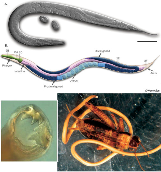
Top: a hermaphrodite adult of Caenorhabditis elegans (Nematoda), about 1 mm long; this species has males and hermaphrodites, but no specialized females (WormAtlas). Bottom left: the friendly smile of the hookworm Ancylostoma caninum (Nematoda), which uses those hooks to attach to the intestine wall of dogs (Brusca 2016). Bottom right: the horsehair worm Paragordius tricuspidatus (Nematomorpha) leaving the husk of its host, a cricket (Hickman & al 2008). As they need to hatch in water, Nematomorphs of that species alter the behavior of their host to induce it to drown itself in the closest pond.
3c2e3. Panarthropoda “all arthropods”: Or rather, one should say, Arthropods plus their closest relations, which usually have some kind of paired limbs. Loosely synonymous with Lobopodia “lobed feet”. Their coelom is very reduced, and replaced by a haemocoel filled with blood (more properly hemolymph) in which organs are directly soaking. A muscular heart ensures blood circulations, but other than it there are no blood vessels.
?3c2e3a. † DINOCARIDIDA “fearsome shrimps”: Wonderfully weird quasi-Arthropods from the seas of the Cambrian. They were the top predators of their environment, with well-developed eyes, relatively large brains, and massive raptorial appendages. Just take † Anomalocaris, with its pair of articulated mouth tentacles, looming with its half meter of length over a mostly cm-sized fauna, or † Opabinia, with its five (!) eyes and mouth at the end of a flexible proboscis. The later † Tamisiocaris even took to whale-style filter-feeding with comb-like appendages. They didn’t have real limbs, but lateral flaps that probably beat up and down, “rowing” in the water.
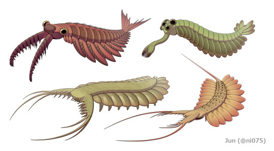
A beautiful selection of † Dinocaridida by Junnn11 (Wikipedia). Top left: † Anomalocaris canadensis, the largest predator of Burgess Shale. Top right: † Opabinia regalis. Bottom left: † Pambdelurion whittingtoni. Bottom right: † Kerygmachaela kierkegaardi. All are from the Cambrian; top row from Burgess Shale (Canada, about 510 million years ago), bottom row from Sirius Passet (Greenland, about 520 million years ago).
3c2e3b. TARDIGRADA “slow walkers”: You know these: “water bears”, those adorable microscopic eight-pawed creatures famous for being incredibly resistant to environmental stresses and dangers by dehydrating and stopping their own metabolism. (Boiling water! Liquid nitrogen! Pure alcohol! Hard vacuum! In reality, no species is resistant to all.) They have, guess what, a muscular pharynx with retractile stylets to pierce and suck fluids from animal or plant cells. They are so small that their muscles only have a handful of cells each, and they can expel eggs or waste only when molting their cuticle.
3c2e3c. ONYCHOPHORA “claw-carriers”: While all the other phyla are partly, mostly, or even exclusively aquatic (and often just marine), Onychophorans are the only animal phylum that is exclusively terrestrial. To be sure, its first representatives in the Cambrian, like Burgess Shale’s Aysheaia, lived in shallow seas; but their descendants are found only in the litter of tropical forests. Known as “velvet worms” because of their soft chitinous cuticle, they have cylindrical bodies walking on up-to-40 pairs of stubby clawed legs. The mouth is flanked by antennae and by massive slime glands that can shoot strings of gluey mucus to entangle preys and predators. Like insects, they breathe through lateral spiracles that allow air directly into the body. They are usually live-bearing, and may even have a placenta.
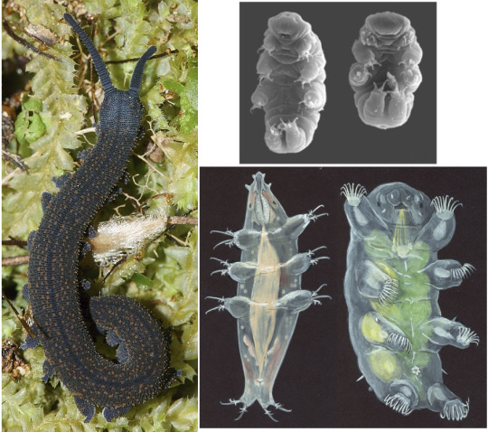
Left: the velvet worm Peripatoides novaezelandiae (Onychophora) (Uwe Schneehagen, Wikipedia). Elsewhere: a variety of Tardigrades: Hypsibius dujardini (top), Milnesium tardigradum (bottom left), and Echiniscoides sigismundi (bottom right) (Animal Diversity Web).
3c2e3d. ARTHROPODA “articulated feet”. Nothing much. Just like, you know, >90% of the whole Animal kingdom by species count. Spiders, scorpions, mites, crustaceans, millipedes, and all sorts of insects are here -- see parts 8 and 9 on that!
3c3. Deuterostomia “mouth second”: The other major part of Bilateria, distinguished from Protostomia by features of embryonal development, chiefly the fact that the blastopore develops into the anus and not the mouth. The two main phyla of Deuterostomes are Echinodermata (starfish, sea urchins, and such) and Chordata (vertebrates and our kin). See part 10 to continue our journey in that direction.
...
Special secret bonus phylum! MONOBLASTOZOA “single-layer animal”. Only one species known, Salinella salve, which was observed exactly once in 1892 in a salt pan in Argentina and then was never seen again, so that today most zoologists doubt it ever even existed. They supposedly have only one kind of cells, forming a single ciliated layer than encloses a gut with a mouth and an anus.
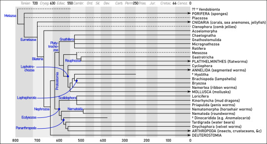
Summary. Dates (mostly from Edgecombe & al 2011 and Parfrey & al 2011) are in millions of years ago. Note how most of the radiation occurs between the Ediacaran (”Ediac.”) and Cambrian (“Cambr.”) periods -- roughly between 600 and 500 million years ago.
Sources
Adl & al (2019), “Revisions to the Classification, Nomenclature, and Diversity of Eukaryotes” (link)
Brusca (2016), Invertebrates (3rd edition), Sinauer
Dunn &al (2018), “Ediacaran developmental biology” (link)
Edgecombe & al (2011), “Higher-level metazoan relationships: recent progress
and remaining questions” (link)
Fernández & Gabaldón (2020), “Gene gain and loss across the metazoan tree of life” (link)
Giribet (2016), “Genomics and the animal tree of life: conflicts and future prospects” (link)
Hickman & al (2008), Integrated Principles of Zoology (14th edition), McGraw-Hill
Ivantsov & Zagrevskaya (2021), “Symmetry of Vendobionta (Late Precambrian Metazoa” (link)
Laumer & al 2019, “Revisiting metazoan phylogeny with genomic sampling of all phyla” (link)
Nielsen (2012), Animal Evolution: Interrelationships of the Living Phyla
Parfrey & al (2011), “Estimating the timing of early eukaryotic diversification with multigene molecular clocks“ (link)
4 notes
·
View notes
Text
CAN YOU MOISTURIZE YOUR SKIN TOO MUCH?
Moisturizer helps keep your skin hydrated and refreshed. It can also reduce the risk of many skin concerns, like extreme dryness or oiliness, fine lines etc. But is it possible to moisturize your skin too much? The short answer is YES, but not as you expect.
Why are we moisturizing the skin in the first place?
Moisturizers help our skin in terms of reducing water loss and keeping irritants out, but isn’t that the job of our skin in the first place? The answer is YES, it is.
How does the skin do that?
Well skin the outermost layer is called the Stratum Corneum. It’s essentially composed of dead skin cells called Corneocytes. There are specialized proteins in between the Corneocytes called Cornea Desmosomes that rivet between them and, with time, degrade. Which allows for shedding of the Corneocytes and that’s what exfoliation basically is.
Microanatomy of stratum corneum depicting the layers of epidermis. From: Sehgal, Virendra. (2018). The impact of microanatomy and changing physiology of stratum corneum the skin barrier on evolution of atopic dermatitis. A visual representation to better explain how you can moisturize your skin too much.
Microanatomy of stratum corneum depicting the layers of epidermis. From: Sehgal, Virendra. (2018). The impact of microanatomy and changing physiology of stratum corneum the skin barrier on evolution of atopic dermatitis.
Now, when the water content of that outer layer drops, that can lead to dry heaped up skin cells. And ultimately impair the health of your skin barrier.
You’ve also got lipids that seal in water and keep irritating things out. Within that milieu, you also have hydroscopic factors that bind water and hold it in place. That’s referred to as natural moisturizing factors.
It’d be all well and good if we weren’t exposed to a lot of things that eat away at that protective layer. Detergent that we are encounter with washing our hands, cleansing cosmetics, makeup, dirt, impurities, pollutants, and all sorts of things settle on the surface of the skin. That protective layer can become more and more damaged, and you can start losing more water.
When our moisture barrier is impaired, we lose more water from the skin. Making us more prone to irritation, acne flares, redness, and all sorts of skin problems.
How does moisturizer work?
Moisturizers employ two strategies:
• The first strategy is to increase the water content in the Stratum Corneum (remember that outermost layer of your skin?) through the inclusion of hydroscopic ingredients. Namely things like Glycerin and Hyaluronic Acid that bind water. This way, they kind of mimic your skin’s natural moisturizing factors.
• The second strategy behind moisturizers is to deposit a water-soluble oily substance on the surface of the skin. Which kind of mimics the skin’s intracellular lipids. That’ll trap water in there, and ultimately prevent water loss. Those ingredients are known as occlusives and one of my favorites is Petrolatum. Why? Because it can reduce water loss out of the skin by 99%.
Moisturizers help address the needs of the skin barrier and everyone can benefit from that, including people who have oily skin.
Can you moisturize your skin too much?
Can you have too much moisture in the skin? The short answer is YES, but it’s not likely because you are using a moisturizer.
There is a tipping point where you can have too much hydration in the Stratum Corneum. And there are quite a few conditions in which that occurs.
The first situation that comes to mind is diaper rash. You must think I’m out of my mind for even bringing up diapers in a blogpost about skincare but hear me out! Diaper rash happens because you have a moist environment in the diaper that overly hydrate the skin. Naturally, the end result is losing even more water and compromising your skin barrier. But there is another situation where a similar process takes place. One that I’m sure you’ve experienced, maskne.
Interestingly enough the remedy for both of these situations is to use an occlusive moisturizer to act as a barrier to water loss, and to prevent friction and further irritation so that your your skin can heal and recover.
The same thing can also happen in anywhere where you have skin on skin contact (e.g. under the armpits, abdominal folds etc.)
Can too much moisturizer make your skin oilier?
That is not true, too much moisturizer will not make your skin oilier. Oil production is governed by your hormones. Which means that it won’t be influenced by putting moisturizer on your skin.
How much moisturizer is too much?
I am hesitant to say that you can’t overdo it with moisturizer because I know there will be inevitably someone out there who goes overboard. Someone that will be putting on moisturizer every minute of the day. And yes that is excessive, and you certainly can always overdo anything. So I’m not advocating for the excessive use of moisturizer.
What I will always encourage people to do is use a moisturizer after cleansing. Because the cleansing action can disrupt some of the lipid barrier, and a moisturizer can help reduce water loss out of the skin. Naturally, if you wash your face twice a day, you should be using a moisturizer twice a day.
If you have eczema or some other dry skin condition. Then you may benefit from putting on moisturizer again at some other point in the day.
When it comes to hands, you can definitely get away with reapplying moisturizer more frequently. Because we wash our hands more often, we are constantly stripping them of moisture. So using a lot of occlusive moisturizer to the hands is recommended.
Conclusion
You can certainly moisturize your skin too much, but it most likely won’t be because of a moisturizer.
I hope you enjoyed this post! It’s an excerpt of this blogpost on my website ;) See you soon~
3 notes
·
View notes
Text
Skin
The skin is actually the largest organ and it actually makes up 15 per cent of your bodyweight. The average person roughly 10kg covering two square metres. Your skin does many things. It:
Contains nerve receptors that allow you to feel touch, pain, and pressure
Helps control fluid and electrolyte balance
Helps control your body temperature
Protects you from the environment
This system acts as a protective barrier between the external environment and the inside of the body, shielding the internal organs against heat, light, injury and infection. In addition, skin plays an important role in regulating body temperature, preventing water loss, producing vitamin D and detecting sensations caused by mechanical stimuli that make contact with or put pressure on the skin.
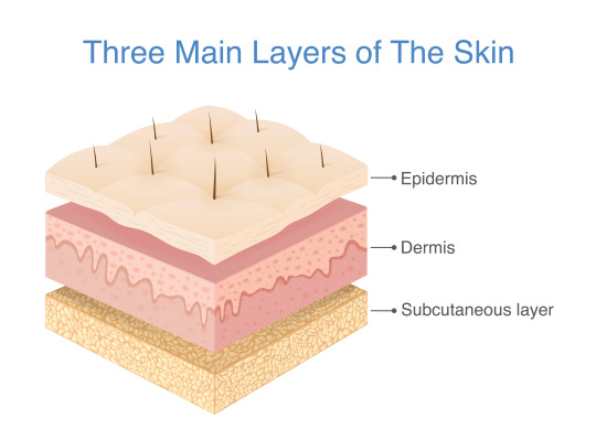
Our outer organ is made up of two main layers. The outer ‘epidermis’ is as thin as a sheet of paper, yet it accounts for most of the skin’s barrier functions. The ‘dermis’ beneath is thicker and carries out a diverse range of roles: collagen and elastin give skin its shape, plumpness and elasticity, over 17 kilometres of blood vessels – enough to bridge the Strait of Gibraltar – and millions of sweat glands regulate body temperature by retaining or releasing heat, exquisitely sensitive nerve receptors enable us to feel our way through life and a standing army of immune cells waits for any foreign intruder.
Your paper-thin epidermis is scratched, squashed and stretched thousands of times a day, but it doesn’t break – at least not easily– or wear out. This is because the wall of the skin is constantly being supplied with new, living bricks: keratinocytes. These cells are made up of the tough protein keratin, which is unbelievably strong: it also forms our hair and nails, as well as the unbreakable claws and horns found in the animal kingdom.
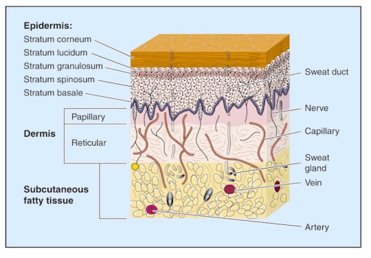
Epidermis
The epidermis is a complex ‘brick wall’ made of cells called keratinocytes, which produce a protein called keratin. The epidermis also contains pigment cells called melanocytes, which produce melanin, Langerhans cells, which present antigens to the immune system, and Merkel cells, which have a sensory function.
Basal layer — the columnar or rectangular cells at the bottom of the epidermis from which new cells are continuously produced. Scattered melanocytes are normally found in this layer.
Squamous cells — flat epithelial cells found on the skin surface. The structure of the skin is described as a stratified squamous epithelium, referring to the way the cells are built up in layers.
Granular layer — flattened cells filled with dark granules containing keratohyaline protein.
Horny layer — stacks of dead cells without nuclei make up the dry or keratinised stratum corneum. The top layer of cells loosens and falls off.
Desmosomes — the structures that stick adjacent keratinocytes tightly together, rather like cement between bricks.
Epidermal appendages include:
Eccrine glands, which produce sweat
Apocrine glands, scent glands found in armpits and groins
Pilosebaceous structures containing hair and sebaceous glands
The dermis
The dermis is made up of connective tissue that supports the epidermis, providing nutrients and protecting it. The papillary dermis is the upper portion beneath the epidermis, and the lower portion is the reticular dermis.
Collagen — a structural protein making up the bulk of the dermis. It is produced by fibroblasts. It is composed of a triple helix of strong fibres.
Elastin — the protein that makes up thin elastic fibres. These are produced by fibroblasts. They return deformed skin to its resting position.
Ground substance — the gel component of the dermis. It contains hyaluronic acid, dermatan sulphate, and chondroitin-6-sulphate (these are anionic polysaccharides or glycosaminoglycans).
Fibroblasts — cells found in the dermis that produce collagen, elastin, ground substance and fibronectin (a glycoprotein).
Nerves — sensory and autonomic fibres with distinct nerve endings for touch, heat, cold, pressure and pain.
Blood vessels — arteries, arterioles, capillaries, venules and veins carrying blood to and from the skin.
Lymphatics — an extensive network of thin-walled vessels that nourish and drain the skin.
Arrector pili muscles — these are attached to hair follicles. Contraction results in goosebumps.
Cellular infiltrations — immune cells around blood vessels, and recruited in great numbers to heal wounds and fight infection. Many skin diseases are characterised by specific patterns of these cells.

Dr Monty, Lyman (2019) 10 remarkable facts about skin. https://www.sciencefocus.com/the-human-body/10-remarkable-facts-about-skin (Accessed: November 10, 2023).
0 notes
Text
Histology (desmosomes) and our histo professor, drawn by yours truly
1 note
·
View note
Text
The morula forms after fertilization and multiple cleavages about 4 days after fertilization. During morula formation compaction occurs, tightening the blastomeres, and desmosomes and gap junctions form. Desmosomes are protein structures that attach cells together
0 notes
Text
Subconscious Problems Among Aids Medical Providers In the COVID-19 Outbreak in China: Mediating Jobs involving Institutional Help along with Strength.
Trademark (Chemical) 08 Azines. Karger AG, Basel.Your remaining atrial rear walls (LAPW) has an important position inside atrial fibrillation, however the fundamental procedure stays uncertain. In our examine, we all looked for to define the particular histological top features of the particular LAPW. Diverse atrial parts ended up dissected through minds of normal Sprague-Dawley rodents and individuals. Haematoxylin/eosin as well as lorrie Gieson discoloration were utilised to review atrial cardiomyocyte agreement as well as bovine collagen submission, correspondingly. Intercellular junctions were looked at by transmitting electron microscopy. As opposed along with other atrial areas, the particular LAPW exhibited Denosumab solubility dmso far more cluttered cardiomyocytes, more substantial intercellular areas and varied myocardial dietary fibre arrangement. The amount involving collagen was significantly greater within the LAPW than in other atrial locations. Oddly enough, desmosomes ended up rare as well as intercellular breaks from the LAPW. In conclusion, specific disarrangement involving cardiomyocytes with an large quantity of bovine collagen exist in the particular LAPW. The particular sparsity of desmosomes in the LAPW could possibly be related to your heterogeneous syndication as well as separation regarding atrial myocytes.To research the relationship between Quiet Cerebral Infarction (SCI) along with intellectual impairment to be able to testify that will Montreal Mental Examination (MoCA) is really a more delicate verification application as opposed to Tiny Mental State Exam (MMSE) to distinguish intellectual deficits in people along with SCI. We all enrolled 196 men and women in Shanghai involving Cina as well as divided these straight into Two organizations: the actual SCI team which include 112 folks (Forty five males along with 67 girls; aged through Forty three to be able to 80 years of age) as well as the manage group which includes 86 people (43 men and also 41 girls; aged through 44 in order to 79 years). We all accumulated data which includes participants' sexual category, age, blood sugar, elimination operate, blood vessels fats, blood pressure, Electrocardiogram (ECG), carotid artery ultrasound examination, MMSE score, and MoCA report. SAS computer software (model Being unfaithful.A single) was used to perform record evaluation in actual exams benefits through 196 individuals. Additionally, a number of logistic regression product had been piled up in order to display screen your unbiased risk factors regarding SCI. Aside from, Device User Characteristic (ROC) evaluation was also used. Multivariate logistic regression investigation demonstrated that irregular ECG along with information on carotid artery plaques were unbiased risk factors involving SCI incidence, even though MoCA standing had been adversely connected with SCI risk. Additionally, MoCA might be more valuable in projecting vascular cognitive problems (VCI) compared to MMSE (0.667 versus 3.626). We all discovered that the particular MMSE had been generally insensitive to detect cognitive incapacity; but the MoCA includes a good harmony involving awareness and also nature in the cut-score involving Twenty-six. All of us learned that your sufferers with SCI may well accompany with VCI as well as the MoCA seemed to be a great screening process check regarding people using SCI, worthy of becoming promoted inside the clinical practitioning upon China inhabitants.
#Autophagy Compound Library#Akt inhibitor#Tremelimumab#Denosumab#Bevacizumab#Trastuzumab#Cetuximab#Nivolumab#Atezolizumab#Pembrolizumab#Ipilimumab#Ramucirumab#Pertuzumab#Rituximab#Adalimumab#Sarilumab#Tocilizumab#Durvalumab#Avelumab#Camrelizumab#HDM201#Abraxane#MI-773#β-Sitosterol#GSK3787#OTS964#SN-011#Enarodustat#Trastuzumab Emtansine#Bindarit
1 note
·
View note
Text
Long-term abuse of a high-carbohydrate weight loss program is because dangerous like a high-fat diet plan for development as well as progression of liver organ injury in a mouse style of NAFLD/NASH.
The end results of these challenges upon spreading, difference as well as phrase involving cell-cell bond compounds had been looked at for the first time in the organotypic type of our skin. Entirely stratified tissue had been subjected to a time span of air starvation as well as up coming reoxygenation. Localized changes in keratinocyte morphology, glycogen retailers and mobile junctions have been witnessed, with additional told apart levels on the skin showing the initial proof o2 starvation. Cell bloating from the granular level had been concurrent together with aquaporin-3 depletion. Your keratinocyte adherens jct meats E-cadherin and beta-catenin had been substantially reduced in a regio-specific manner Zelavespib cell line during the entire skin following oxygen lack. As opposed, P-cadherin along with the desmosomal healthy proteins desmoplakin and desmoglein-1 ended up refractory to oxygen deprival. Relative to normoxic regulates, hypoxic tissues exhibited greater mRNA quantity of a transcriptional repressor Slug; nonetheless, mRNA amounts of the connected transcriptional issue Snail ended up unaffected. Just about all cell and also molecular alterations ended up relatively easy to fix after reoxygenation. These kinds of benefits demonstrate that fresh air lack and also reoxygenation exert differential consequences upon skin bond healthy proteins as well as advise a story position for cadherins, beta-catenin, and Slug in hypoxia-induced junctional adjustments developing in stratified squamous epithelium.Goal: Case study had been targeted to gauge the particular effect of gender in still left ventricular (LV) upgrading throughout metabolic affliction (MetS). Methods as well as results: Many of us signed up 200 themes with no diabetes mellitus as well as overt heart diseases, never addressed with anti-hypertensive drug treatments as well as statins: 58 men and also Forty five ladies using MetS harmonized through grow older, sex and also Twenty-four they would systolic and also diastolic hypertension (British petroleum) along with 58 men as well as Forty five females without MetS. The actual individuals underwent blood vessels checks, Twenty four our own BP monitoring, LV echocardiographic evaluation. LV muscle size indexed by nine(Only two.Seven) has been substantially higher in males and some women together with MetS as compared to with no MetS. Weighed against women with no MetS, ladies with MetS had considerably greater rear walls thickness and also comparable wall membrane width, higher prevalence regarding LV concentric remodeling/hypertrophy minimizing search engine spiders involving LV diastolic perform, although all these details are not drastically various between men using as well as with out MetS. MetS had been an independent predictor of comparative wall structure fullness and LV size list in females, but not in men. Conclusion: The effect of MetS in LV remodeling is really a lot affected by sex: the effects of MetS tend to be more obvious in women, along with continuing development of LV concentric hypertrophy/remodeling as well as preclinical diastolic dysfunction. (D) 2012 Elsevier B. Sixth is v. Most rights set aside.Quantifying testicular homogenization-resistant spermatid mind (HRSH) is really a highly effective signal involving spermatogenesis. These types of matters possess typically been recently performed personally using a hemocytometer, but this technique might be frustrating along with not impartial.
0 notes
Text
IJMS, Vol. 23, Pages 16078: Rumen Epithelial Development- and Metabolism-Related Genes Regulate Their Micromorphology and VFAs Mediating Plateau Adaptability at Different Ages in Tibetan Sheep
The rumen is an important hallmark organ of ruminants and plays an important role in the metabolism and immune barrier of Tibetan sheep on the Plateau. However, there are few studies on rumen development and metabolism regulation in Tibetan sheep at different ages. Here, we comprehensively analyzed the immune function, fermentation function, rumen epithelial micromorphology and transcriptome profile of Tibetan sheep at different ages. The results showed that the concentration of IgG decreased and the concentration of IgM increased with age (p < 0.05), and the highest concentration of IgA was observed at 1.5 and 3.5 years of age. In terms of rumen fermentation characteristics, VFAs of 4-month-old lambs were the highest, followed by VFAs and NH3-N of Tibetan sheep at 3.5 years of age. Hematoxylin-eosin staining and transmission electron microscopy section examination of rumen epithelial tissue showed that the rumen papilla width increased with age (p < 0.001), the thickness of the stratum corneum decreased, the cells in the stratum corneum showed accelerated migration and the thickness of the rumen muscle layer increased (p < 0.001). Desmosomal junctions between the layers of rumen epithelium increased at 1.5 and 3.5 years old, forming a compact barrier structure, and the basal layer had more mitochondria involved in the regulation of energy metabolism. #RNA-seq analysis revealed that a total of 1,006 differentially expressed genes (DEGs) were identified at four ages. The DEGs of Tibetan sheep aged 4 months and 6 years were mainly enriched in the oxidation–reduction process and ISG15-protein conjugation pathway. The 1.5 and 3.5-year-olds were mainly enriched in skeletal muscle thin filament assembly, mesenchyme migration and the tight junction pathway. WGCNA showed that DEGs related to rumen microbiota metabolite VFAs and epithelial morphology were enriched in “Metabolism of xenobiotics by cytochrome P450, PPAR signaling pathway, Butanoate metabolism pathways” and participated in the regulation of rumen epithelial immune and fermentation metabolism functions of Tibetan sheep at different ages. This study systematically revealed the regulatory mechanism of rumen epithelial development and metabolism in the plateau adaptation of Tibetan sheep, providing a new approach for the study of plateau adaptation. https://www.mdpi.com/1422-0067/23/24/16078?utm_source=dlvr.it&utm_medium=tumblr
0 notes
Text
Randomized Phase II Research together with Cetuximab in Combination with 5-FU and also Long-chain-fatty-acid-CoA ligase or Carboplatin as opposed to Cetuximab in conjunction with Paclitaxel and Carboplatin to treat Sufferers along with Relapsed as well as Metastatic Squamous Cell Carcinoma with the Neck and head (CETMET Trial)
Aim Well-designed dyspepsia (FD) is an extremely frequent functional digestive condition, the particular pathophysiology being badly understood. We hypothesised which damaged digestive tract buffer perform is mixed up in onset as well as endurance of the condition by simply inducing low-grade irritation. Consequently, the goal was to consider duodenal mucosal strength and also low-grade inflammation inside patients along with FD. Style Duodenal biopsy examples have been purchased from 20 patients using FD fulfilling the actual Rome 3 criteria and also 20 age- along with gender-matched healthy volunteers. Transepithelial electric powered resistance (TEER) and paracellular leaks in the structure had been tested inside Ussing spaces. Term regarding cell-to-cell bond protein has been evaluated through real-time PCR, developed mark and/or immunofluorescence. Numbers of mast tissues, eosinophils and intraepithelial lymphocytes had been examined by immunohistochemistry. Final results Individuals using FD displayed reduced TEER and also elevated paracellular passageway compared with healthy regulates, that's an indication of impaired mucosal strength. Additionally, unusual phrase involving cell-to-cell adhesion healthy proteins at the a higher level tight junctions, adherens junctions and also desmosomes has been demonstrated. Furthermore, sufferers had been classified by the presence of low-grade irritation, as exhibited by #Link# improved infiltration of mucosal mast tissue as well as eosinophils. A substantial organization between the term a higher level several cell-to-cell bond protein, the actual level involving increased permeability along with the seriousness of low-grade infection was found. A conclusion These bits of information concern your traditional model that sufferers with FD demonstrate no constitutionnel alterations in the stomach tract. We recommend that damaged digestive tract barrier purpose is often a pathophysiological procedure throughout FD. Hence, restoration regarding intestinal tract buffer ethics might be a potential restorative target for the treatment of people with FD.ObjectivesThe target ended up being to see whether several steps involving urgent situation department (ED) excitedly pushing are generally connected with an critical indicator associated with top quality and also safety: time and energy to reevaluation of youngsters using reported really abnormal triage essential indications. MethodsThis was obviously a retrospective cross-sectional review of people using severely irregular crucial indicators assessed in triage on the 2.5-year time period (September One, 2006, to be able to May possibly #Link# A single, 09). Cox proportional threat examination was utilized to discover fee percentages with regard to time to severely excessive important indication reassessment, while manipulated for possible confounders. ResultsIn this A couple of.5-year trial, Nine,976 people using really irregular #Link# important indicators throughout triage (addressing Three.9% regarding 254,408 sessions) ended up placed in normal Male impotence rooms along with electric notifications motivating important indication reassessment following One hour. All round, the mean time to reassessment had been Eighty four units.
0 notes
Photo
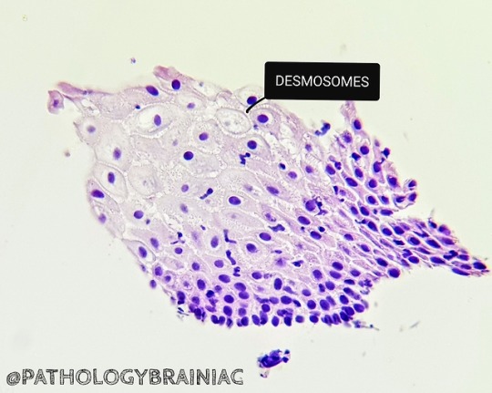
Desmosomes are one of four intercellular junctions present on the lateral side of neighboring polarized epithelial cells. Desmosomes mechanically integrate adjacent cells by coupling adhesive interactions mediated by desmosomal cadherins to the intermediate filament (keratin) cytoskeletal network.
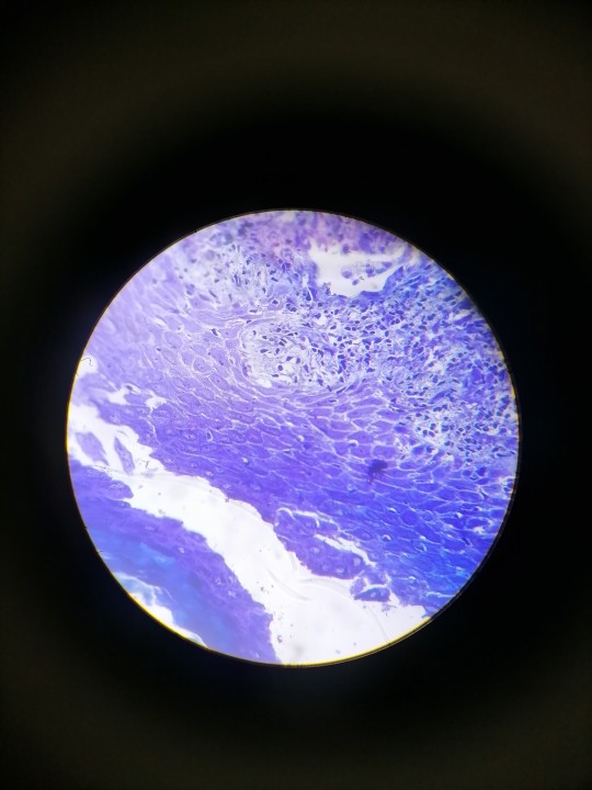
*** Esophageal squamous epithelium.
#desmosome#junction#cell#network#keratin#epithelium#adhesion#interaction#cadherin#filament#medicine#medical#pathology#dematology#esophagus#nucelus#learning#motivation#medblr#pathblr
2 notes
·
View notes
Photo
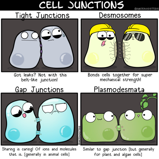
Some cell junction functions!
#science#biology#scicomm#studyblr#studyspo#studyhard#sciart#stem#cells#microbiology#desmosomes#plasmodesmata
669 notes
·
View notes
Last Seen Blogs

plum-blossoms-in-snow
Atlas

3312milits-blog
milla_vagg

glebvagnov
a new wind blows

the-kingshound
The King's Hound

thelibrarybookdragonishere
The Library BookDragon Is Here