#neural crest cell
Text
I….AM…..GOING….TO…….GRADUATE!!!!!!!!
#manifesting manifesting manifesting#my mental health has taken its monthly dive exactly a week before my thesis is fucking due#I NEED to do my thesis I need it to be DONE so I can fucking graduate#and I have so many complicated feelings about it but I’m not going to think about that I’m not going to cry I’m just going to write about#melanoblast development and neural crest diffeeentation of cells in snakes#at all costs I am graduating from college in 3 weeks
9 notes
·
View notes
Text


☆ man's best friend
☆ how to stop dog begging behavior - tractive | 4x20 - the rapture | 9x22 - stairway to heaven | 15x08 - our father, who aren't in heaven | 6x21 - let it bleed | 050612-274-wolf - ron niebrugge ; herakles - euripides ; 8x07 - a little slice of kevin | the archaeology of dog domestication - bgillard | how to teach your dog to walk to heel in seconds - will atherton canine training | violence happening location #38 - moisés mahiques | 9x09 - holy terror | unidentified photo source, earliest trace posted 07.16.14 by catherine zhang | two dogs eating from one bowl - nick onken | 150317.jpg - borzoikavka | the thorn merchant - yusef komunyakaa ; 10x22 - the prisoner | desperation sits heavy on my tongue - tullipsink | 6x19 - mommy dearest | ecstasy of saint teresa - gian lorenzo bernini ; photo courtesy of findingharmonyinartanddesign | 7x21 - reading is fundamental | 8x17 - goodbye stranger | 'i think about this a lot' tumblr post - 1rakus | quote by vàzaki nada ; unidentified photo source, earliest trace posted 09.30.12 by pam bliss | tecumseh fitch : the domestication syndrome and neural crest cells - university of california television (UCTV) | nasty little thoughts 1/? - notideeart | chosen - zeppelinmoon | bayerischer wald - wim berlijn | the plague dogs (1982) | creamthing has a vision- a tombstone that looks like him - samsketchbook | can old dogs learn new tricks? - malcolm weir, dvm, msc, mph; lynn buzhardt, dvm, vca animal hospitals ; 15x18 - despair
#cas.art#cas supernatural#cas web weaving#web weaving#web weave#spn web weaving#supernatural#spn art#parallels#supernatural web weaving#meta#lgbt castiel#destiel#animal death#spnedit#cas studies#dogs#spn fanart#dognatural#cannibal#hiii. i need to go eat now. runs away. i hope u guys like.it i feel insane<333 hope it Makes Sense#ok ily guys byeeee
133 notes
·
View notes
Text
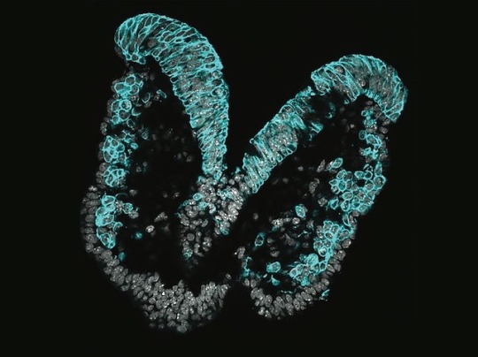
Extrusion Zone
Early in embryo development, neural crest cells (NCC) leave their origin in a process called delamination and migrate to become various cell types. Here, live time lapse imaging reveals that, and how, mammalian NCC are extruded in the process of delamination
Read the published research article here
Image from work by Emma Moore and colleagues
Stowers Institute for Medical Research, Kansas City, MO, USA
Image originally published with a Creative Commons Attribution – NonCommercial – NoDerivs (CC BY-NC-ND 4.0)
Published in bioRxiv, March 2024 (not peer reviewed)
You can also follow BPoD on Instagram, Twitter and Facebook
#science#biomedicine#immunofluorescence#biology#embryo development#developmental biology#cell fate#butterfly
6 notes
·
View notes
Text
in that fox-taming experiment, iirc researchers noticed that the friendlier fox lineages started accumulating neotenous characteristics—floppy ears, big eyes, etc. this implies an early stop to their development, before mature skittishness sets in.
but there was also an increased prevalence of white spots around the digits and face, like modern dogs and cats and horses have, but that’s not something that develops after birth. pigment cells are well in place before that.
pigment cells develop from the neural crest, which also gives rise to various parts of the nervous system… is domestication the result of basic nerve restructuring? not just the obvious shift in behavior, but something much deeper?
gotta read some articles. will report back.
16 notes
·
View notes
Text
I wanted to add facial muscles to the anatomy of crocodiles in Chima, but reptile skin is way different than mammalian skin...
Maybe I could make the Chi alter the neural crest cells of the crocodiles?? Chi alters the anatomy of those who consume it, so it would make sense for mammal-like traits to appear on non-mammalian tribes I guess
Like,, idk, maybe Chi made the crocodiles evolve a new type of muscle?? Especially because their faces in Chima look wildly different from the actual animal
If you're confused about how crocodiles in Chima are able to snarl and frown or make slight facial expressions, then here's the answer lol
4 notes
·
View notes
Text
Title: The Transformative Power of Long Distance Hiking on Mind and Body

Introduction:
Long distance hiking has emerged as a popular activity for individuals seeking physical and mental rejuvenation. The effects of extended hikes on both physiology and mental clarity are truly remarkable. In this blog, we will explore the profound impact that long distance hiking can have on an individual's overall well-being, with a particular focus on its potential to alleviate mental health issues such as anxiety.
Physiological Effects:
Engaging in long distance hiking offers numerous physiological benefits. Firstly, it improves cardiovascular health by challenging the heart and increasing blood flow throughout the body. The sustained physical exertion involved in hiking for extended periods strengthens the heart muscle, leading to improved endurance and overall fitness levels.
Additionally, long distance hiking promotes muscular strength and tone. The repetitive movements required during hikes engage various muscle groups, resulting in increased strength and flexibility. Over time, hikers experience enhanced muscle definition and improved overall physical performance.
Mental Clarity:
One of the most remarkable aspects of long distance hiking is its ability to provide mental clarity and focus. When immersed in nature and disconnected from the distractions of everyday life, hikers often find themselves experiencing a heightened sense of awareness and tranquility. The rhythmic motion of walking, combined with the soothing sounds of nature, creates an environment conducive to deep introspection and reflection.
As hikers traverse through breathtaking landscapes, they are able to detach from the stresses and worries of their daily lives. This detachment allows them to gain a fresh perspective on their challenges and concerns. Many hikers report feeling a sense of peace and serenity while on the trail, which can lead to reduced stress levels and improved overall mental well-being.
Cognitive Growth:
The benefits of long distance hiking extend beyond physical and mental rejuvenation. Scientific research suggests that engaging in activities like hiking stimulates neurogenesis, the process by which new brain cells are formed. This phenomenon has been linked to improved cognitive function, memory retention, and problem-solving skills.
When individuals embark on long distance hikes, they are exposed to new environments, challenges, and stimuli. This exposure prompts the brain to adapt and form new neural connections, ultimately enhancing cognitive abilities. Hikers often find that their creativity and critical thinking skills are sharpened as a result of these experiences.
Case Studies:
Numerous case studies have highlighted the positive impact of long distance hiking on individuals struggling with mental health issues, particularly anxiety. For example, Sarah, a 35-year-old woman diagnosed with generalized anxiety disorder, embarked on a month-long hike along the Pacific Crest Trail. Throughout her journey, she noticed a significant reduction in her anxiety symptoms. Sarah attributed this improvement to the combination of physical activity, immersion in nature, and the opportunity for self-reflection that the hike provided.
Scientific Proof:
Scientific evidence supports the notion that spending time in nature, such as during a long distance hike, can significantly reduce levels of cortisol, the stress hormone, in the body. A study published in the Journal of Environmental Psychology found that individuals who engaged in nature-based activities experienced lower cortisol levels compared to those who did not. This suggests that immersing oneself in nature can have a calming effect on the mind and body, reducing stress and anxiety.
Conclusion:
In conclusion, the amazing effects of long distance hiking on both physiology and mental clarity cannot be understated. From improved cardiovascular health and muscular strength to enhanced cognitive function and mental well-being, the benefits of embarking on a long hike are numerous. For individuals grappling with mental health issues, such as anxiety, the transformative power of nature can provide a much-needed respite and pave the way for long-term healing and growth. So, lace up your hiking boots, embrace the wonders of the great outdoors, and embark on a journey that will not only challenge your body but also nourish your mind and soul.
#self impowerment#self importance#self improvement#mental health#self identity#stress#self introspection#self care#resilience#self confidence#anxiety#cognition
0 notes
Quote
Mechanisms specifying cancer cell states and response to therapy are incompletely understood. Here we show epigenetic reprogramming shapes the cellular landscape of schwannomas, the most common tumors of the peripheral nervous system. We find schwannomas are comprised of 2 molecular groups that are distinguished by activation of neural crest or nerve injury pathways that specify tumor cell states and the architecture of the tumor immune microenvironment. Moreover, we find radiotherapy is sufficient for interconversion of neural crest schwannomas to immune-enriched schwannomas through epigenetic and metabolic reprogramming. To define mechanisms underlying schwannoma groups, we develop a technique for simultaneous interrogation of chromatin accessibility and gene expression coupled with genetic and therapeutic perturbations in single-nuclei. Our results elucidate a framework for understanding epigenetic drivers of tumor evolution and establish a paradigm of epigenetic and metabolic reprograming of cancer cells that shapes the immune microenvironment in response to radiotherapy.
Epigenetic reprogramming shapes the cellular landscape of schwannoma | Nature Communications
0 notes
Text
Single-cell and spatial transcriptomics of the avian embryo tailbud
Vertebrate body axis formation initiates during gastrulation and continues within the tail bud at the posterior end of the embryo. Major structures in the trunk are paired somites, which generate the musculoskeletal system, the spinal cord - forming part of the central nervous system, and the notochord, with important patterning functions. The specification of these different cell lineages by key signalling pathways and transcription factors is essential, however, a global map of cell types and expressed genes in the avian trunk is missing. Here we use single-cell RNA sequencing and RNA tomography to generate a molecular map of the emerging trunk and tailbud in the chick embryo. Single cell RNA-sequencing (scRNA-seq) identifies discrete cell lineages including somites, neural tube, neural crest, lateral plate mesoderm, ectoderm, endothelial and blood progenitors. In addition, high-throughput RNA-seq of sequential tissue sections provides a spatially resolved, genome-wide expression dataset for the avian tailbud and emerging body, comparable to other model systems. Combining the single-cell and spatial datasets, we identify spatially restricted genes, focusing on somites and early myoblasts. Thus, this high-resolution transcriptome map incorporating cell types in the embryonic trunk can expose molecular pathways involved in body axis development. http://dlvr.it/T2NPYT
0 notes
Text
SOX10 loss sensitizes melanoma cells to cytokine-mediated inflammatory cell death
The transcription factor, SOX10, plays an important role in the differentiation of neural crest precursors to the melanocytic lineage. Malignant transformation of melanocytes leads to the development of melanoma, and SOX10 promotes melanoma cell proliferation and tumor formation. SOX10 expression in melanomas is heterogeneous, and loss of SOX10 causes a phenotypic switch towards an invasive, mesenchymal-like cell state and therapy resistance; hence, strategies to target SOX10-deficient cells are... http://dlvr.it/SxZZBw
0 notes
Text
Microtia Ear Reconstruction Guide by Dr. Parag Telang
Many people have abnormal size of ears. The oversize or smaller size of the ears than the normal look grabs attention in an inappropriate way. It can hamper one’s self-confidence. These can be corrected through an ear surgery called microtia ear surgery.
In this blog, Dr. Parag Telang, known as the best ear surgeon in Mumbai, will share a surgical guide on microtia ear reconstruction. So read the blog carefully.
What is Microtia?
The absence or underdevelopment of the external ear characterizes a congenital malformation called microtia. Due to the obvious differences in appearance, people with microtia frequently experience physical, emotional, and social difficulties.
Causes of Microtia
Even though the exact origins of microtia are unknown, environmental and genetic factors are thought to be the main contributors to this ear impairment. Only 5% of patients are estimated to have genetic characteristics as a contributing factor. Many theories have explained the causes of microtia during fetal development. Some of them are underneath:-
Disruption of neural crest cells, vascular disruption, and altitude.
Drinking alcohol, Using drugs, and coffee was taken by the woman during her pregnancy.
Side-effects of drugs, including Thalidomide and Accutane
Grade of Microtia
Grade 1- A little ear, frequently with a constrained or even absent ear canal.
Grade 2- An improperly formed outer ear, usually in the top half, with an often constricted or occasionally absent ear canal.
Grade 3: The most prevalent kind of ear disease is characterized by small, abnormally formed ears with an absent ear canal.
Grade 4- A condition called anotia in which the ear and ear canal are missing.
Understanding Microtia and Its Impact
Microtia may affect one ear unilaterally, or both ears may be affected bilaterally. It is generally divided into four grades based on the deformity's severity. While grade II microtia only has a partial ear structure, grade I microtia involves a small ear with a regular form and placement. A small, undeveloped ear characterizes grade III microtia, and the lack of an external ear characterizes grade IV anotia.
Microtia has effects that go beyond appearance. Functional difficulties brought on by ear abnormalities include hearing loss, trouble locating sound sources, and poor sound transmission. Additionally, because of the obvious difference, people with microtia frequently experience emotional and psychological distress, which could result in low self-esteem and social anxiety.
Microtia Ear Reconstruction: The Surgical Guide
Microtia ear reconstruction is an invasive procedure that aims to create a natural-looking ear while enhancing the ear’s functionality. A multidisciplinary team with members who specialize in audiology, psychology, reconstructive surgery, and otology is needed to complete the treatment, which has numerous stages. Taking a closer look at the surgical guide's key procedures
Step 1: Preoperative Assessment
A thorough analysis of the patient's situation comes first. This entails evaluating the degree of microtia, determining the patient's hearing capacity, and discussing the patient's expectations and intended results. This stage also includes getting any necessary imaging, such as CT or MRI scans, to prepare the surgical strategy.
Step 2: Rib Cartilage Harvesting
Due to its stability and capacity to replicate the shape of a natural ear, rib cartilage is frequently chosen as the material for ear restoration. During this phase, a small incision is made in the patient's chest to harvest a piece of rib cartilage. The wound is then stitched up, barely leaving a mark.
Step 3: Framework Fabrication
The harvested rib cartilage is painstakingly cut and molded to imitate the framework of the absent ear. According to the microtia surgeon, the patient's unaffected ear or the desired result must match the measurements and proportions. The new ear is built on top of this framework.
Step 4: Pocket Creation
To fit the framework, the microtia surgeon carves a pocket beneath the patient's skin at this stage. The pocket is meticulously sculpted to ensure that the reconstructed ear is positioned and oriented correctly.
Step 5: Framework Insertion
The framework is put into the pocket after it has been formed and fastened using sutures or minuscule screws. The surgeon makes sure the framework is placed organically and symmetrically.
Step 6: Skin Grafting
In the following stage, a tiny layer of skin is removed, frequently from the patient's scalp, to cover the framework and create a natural texture. The skin graft is meticulously sutured in place to create an aesthetically pleasing contour.
Step 7: Postoperative Care
The patient is closely watched during the healing process after surgery. For optimum healing and best results, pain management, wound care, and follow-up sessions are crucial. In accordance with the patient's requirements, extra operations such as earlobe rebuilding.
Benefits of Microtia Ear Reconstruction Surgery
The utilization of the patient's rib cartilage prevents transplant rejection.
Outcomes that look natural.
Surgery can also be used to treat hearing issues.
Improves facial proportion and attractiveness.
Helps people build their self-esteem and confidence.
The ideal age for Microtia Surgery
An ideal age for microtia surgery is 9 years. Before that, the child’s ear is still developing, so it is not advisable to do the surgery before its full formation.
If someone is looking for an expert for ear reconstructive procedures, they can consult with Dr. Parag Telang, the best microtia surgeon in Mumbai, at Designer Bodyz.
0 notes
Text
Applications of Dental Pulp Stem Cells in Tissue Engineering
Oral tissues and organs are connected functionally in the oral cavity, which is a complex structure. Dental pulp stem cells have therapeutic potential in the regeneration of dental, periodontal, and oral systems.
Dental pulp stem cells (DPSCs) are neural crest-derived cells with a high capacity for differentiation across numerous cell lineages. The regeneration of salivary glands and bone deformities appears to be feasible by employing DPSCs in oral-related structures.
Read more:- https://kosheeka.com/applications-of-dental-pulp-stem-cells-in-tissue-engineering/
#Oraltissues #oralcavity #Dentalpulpstemcells #DPSCs #biotechcompany #stemcells #exosomes #stemcellresearchcenter #primarycells #regenerativemedicine #bioengineering #customstemcellprovider
#research#cellculture#kosheeka#cell culture#biotech#primarycells#cell biology#biotechnology#primary cells#cellbiology
0 notes
Photo
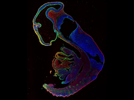
Supporting Development
Like students graduating into the wide world, cells in an embryo sprout from a single collection, called the neural crest, and adventure on as bold early elements of various body structures. Mishaps can cause significant physical abnormalities, so researchers are examining the role of hyaluronan, a key component in the extracellular matrix, which provides support for much of the process. Previous studies of enzymes known to regulate its presence haven’t pinpointed its role. A new approach examined another enzyme, TMEM2 (green in the mouse embryo pictured), which acts on the surface of cells to break down hyaluronan (red, present where TMEM2 levels are low). Like a great teacher, TMEM2 was essential for breaking down barriers and creating an environment conducive to successful cell migration and thriving. Identifying the key regulators and actors of the process might help clear the way for cells, and ultimately people, to fulfil their potential.
Written by Anthony Lewis
Image from work by Toshihiro Inubushi and colleagues
Department of Orthodontics and Dentofacial Orthopedics, Osaka University Graduate School of Dentistry, Osaka, Japan
Image originally published with a Creative Commons Attribution 4.0 International (CC BY 4.0)
Published in PLOS Genetics, July 2022
You can also follow BPoD on Instagram, Twitter and Facebook
#science#biomedicine#extracellular matrix#immunofluorescence#neural crest#embryonic development#developmental biology#hyaluronic acid
4 notes
·
View notes
Text
Postdoctoral Fellow in Cell fate, plasticity and reprogramming
Inserm
Interested in mechanisms regulating cell fate and plasticity during development and regeneration?
Want to do good and fun science in beautiful Paris?
See the full job description on jobRxiv: https://jobrxiv.org/job/inserm-27778-postdoctoral-fellow-in-cell-fate-plasticity-and-reprogramming/?feed_id=45645
#ScienceJobs #hiring #research
Paris #France #PostdoctoralFellow
0 notes
Text
How to assemble a complete jaw
The skeleton, tendons, and glands of a functional jaw all derive from the same population of stem cells, which arise from a cell population known as neural crest. To discover how these neural crest-derived cells know to make the right type of cell in the right location, researchers focused on a particular gene, Nr5a2, that was active in a region of the face that makes tendons and glands, but not…
View On WordPress
0 notes
Text
Three Germ Layers
An embryonic disc is made up of three germ layers. The name of these three embryonic gem layers are ectoderm, endoderm and mesoderm. This three germinal layer disc is formed during third week of intrauterine life. After fertilization cleavage take place in which eight cell stage is converted into sixteen cell stage called morula.
Then, formation of blastocyst take place. This blastocyst differentiate into inner cell mass and outer cell mass. Outer cell mass is known as trophoblast.
During implantation this trophoblast penetrates to uterine epithelium and differentiates into cytotrophoblast & syncytiotrophoblast.
During 2nd week of development , few cells of inner cell mass become flatten and form primitive endoderm. The remaining cell of inner cell mass become columnar & form primitive ectoderm.
Few cells of primitive ectoderm multiplies and form primitive streak. These primitive streak cells migrated between ectoderm & endoderm & form intraembryonic mesoderm. The cells of trophoblast multiply and form extraembryonic mesoderm.
Cavities developed in extraembryonic mesoderm and form extraembryonic coelom which differentiates into somatopleuric and splanchnopleuric layer.
youtube
3 Germ Layers details
1. Ectoderm: The ectoderm is the outermost of the three germ layers. It gives rise to the skin, hair, nails, and the nervous system, including the brain and spinal cord.
2. Mesoderm: The mesoderm is the middle of the three germ layers. It gives rise to the muscles, bones, cartilage, and connective tissue, as well as the circulatory system, including the heart and blood vessels. The mesoderm also gives rise to the kidneys, adrenal glands, and reproductive organs.
3. Endoderm: The endoderm is the innermost of the three germ layers. It gives rise to the lining of the gastrointestinal tract, the liver, pancreas, and the respiratory system, including the lungs. The endoderm also gives rise to the thyroid gland and the bladder.
Additionally, it is important to note that during embryonic development, the three germ layers interact with each other and form various structures such as the notochord, which provides structural support for the developing embryo and also gives rise to the vertebral column. The ectoderm and mesoderm also form the neural crest, which gives rise to several important structures such as the cranial and spinal nerves and adrenal medulla.
It is also important to note that the three germ layers interact with each other throughout the lifespan of an organism. For example, the ectoderm and mesoderm interact to form the dermis and epidermis of the skin, while the mesoderm and endoderm interact to form the lining of the digestive tract and the organs within it.
In the study of anatomy and physiology, medical students will learn how the three germ layers interact with each other to form the various tissues and organs in the body, and how these tissues and organs interact with each other to maintain homeostasis, or balance, within the body. Understanding the three germ layers is essential for medical students, as it lays the foundation for understanding the complex processes that occur within the human body. It helps medical students understand how the various organs and tissues interact with each other and how disruptions in these interactions can lead to disease.
In addition to anatomy and physiology, the three germ layers also have important implications for medical treatments and procedures. For example, knowledge of the ectoderm and its derivatives can help in the treatment of skin conditions, while knowledge of the mesoderm and its derivatives can aid in the treatment of musculoskeletal and cardiovascular conditions. Understanding the endoderm and its derivatives is important for the treatment of digestive, respiratory, and endocrine conditions.
In conclusion, the three germ layers are a fundamental concept in the study of anatomy, physiology, and medicine. Medical students must have a thorough understanding of the three germ layers in order to fully understand the complexity of the human body and to provide effective medical care to their patients.
Watch full Video On youtube Three Germ Layers
1 note
·
View note
Text
Quanta Magazine
With self-generated gradients of chemicals and physical tension, cells in the body steer themselves to vital destinations.
ToKTutor's insight:
may2023 Q2 Neural crest migration, self-generated gradients & solving mazes: new explanations about how cells 'know' where to go within the body...
0 notes
Last Seen Blogs

ecksnohhs
VAGABOND

krunchie-konfections
Welcome.

hello-airiin
Suzuki Arin

vergilsama922
Creator of the Hope Universe

sl-real-estate
SL Real Estate