#are oil glands part of the integumentary system
Text
What is an oil gland? How does the sebaceous gland (lipoma) pass? 2023
New Post has been published on https://bankakredin.com/what-is-an-oil-gland-how-does-the-sebaceous-gland-lipoma-pass-2023/
What is an oil gland? How does the sebaceous gland (lipoma) pass? 2023
Sebaceous glands , medical term lipomaThey are fat-filled and usually small, tumor-like formations that occur in any part of the body. The sebaceous glands, which can be of various sizes, are surrounded by a capsule and are visible in white-yellow colors. While fat formation can be observed at any age in adulthood, the incidence of this problem in children is quite low.
Sebaceous glands located in visible parts of the body are generally undesirable because they disturb the individual visually. Methods such as squeezing these glands at home and emptying them with a needle both pose a risk of infection and cause scarring in the area where the oil gland is located. For this reason, the procedures to be performed for the removal of the oil glands must be carried out by the physician in a health institution using appropriate equipment.
What is an oil gland?
The encapsulation of the fat deposits in the body by settling under the skin results in the formation of sebaceous glands, also called lipomas. These glands, which can also be described as a simple form of tumor, are generally benign and do not cause any health problems, except for disturbing people in terms of appearance.
While sebaceous glands can be seen in all parts of the body, it most commonly occurs on the face, shoulders, scalp, genital area, neck and back. Many have a soft texture and feel as if they are moving when pressed by hand. This is because they are not fully integrated with the skin. Although sebaceous glands are a problem that can be seen in people of all ages, it is rarely seen in children.
They usually do not cause pain. Pain in these types of glands, It is usually a condition that indicates that the sebaceous gland requires medical attention. Fat cells, which grow and multiply over time, like all cells, can in some cases cause the size of the sebaceous glands to get out of control and to overgrow. In these cases, it is necessary to remove the growing oil glands with the help of surgical operations.
What are the symptoms of sebaceous glands?
The first and most characteristic symptom of sebaceous glands is swelling in the tissue where the gland develops. This swelling can grow slowly and continuously. While the oil glands in the face vary from the size of a pinhead to a few millimeters, the swellings that occur in areas such as the back and neck are larger and deeper.
Again, in the formation of under-eye oil glands on the face and oil gland formation on the eyelid, it is possible that the oil mass inside can be easily seen due to the fact that the gland is white-yellow in color and the skin covering it is very thin. In deep sebaceous glands formed in other parts of the body, since the gland is located in the lower layers of the skin, only swelling is felt, the fat layer cannot be seen from the outside.
They usually do not cause pain. But if the meringue is hit, In case of infection or malignancy, the sebaceous glands may cause pain or discharge over time. Especially in cases of rapid growth, color change, redness and severe pain, lipomas should be removed with the help of surgical procedures and sent for pathological examination.
What causes oil glands?
The exact cause of the formation of oil glands is not known. However, there are some issues that are known to increase the probability of occurrence. Some of the factors that predispose to the formation of sebaceous glands are as follows:
Genetic predisposition
Impact of the skin
High cholesterol and triglyceride levels
metabolic diseases
Unhealthy eating
advanced age
Insulin resistance and diabetes
Liver diseases
Obesity
What are the types of oil glands?
Oil glands are examined under 3 different groups according to their structures. These are as follows:
Sebaceous cysts: Sebaceous cysts , which are commonly seen on the scalp, are not completely glandular, but consist of a fluid fat mass and a very thin layer of skin covering it.
Benign adipose tissue tumors: Adipose tissue tumors , which have a glandular structure commonly seen in the neck and back regions, are generally benign and fall into this group. The fat mass in it is light yellow in color and has a hard structure. Just like other types of cysts, they can reach large sizes and therefore need to be surgically removed.
Malignant adipose tissue tumors: Masses formed as a result of cancerization of the subcutaneous fatty tissue are called malignant adipose tissue tumors. The fluid in these tumors, also called liposarcoma, has a yellow to grayish color. This rare type of sebaceous gland is softer and must be removed with the help of surgical operations.
Apart from the above grouping, the under-eye oil glands are also divided into three. These are examined under 3 subheadings: thin layers of fat called xanthelasma, which occur due to high cholesterol, small round fat cysts called syringoma, and miliums that are smaller than syringoma and have a fainter appearance.
How is the diagnosis of sebaceous gland made?
Visible sebaceous glands can only be detected by physicians by physical examination. Sebaceous cysts usually have a round structure, a spot in the middle and a slightly reddened appearance. In many types of sebaceous glands, the fat mass under the skin is displaced when pressed by hand. A fixed swelling that does not move easily may indicate a different disease.
Although it may vary according to the region where it occurs, lipomas generally have a soft structure and may change shape when pressed during manual examination. In the diagnosis of larger and hard sebaceous glands, the diagnosis can be supported by ultrasonographic imaging in order not to be confused with different diseases with similar appearance.
How is the oil gland treatment done?
Almost 99% of the sebaceous glands are benign and do not tend to become cancerous. However, especially the oil glands in visible parts of the body such as the face and neck do not look aesthetically pleasing and cause discomfort in patients. In addition, although adipose tissue tumors in the back region do not pose any risk to health, they can cause pain due to pressure in situations such as leaning back and using a backpack.
Infection may develop as a result of blows in some oil glands or spontaneously, and accordingly symptoms such as pain, redness, discharge and fever may occur. In all these cases, the glands should be removed with the help of a simple surgical operation, also known as oil gland removal.
This operation is performed under local anesthesia. usually completed in less than half an hour. It does not require hospitalization, patients can be discharged on the same day and return to their daily lives the next day.
In cases where the sebaceous glands do not cause any discomfort to the individual in terms of aesthetic appearance or health, they may not need to be removed. However, it should be kept in mind that these masses may be malignant, albeit very rarely. For this reason, in some cases, a biopsy can be taken for masses that are suspected to be cancerous or that tend to grow rapidly.
By performing a pathological examination of this sample, it can be determined whether the mass is benign or malignant, and the next treatment process can be planned according to this result. If you have sebaceous glands in any part of your body and are wondering how the sebaceous glands go away, you can go through a doctor’s control by applying to a health institution and get rid of the sebaceous glands that bother you.
oil gland,are oil glands part of the integumentary system,are oil glands located in the epidermis,are oil glands in the epidermis,are oil glands,where are oil glands in eyes,where are oil glands located,what are oil glands called,where are oil glands located in the skin,where are oil glands on face,where are oil glands found,can oil glands be removed,bacterial infection of an eyelid oil gland codycross,what is an oil gland cyst,an inflammation of oil gland caused by a bacteria,what causes an infected oil gland,an oil gland is called,can oil glands cause acne,do chickens have an oil gland,can you pop a clogged oil gland,
#an inflammation of oil gland caused by a bacteria#an oil gland is called#are oil glands#are oil glands in the epidermis#are oil glands located in the epidermis#are oil glands part of the integumentary system#bacterial infection of an eyelid oil gland codycross#can oil glands be removed#can oil glands cause acne#can you pop a clogged oil gland#do chickens have an oil gland#oil gland#what are oil glands called#what causes an infected oil gland#what is an oil gland cyst#where are oil glands found#where are oil glands in eyes#where are oil glands located#where are oil glands located in the skin#where are oil glands on face
0 notes
Text
18th May 2024
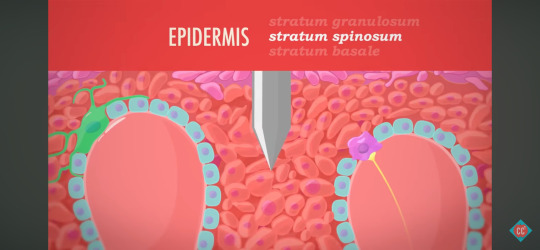
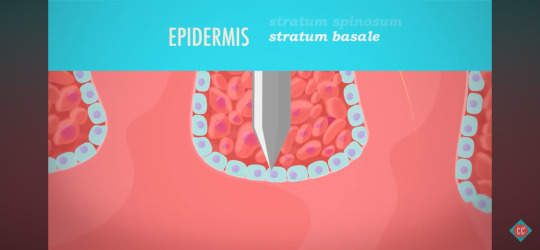
CC: A&P - Skin Deep Terminology
Integumentary System - The organ system that includes your skin, hair, nails, sweat glands and oil glands.
Friction Ridges - Structures supported by dermal papilae, also known as fingerprints.
Keratinocytes - The main type of cells in the epidermis, these make a lot of the protein keratin which makes the skin durable.
Melanocytes - The spider shaped cells in the epidermis that are responsible for producing the brown pigment melanin.
Tactile Cells (Merkel Cells) - Epidermal cells that work with neurons to provide sensation for light touch.
Dendritic Cells (Langerhans Cells) - Epidermal cells that are part of the immune system, and try to gobble up invading bacteria before they can break into the deeper layers of the skin.
Statified Squamous Epithelium - The tissue type found in the epidermis.
Thin Skin - The type of epidermis with just four named layers, and found everywhere on the body other than the palms of the hands and soles of the feet.
Thick Skin - The type of epidermis with five named layers, and only found on the palms of the hands, and the sole of the feet.
Epidermis - The most superficial of the three main layers of the skin.
Stratum Corneum - The most superficial layer of the epidermis, made of 20 - 30 layers of dead keratinocytes that eventually slough off, its name means "horny layer".
Stratum Lucidum - The so called "clear layer" of the epidermis, these 2 - 3 layers of keratinocytes are only found in thick skin on the palms of the hands and soles of the feet.
Stratum Granulosum - This layer of epidermal cells looks grainy because the cells are producing large amounts of keratin before they die.
Stratum Spinosum - This layer of epidermal cells is held together by protein fibers, and is the layer just superficial to the layer where active cell division is occuring.
Stratum Basale - This is the deepest layer of the epidermis, is made of just one layer of cells that are actively diving to produce cells that get pushed out to the other layers.
Dermis - The middle of the three main layers of the skin, where you find blood vessels, sweat glands, and most touch receptors.
Dermis - The layer of the skin just deep to the epidermis, containing blood vessels, and making skin elastic.
Papilary Layer - Made of loose areolar connective tissue, this is the layer of the dermis just deep to the epidermis.
Reticular Layer - The dermal layer just deep to the papilary layer, made of dense irregular connective tissue.
Hypodermis - The deepest of the three main layers of the skin, made mostly of adipose tissue.
0 notes
Text
Integumentary system: Function, parts, and conditions
New Post has been published on https://petn.ws/OMqK8
Integumentary system: Function, parts, and conditions
The integumentary system is a complex and vitally important organ system in the human body. It comprises the skin, hair, nails, and glands that produce sweat and oil. These tissues work together to protect the body from infection and injury and regulate bodily processes. The skin is the first line of defense against the outside […]
See full article at https://petn.ws/OMqK8
#OtherNews
0 notes
Text
Four Essential Functions of the Skin

First impressions go a long way, and the initial aspect people are likely to notice about someone else is their skin. As a culture, we’re seemingly obsessed with improving or altering some aspect of this most visible surface, whether that means tanning, treating acne symptoms, injecting dermal fillers to make it look youthful and plump, or applying topical products. Dr. Marguerite Germain and her team at Germain Dermatology specialize in medical and cosmetic dermatology in the Mt. Pleasant area in South Carolina. She explains that the skin has several important jobs that are necessary to maintain good health, meaning that any abnormal occurrences on the skin require attention.
Temperature
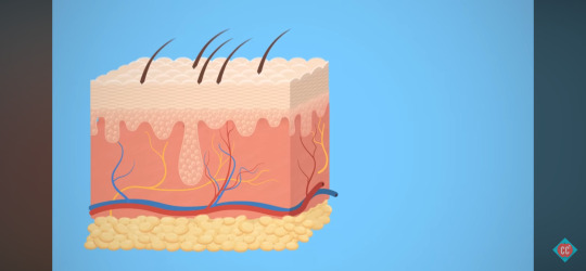
The skin is our largest and heaviest organ, consisting of three layers: the epidermis, the dermis, and subcutaneous fat. It is one aspect of the integumentary system, which also includes the hair and nails. Among the many physiological functions governed by this outer wrapping is thermal regulation: It acts as a thermostat, which triggers responses that keep a body cooler in hot weather or warm in colder climates. It can automatically adjust to temperature changes large and small.
The second layer of skin, the dermis, is full of red blood cells, hair follicles, oil, and other substances that help release or retain heat to bring the body to a stable temperature. Specialized glands in the skin create sweat, which lowers the body’s temperature by producing moisture and removing excess heat through evaporation. To accomplish all of this, the skin communicates with a part of the brain known as the hypothalamus, which senses when temperatures are too low or high.
Sensory
Nerve endings and receptors, which are abundant in the skin, allow people to have a sense of touch, detect vibration, sense different temperatures, experience pain and pleasure, and otherwise physically connect with the external world. By relaying messages through the nervous system, the skin enables the brain to receive information about a person’s immediate surroundings.
Nutrient Retention and Excretion
The upper layer of skin is basically waterproof, which works two ways. One of its vital roles is regulating the internal balance of moisture, electrolytes, and nutrients to prevent dehydration. It utilizes direct sunlight to manufacture vitamin D (which is vital for maintaining healthy bones) and assists the excretory system by eliminating metabolic waste, such as ammonia and uric acid, through the sweat glands.
Protection
The skin has a strong, yet flexible and elastic quality that provides structure and mechanic support for other organs and systems. The thick outer layer of skin acts as a barrier and the first line of defense, protecting the body against irritants and environmental threats such as germs, microorganisms, bacterial invasion, and toxic materials. It’s also necessary as a shield to cushion the internal organs against pressure and trauma, allowing people to move around freely.
Skin contains Langerhans cells, which are part of the immune system and prevent antigens from invading the body. Through a process of healing, it regenerates itself naturally when it is wounded. The fat beneath the skin acts as padding for the muscles, tissues, and bones.
As any person with fair skin who is prone to burning easily can tell you, sun damage is nothing to scoff at. Skin cancer—which is usually directly linked to intense or prolonged exposure to ultraviolet (UV) radiation from the sun—is the most common type of cancer in the United States. The skin takes most of the beating when it comes to contact with UV radiation. It protects against damage by building up a shield consisting of a darker pigment known as melanin. This pigment absorbs the harmful radiation and prevents it from damaging healthy cells. Of course, this is not a perfect defense, as cosmetic and medical issues can develop over time due to sun exposure. Regularly scheduled appointments with a dermatologist can help to identify problems early, making successful treatment more likely.
To discover more about dermatology, contact board-certified dermatologist Dr. Marguerite Germain and her team at Germain Dermatology. Call 843-881-4440 or submit information via an online contact form for further details.
1 note
·
View note
Text
Body Systems
Integumentary System
-- includes skin, nails, hair, sweat and oil glands, and mammary glands
-- function is to protect the body from the outside world, retain body fluids, protect from disease, regulate body temperature, and eliminate waste
Musculoskeletal System
-- includes bones and muscles
-- system allows movement and supports and protects the internal organs
Respiratory System
-- made up of nose and nasal cavity, pharynx, larynx, trachea, bronchi, and lungs
-- supplies blood with oxygen
Cardiovascular System
-- made up of the heart, arteries, and veins
-- distributes oxygen, removes waste products, and provides temperature control
Lymphatic System
-- made up of lymph nodes, ducts, tissues, capillaries, and vessels
-- transports lymph fluid to the circulatory system
Digestive System
-- begins at the mouth and goes to the anus
-- eats, digests, and eliminates food waste
Urinary System
-- made up of kidneys, ureter, bladder, and urethra
-- filters blood by removing waste
-- controls the amount of salt and water in the body
Reproductive System
-- vary by sex
-- work together to make hellions (babies)
Nervous System
-- divided into central and peripheral parts
-- includes the brain, the spine, nerves, and neurons
-- coordinates muscules, monitors organs, gets input from senses, initiates action
Auditory and Ocular Systems
-- seeing and hearing
-- both are sensory organ systems
-- includes all hearing organs and bones in the auditory system, as well as ocular organs such as eyelids and tear ducts
Immune System
-- made up of all molecular and genetic components that defend the body from foreign organisms
-- create antibodies and releases them into the blood to fight off pathogens
#medblr#studyblr#notes#my notes#medical notes#medblr notes#med notes#biology#anatomy and physiology#biology notes#anatomy#physiology#physiology notes#anatomy notes#body systems#organ systems#body system notes#organ system notes
149 notes
·
View notes
Text
St. John’s Wort: Hypericum perforatum

• Family: Hypericaceae (St. John’s Wort family)
• Preparations/dose:
Aerial parts are used medicinally.
Tincture - 1:2 or 1:5: 40-60 drops, three to four time per day //
Tea - Add 2 tsp. dried flowers/buds to 8oz. hot water, steep 30-40 minutes. Take 4oz. three to four times per day //
Capsules - take a capsule containing 350mg of standardized extracted three times per day //
Infused oil - applied topically as needed.
• Herbal actions: antidepressant, nervine tonic, antiviral, vulnerary, antimicrobial (topically), antiinflammatory, astringent
• System affinities: nervous system (depression, anxiety, nervousness), immune system (antiviral, antibacterial), integumentary system (wound healing).
• Safety: Pregnancy Category B1: No increased risk of teratogenic effects noted with limited use during pregnancy; minor adverse effects/teratogenic effects noted with high dose use in mice, however how this translates to effects on human fetus’ is unknown (Mills & Bone, 2005).
Lactation Category CC: may be used during breastfeeding, however it should be used cautiously and under the supervision of a healthcare provider. Mills and Bone (2005) state because SJW penetrates through the blood-brain barrier poorly, it is likely that it would cross the breast milk compartment poorly as well.
• Other notes:
The common name “St. John’s wort” comes from the traditional flowering and harvesting on St. John’s Day. The translation from Greek of Hypericum is above (hyper) and picture (eikon). This name originated from the tradition of warding off evil with this plant by hanging plants over religious icons on St. John’s Day.
Though modern herbalism projects it as the “depression” herb, it is useful for other things as well historically. It has been described as the arnica for nerves. The aerial flowering parts of SJW have been used in traditional European medicine for centuries to treat neuralgia, anxiety, neurosis, and depression,
How to positively identify Hypericum perforatum:
When the leaves are viewed up close (held up towards light) they have many tiny little spots on them like pinholes (“perforated” like the name perforatum). These spots are actually translucent glands, not holes.
When the flower heads are pinched and squeezed in the fingers, a red pigment can be seen left on the fingers. This has a very distinct SJW fragrance. When making SJW tincture, the tincture almost immediately turns red once alcohol is poured over the plant. This is in part due to the compound hypericin.
//
Organoleptic notes:
Have used SJW extensively in the past for mental health/SADD support, with mixed results. It seemed to help somewhat, but perhaps not more than the placebo effect after a few months of use. In hindsight, this may be because I don’t make enough serotonin without supplementation, and without adequate levels of serotonin, I won’t benefit from SJW’s SSRI effect. Would like to try SJW again after some more time regularly supplementing with nettle.
Class discussion: helps balance depleted neurotransmitters. Feeds into 5-HTP substrate > serotonin > melatonin. Helps with focus, insomnia. Supports metabolism (cytochrome P450). Mobile, clearing.
–
BMMC 2: Postpartum
Feb 2018
8 notes
·
View notes
Text
300+ TOP DERMATOLOGY Objective Questions and Answers
DERMATOLOGY Multiple Choice Questions :-
1. Which of the following would be prescribed for acne?
A. Actiq
B. Actonel
C. Accu-Check
D. Accutane
Ans: D
2. An absence of pigment in the skin is called
A. acanthosis nigricans
B. albinism
C. melanism
D. xanthoderma
Ans: B
3. A burn which involves 2 layers of the skin and estroys the nerves and blood vessels, but does not go down to muscle or bone is a
A. first-degree burn
B. second-degree burn
C. third-degree burn
D. full-thickness burn
Ans: B
4. Death of tissue associated with loss of blood supply to the affected area is called
A. cellulitis
B. eczema
C. gangrene
D. psoriasis
Ans: C
5. An acute eruption of intensely itchy papules or wheals is called
A. acne vulgaris
B. pityriasis rosea
C. psoriasis
D. urticaria (hives.
Ans: D
6. Moles with the potential to develop into malignant melanoma are
A. intradermal nevi
B. dysplastic nevi
C. giant nevi
D. verrucae
Ans: B
7. The type of cyst contains yellowish sebum and is commonly found on the scalp, vulva, and scrotum.
A. papule
B. sebaceous cyst
C. ulcer
D. vesicle
Ans: B
8. Excessive hair on the face or body, especially in women, is called:
A. albinism
B. atrichia
C. alopecia
D. hirsutism
Ans: D
9. The half-moon shaped, white area located at the base of a fingernail is called the
A. basal layer
B. cuticle
C. lunula
D. stratum
Ans: C
10. An epidermal growth caused by a virus (wart. is called a
A. impetigo
B. melanoma
C. nevus
D. verruca
Ans: D
DERMATOLOGY MCQs
11. Yellowing of the skin is indicative of
A. hyperbillirubinemia
B. hyperuricemia
C. hyperkalemia
D. hyporeninemia
Ans: A
12. Which of the following is a combining form meaning skin?
A. adip/o
B. cutane/o
C. pachy/o
D. xanth/o
Ans: B
13. A chronic dermatitis of unknown etiology in patients with a history of allergy is called
A. actinic dermatitis
B. atopic dermatitis
C. stasis dermatitis
D. seborrheic dermatitis
Ans: B
14. The outermost layer of skin is the
A. dermis
B. endodermis
C. epidermis
D. hypodermis
Ans: C
15. Of the three layers of the skin, which is the thick, fat-containing layer?
A. dermis
B. epidermis
C. epithelium
D. subcutaneous tissue
Ans: D
16. The brown-black pigment of the skin that is transferred to other epidermal cells and gives the skin its color is called
A. albumin
B. collagen
C. keratin
D. melanin
Ans: D
17. Which of the following is transcribed correctly?
A. The patient was given metronidazole for rosacea and Lamisil for onychomycosis.
B. The patient was given metronidazole for roseola and Lamisil for onychomycosis.
C. The patient was given metronidazole for roseola and Lamisil for onychomycosis.
D. The patient was given metroprolol for rosacea and Lamisil for onychomycosis.
Ans: A
18. Apocrine glands produce
A. mucus
B. sebum
C. sweat
D. keratin
Ans: C
19. Which of the following infections is also known as ringworm?
A. folliculitis
B. herpes simplex
C. impetigo
D. tinea corporis
Ans: D
20. Another term for itching is
A. dermatitis
B. keratosis
C. petechiae
D. pruritus
Ans: D
21. The skin, hair, nails, and glands all make up this system of the body.
A. integumentary system
B. lymphatic system
C. musculoskeletal system
D. nervous system
Ans: A
22. Clotrimazole and nystatin are both
A. topical antifungals
B. anti-itch creams
C. topical antibiotics
D. used to treat eczema
Ans: A
23. Which skin neoplasm is associated with an increase in the growth of cells in the keratin layer of the epidermis caused by pressure or friction?
A. callus
B. keloid
C. keratosis
D. leukoplakia
Ans: A
24. In this condition, there is a scaly dermatitis affecting parts of the skin that are supplied by oil glands.
A. chronic dermatitis
B. contact dermatitis
C. eczema
D. seborrheic dermatitis
Ans: D
25. Which of the following is a fungal infection?
A. lichen planus
B. keratosis
C. suborrhea
D. tinea capitis
Ans: D
26. A groove or a crack-like sore is called a (an.
A. fissure
B. nodule
C. polyp
D. ulcer
Ans: A
27. Which of the following is transcribed correctly?
A. This 58-year-old woman had a biopsy proven melanoma. Clarks level 1, on the left cheek.
B. This 58-year-old woman had a biopsy-proven melanoma. Clark's level 1, on the left cheek.
C. This 58-year-old woman had a biopsy proven melena. Clark level 1, on the left cheek
D. This 58-year-old woman had a biopsy-proven melanoma. Clark level 1, on the left cheek. (Correct Ans: D
28. Follicular dilation involving the nose and portions of the cheeks, erythema, papules, and pustules are classic signs of this dermatologic disorder.
A. acne cosmetica
B. acne pustulosa
C. acne rosacea
D. acne vulgaris
Ans: C
29. A skin disorder most often caused by the herpes virus and consisting of red lesions that look like targets is
A. candidiasis
B. erythema multiforme
C. hirsutism
D. keratosis pilaris
Ans: B
30. The vascular layer of skin is the
A. dermis
B. epidermis
C. stratum corneum unguis
D. hypodermis
Ans: A
31. When scraping for demodex, it may be helpful to pinch the skin while you scrape.
A. True
B. False
Ans: A
32. Impetigo is confined to the glabrous areas of a dog's body
A. True
B. False
Ans: A
33. What is the growth phase of a hair follicle?
A. Anagen
B. Catagen
C. Growagen
D. Telogen
Ans: A
34. What is one way to distinguise ARF from FAD?
A. Excoriations
B. Papules
C. GI Signs
D. Scratching of muzzle
Ans: C
35. Atopy is curable
A. True
B. False
Ans: B
36. Animals with fur have what type of follicles?
A. Simple
B. Intermediate
C. Compound
D. Fuzzy
Ans: C
37. When is a dog "cured" of demodex?
A. Disease free for 1 year
B. After 2 negative scrapes
C. Never
D. After 1 negative scrape
Ans: A
38. Dermatophilosis is zoonotic
A. True
B. False
Ans: A
39. Accral lick dermatitis can be a behavioral problem
A. True
B. False
Ans: A
40. Superficial pyoderma is a secondary disease process.
A. True
B. False
Ans: A
41. Eosinophillic Granuloma Complex is a disease in and of itself.
A. True
B. False
Ans: B
42. FAD is what type of reaction?
A. Type I (Missed.
B. Type II
C. Type III
D. Type IV (Missed.
Ans: A,D
43. All in contact dogs should be treated in a scabies case.
A. True
B. False
Ans: A
44. Canine FAD lesions are usually found on the rump, feline are on the head.
A. True
B. False
Ans: A
45. Failure to treat localized demedocosis will cause the dog to develop generalized demedocsis later on.
A. True
B. False
Ans: B
46. What is the resting phase of a hair follicle?
A. Anagen
B. Catagen
C. Relaxagen
D. Telogen
Ans: D
47. Notoedres can only infect cats
A. True
B. False
Ans: B
48. Wetting an area down with alchohol before plucking hairs for culture will decrease inaccurate tests.
A. True
B. False
Ans: A
49. What is the signalment for pemphigus foliaceus?
A. Any age
B. Elderly animals
C. Very young puppies
D. Young to middle aged adults
Ans: D
50. Otitis externa is very common in dogs with scabies.
A. True
B. False
Ans: B
51. Juvenile Cellulitis can appear in days
A. True
B. False
Ans: A
52. Age of onsent for puppy pyoderma
A. 2-9 Months
B. 3-6 months
C. 6 months- 1 year
D. 8 weeks-6 months
Ans: A
53. What is the transition phase of a hair follicle?
A. Telogen
B. Catagen
C. Zenagen
D. Anagen
Ans: B
54. In atopy, the allergens are absorbed via the skin
A. True
B. False
Ans: A
55. Puppies grow out of their puppy coats
A. True
B. False
Ans: B
56. When culturing a pustule, the area must first be cleaned
A. True
B. False
Ans: A
57. Juvenile Cellulitis in dogs usually affects what part of the body
A. Face
B. Glabrous areas
C. Body wide
D. Axilla
Ans: A
58. What is the signalment for juvenile cellulitis
A. 6 month
B. 1 year
C. 3-16 week old puppies
D. 8-12 weeks
Ans: C
59. Demodex mites live where?
A. Stratum Corneum
B. All skin layers
C. In the follicle
D. On the hair shaft
Ans: C
60. FAD and insect hypersensitivity in horses are both similar not only in their presentation, but also in their "cure".
A. True
B. False
Ans: A
DERMATOLOGY Objective type Questions with Answers
61. Demodectic mange is pruritic
A. True
B. False
Ans: B
62. It is necessary to clean the environment to eliminate ringworm
A. True
B. False
Ans: A
63. Which AB is responsible for atopy?
A. IgA
B. IgE
C. IgG
D. IgW
Ans: B
64. If not pruritic, linear granulomas should be treated how
A. Prednisone
B. Prednisolone
C. Scientific Neglect
D. Cyclosporin
Ans: C
65. Where are scabies mites found?
A. Hair Follicles
B. Stratum Corneum
C. Sub-epidermal
D. Throughout the skin layers
Ans: B
66 Surface irritation and inflammation caused by frictional trauma of skin on skin
A. Skin Fold Dermatitis
B. Furunculosis
C. Alopecia
D. Vesicle
Ans: A
67. Small elevation of skin containing purulent material
A. Papule
B. Pustule
C. Vesicle
D. Cyst
Ans: B
68. An erosion exposes the dermis underneath
A. True
B. False
Ans: B
69. An ulcer is:
A. Shallow Epidermal defect
B. Break in epidermis with exposure of dermis
C. Not good for coffee drinkers
D. A primary problem
Ans: B
70. Alopecia is:
A. Full or partial hair loss
B. Difficult for men to deal with
C. Only full hair loss
D. Never where you want it to be
Ans: A
71. Pyotraumatic Dermatitis is also known as a "hot spot"
A. True
B. False
Ans: A
72. Excessive scaling is:
A. Gross
B. Seborrhea
C. Dandruff
D. Epidermal Collarette
Ans: B
73. A vesicle is a sharply circumscribed lesion containing fluid
A. True
B. False
Ans: A
74. What is another term for intertrigo?
A. Furunculosis
B. Pyotraumatic Dermatitis
C. Skin Fold Dermatitis
Ans: C
75. Circumscribed, developmental skin defect
A. Nevus
B. Macule
C. Papule
D. Pustule
Ans: A
76. Scale is an accumulation of fragments of stratum corneum
A. True
B. False
Ans: A
77. Inflammation secondary to rupture of a hair follicle
A. Furunculosis
B. Pustule
C. Vesicle
D. Plaque
Ans: A
78. Remnants of a pustule, vesicle, or bulla can be
A. Epidermal Collarette
B. Scale
C. Plaque
D. Cyst
Ans: A
79. Circumscribed lesion that is raised and consists of edema is urticaria
A. True
B. False
Ans: A
80. What is a bulla?
A. Taunted by a bulla fighta
B. A large vesicle
C. Hives
D. A large cyst
Ans: B
81. A comedone is a primary problem only
A. True
B. False
Ans: B
82. Actinic means related to chemically active rays of the electromagnetic spectrum.
A. True
B. False
Ans: A
83. Accumulation of keratin and follicular material that adheres to hair shaft
A. Impetigo
B. Scales
C. Follicular Cast
Ans: C
84. Thickening of the epidermis and/or dermis
A. Callus
B. Lichenification
C. Scaling
D. Lacking in sensitive people
Ans: B
85. Circumscribed, non-palpable area characterized by color change
A. Patch
B. Papule
C. Macule
Ans: C
86. A patch is a large macule
A. True
B. False
Ans: A
87. "Scratch" is a lay term for excoriation
A. True
B. False
Ans: A
88. Dilated hair follicle containing cornified cells and debris
A. Comedone
B. Pustule
C. Furunculosis
Ans: A
89. A cicatrix is:
A. benign
B. a lesion
C. a scar
Ans: C
90. A cyst is a closed sack of pouch under the skin.
A. true
B. false
Ans: A
91. Ecchymosis means:
A. A condition of the ear
B. Skin discoloration
C. Excessive sweating
Ans: B
92. Eczema is an inflammatory condition of the skin.
A. True
B. False
Ans: A
93. Erythema:
A. blood condition
B. red
C. lack of pigmentation
Ans: B
94. Gangrene is necrosis (dead. tissue due to decomposition.
A. True
B. False
Ans: A
95. Herpes is:
A. An S.T.D.
B. a cold sore or fever blister
C. an invasion of pyogenic bacterium
Ans: B
96. Herpes Zoster is most commonly known as:
A. shingles
B. an enlarged fever blister
C. blue colored skin
Ans: A
97. Cyanoderma means:
A. red skin
B. blue skin
C. yellow skin
Ans: B
98. Impetigo is a contagious bacterial skin infection with pustules that rupture.
A. True
B. False
Ans: A
99. Kaposi's Sarcoma is a cancer associated with:
A. Smokers
B. A.I.D.S.
C. Sun exposure
Ans: B
100. A Laceration is:
A. a pathological change in tissue
B. torn skin
C. laser treatment for skin disease.
Ans: B
101. Metastasis means to:
A. spread infection from one person to another
B. move or spread through the blood stream or lymph nodes
C. abnormal breast condition
Ans: B
102. Benign means non-cancerous.
A. True
B. False
Ans: A
103. A 1st degree burn is:
A. the most severe type of burn
B. superficial burn injuring the top layer of skin
C. the first time the patient has been burned.
Ans: B
104. ___________ means itching caused by dry skin, parasitic infection or disease.
A. Scabies
B. Herpes Zoster
C. Pruritis
Ans: C
105. Tinea ________ means ring worm, athlete's foot.
A. purpura
B. impetigo
C. corporis
Ans: C
106. Scabies is a contagious skin condition caused by parasitic mites.
A. True
B. False
Ans: A
107. Pediculosis means:
A. Inflammatory condition of the skin.
B. Infestation with lice
C. Irritated and peeling feet.
Ans: B
108. Petechiae is a small _____________ spot on the skin.
A. hemorragic
B. raised
C. discolored
Ans: A
109. Urticaria means:
A. allergic reaction of the skin
B. thickened skin
C. dead tissue
Ans: A
110. SLE stands for_________________ and is an autoimmune disease.
A. squamous laceration ecchymosis
B. systemic lupus erythematosus
C. septic lymphodic edema
Ans: B
111. Similar to a vescle but larger usually more than 5mm in diameter, consisting of clear fluid accumulated within or below the epidermis.
A. Vesicle
B. Pustule
C. Bulla
D. Cyst
Ans: C
112. Dried exudate on the surface of the skin.
A. Plaque
B. Crust
C. Scale
Ans: B
113. A localised area of colour or textural change in the skin
A. Macule
B. Ecchymosis
C. Freckle
Ans: A
114. Alopecia is the absence of hair?
A. True
B. False
Ans: A
115. Atrophic skin is thin, translucent and wrinkled with easily visable blood vessels
A. True
B. False
Ans: A
116. An acute abcess formation in adjacent hair follicles.
A. Carbuncle
B. Furuncle
C. Folliculitis
Ans: B
117. A compressible papule or plaque of dermal oedema, red or white in colour.
A. Urticaria (Missed.
B. Erythema
C. Wheal (Missed.
D. Purpura
Ans: A,C
118. A pustule is a visible collection of pus in a blister. Pustules can be seen in psoriasis
A. True
B. False
Ans: A
119. A purulent inflammation of the skin and subcutaneous tissue
A. Erythema
B. Ecchymosis
C. Cellulitis
Ans: C
120. A small solid elevation of the skin, generally defined as less than 5mm, maybe flat or domeshaped.
A. Nodule
B. Papule
C. Macule
Ans: B
121. Irritants cause more contact dermatitis than allergens do?
A. True
B. False
Ans: A
122. Contact dermatitis, which of the following are common irritants?
A. Water (Missed.
B. temperature extremes (Missed.
C. Frictional abrasives (Missed.
D. Nickel
Ans: D
123. Atopic eczema induces lichenification
A. True
B. False
Ans: A
124. Treatments used to treat Atopic eczema
A. Emollients (Missed.
B. Antihistamines (Missed.
C. Hot baths
D. Topical steroids (Missed.
E. Antibiotics (Missed.
F. Wear wool clothing
Ans: C
125. Types of Eczema
A. Discoid eczema (Missed.
B. Seborrhoeic Dermatitis (Missed.
C. Venous eczema (Missed.
D. Arthiritic dermatitis
E. Lichen simplex chronicus (Missed.
F. Lichen striatus (Missed.
Ans: D
DERMATOLOGY Questions and Answers pdf Download
Read the full article
0 notes
Text
Turmeric Curcumin Supplement
Who does not want a flawless skin? Who would not want his or her acne to vanish right away? Who would not want to own a magic wand that can give you back your baby soft skin? Being the exterior most covering of your body structure; skin is exposed to all the troubles that the surroundings pose to you. The skin health is not only affected by external environment but the functions going inside your body.
Skin is the largest organ of your integumentary system. It has up to seven layers of ectodermal tissue. Ectoderm is one of the three primary germ layers in the very early embryo. The other two are mesoderm and endoderm. Your skin guards the underlying bones, muscles, ligaments and internal organs of the body. Here is a surprising fact. Your skin or the human skin in general is very similar to that of a pig. Your skin may appear hairless but nearly all of it is covered with hair follicles. In Latin, the cutis refers to skin and hence the origin of ‘cutaneous’ takes place from the Latin word ‘cutis’. Due to its presence all over the body; skin plays an important role in providing immunity to your body against pathogens and excessive water loss. The little insight to various functions of skin is given below:
Function of Protection: It acts as a physical or anatomical barrier to pathogens who try to enter your body. Specific cells called Langerhans cells in skin are a part of the adaptive immune system of your body i.e. the immune system which is composed of highly specialized, systemic cells and processes that eliminate harmful microbes or prevents their growth.
The perspiration through your skin is effective in limiting bacterial growth as lysozyme present in perspiration breaks the bonds within the cell walls of bacteria.
Function of Sensation: A variety of nerve endings are present in skin that react to heat, cold, touch, pressure, vibration, injury to tissue and other external stimuli. The sensations from these nerves’ endings travel to spinal cord and brain.
Function of Heat Regulation: Sweat glands and dilated blood vessels help in heat loss while constricted blood vessels work to reduce cutaneous blood flow and conserve heat. According to the temperature in the surroundings; the muscles controlling the hair shafts on skin adjust the angle of hair shafts to change the degree of insulation provided by hair.
Function of Control of Evaporation: Skin is relatively dry and semi-impermeable to act as a barrier to minimize fluid loss.
Function of Storage and Synthesis: Your skin acts as a centre for storage of lipids and water.
Function of Absorption of oxygen: The outer most 0.25 to 0.40 mm of skin cells of your body are highly dependent on external oxygen as gases like oxygen, nitrogen and carbon dioxide can diffuse into epidermis in small amounts. This absorption of oxygen is limited to the external skin cells in humans and the contribution to total respiration is negligible.
Function of Water Resistance: The skin acts as a water resistant barrier. Due to this function, essential nutrients are not washed out of the body. The nutrients and oils that help in hydrating the skin are covered by the outer most layer of skin i.e. epidermis. If epidermis is not there; the oils that flow in the inner layer of skin i.e. dermis may be affected by water.
There may also be other functions of your skin apart from the ones explained above. Therefore keeping structure of skin intact and keeping it functioning properly is highly essential. Since every organ needs assistance with age to maintain its integrity and functions; the skin should be fed with its requirements. Products with turmeric curcumin supplement due to turmeric anti inflammatory action can help you.
Take out Your Love For Yoga: There are certain yoga poses which can improve circulation of blood and nutrients to the skin. When the circulation of blood and nutrients increases to skin; it gains its natural glow and shine. Along with enhancement in external appearance; the functions of skin occur without any trouble.
Use the Colourful Fruit and Vegetable Stall to Get that Glow: Winter is right here in India. There would be variety of fruits and vegetables available which can provide the needed glow on your skin. The carrots which are seasonal in winter contain beta-carotene and lycopene that protect your skin from the damage by ultra violet rays to the skin. Though the sun is not that harsh in winter; the presence of UV rays in sunlight is still there. The antioxidants and potassium in carrot keep dry skin and uneven tone at bay. Spinach is also seasonal in winter and contains iron, vitamin C and A. Healthy iron levels ward away paleness from your complexion and vitamins help to repair the skin cells. Oranges available in winter also nourish skin.

Use Oxyrich Capsules and Oxyrich Juice: Oxyrich Juice is a turmeric curcumin supplement and contains aloevera, Himalayan berry and curcumin that rejuvenate skin, hair and nails; purify blood and improve digestion of food so that it is reflected on skin. Himalayan berry has nearly 1000 times more vitamin C than that in known sources of vitamin C which is highly beneficial for skin health. The juice helps your skin fight against ageing and reduces acne and pigmentation marks on skin. Oxyrich Capsules should be consumed with this Oxyrich Juice. These capsules are efficient turmeric curcumin supplements and contain Himalayan berry, green tea and curcumin which enhance the action of Oxyrich Juice and are effective in improving the texture and reducing the dryness of skin by them. These capsules prevent unwanted inflammation and pain in acne, psoriasis, eczema and other skin complaints and diseases. Curcumin in turmeric is especially responsible for turmeric anti inflammatory action.
Keep up With Healthy Fluids: Consume water and water-rich fruits and vegetables. Find ways to add water to your routine. Drink curcumin enriched best turmeric teas like Daily Detox Tea. This Daily Detox tea has Assam green tea, Tulsi, fennel, ginger, Ashwagandha, fenugreek, lemon fruit and curcumin which cleanse and detox you internally so that you can shine and glow externally. The curcumin content of Daily Detox Tea makes it among the best turmeric tea. Internal detoxification and nourishment along with external care can keep the largest organ of your body functioning properly!
#turmeric#curcumin#anti inflammatory#capsules#supplement#tea#turmeric tea#turmeric supplement#curcumin supplement
0 notes
Text
Part.-2
1. Description of the body tissues, components and functions of the integumentary and skeletal muscular system. Body Tissues Connective Tissue: Most widely distributed of the tissues and most abundant. Connective tissues perform many functions including support, protection, strengthens other body tissues. The following tissues are found in the human body: ordinary loose connective tissue, adipose tissue, reticular connective tissue, dense fibrous tissue, cartilage, bone, blood, and lymph, which are all considered connective tissue. Connective tissue has cells that are widely separated by a matrix. Epithelial Tissue: Epithelial tissue covers the body surface and forms the lining for most internal cavities. The major function of epithelial tissue includes protection, secretion, absorption, and filtration. The skin is an organ made up of epithelial tissue which protects the body from dirt, dust, bacteria and other microbes that may be harmful. Cells can be thin, flat to cubic to elongate. There are different types of epithelial tissue: Simple epithelium that has a single layer of cells that function in diffusion, osmosis, filtration, secretion, and absorption. Stratified epithelium consists of two or more layers of cells that protect underlying tissues in locations where there is considerable wear and tear. Pseudo stratified epithelium consists of a single layer of cells. A secreted covering of mucus traps foreign particles and the upward motion of the cilia carries the mucus to the back of the throat, where it may be swallowed or expelled. Glandular epithelium that consists of a single cell or a group of cells that secret substance into tubes, onto a surface, or into the blood. Muscle Tissue: Consists of elongated cells called muscle fibers that use ATP to generate force. There are three types of muscle tissue: Skeletal muscle which is a voluntary type of muscle tissue that is used in the contraction of skeletal parts. Smooth muscle found in the walls of internal organs and blood vessels. It is an involuntary type. And the cardiac muscle found only in the walls of the heart and is involuntary in nature. Muscular tissue has several functions which include: producing body movements, stabilizing body positions, storing substances within the body, moving substances within the body, generating heat. This tissue has four properties that enable it to function and contribute to homeostasis, which are electrical excitability, contractility, extensibility and elasticity. Nervous Tissue: consists of two cell types, neurons and neuroglia, and is present in the brain and spinal cord. It’s composed of specialized cells which not only receive stimuli but also conduct impulses to and from all parts of the body. Components and functions of the Integumentary System: Functions • Protection: the skin protects the underlying structures by providing a physical barrier against the external environment. This can include anything from pathogens to abrasions. • Blood reservoir because the dermis has an extensive network of blood vessels that carry form 8 to 10% of the total blood flow in a resting adult. • Thermoregulation: The skin helps to regulate temperature by perspiring when the body becomes too hot. When the body has become too cold, blood vessels near the surface of the skin constrict to prevent heat from leaving the body. • Sensation: Nerve fibers that run throughout the layers of the skin help us to sense touch, temperature, pressure, and pain. • Elimination: The skin is one of our eliminatory organs. Perspiration removes natural metabolic wastes from the body. (Traditionally, sweating has been an important part of a health regimen.) • Vitamin D production is another important part of the skin’s functions. Compounds Sebaceous or oil glands: secrete sebum, an oily substance that lubricates the hair within the follicle as well as the skin itself. If the oil glands fail to discharge, the secretions collect and form “whiteheads” or “blackheads”. Associated hairs: Humans have hair on practically all parts of the body. It provides various forms of protection. For example, eyebrows and eyelashes protect the eyes from sunlight, particles, and even perspiration. Hair within the nasal cavity prevents dust and foreign objects like insects from entering. Nails: Fingernails and toenails are keratinized cells formed from the epidermis. Nails consist of two parts, the root from which the nail grows and the body, which is visible externally. Nails also serve protective purposes. Soporiferous or sweat gland: coiled tubule within the dermis that straightens out near its opening called a sweat pore. Components and functions of the skeletal muscular system: 1. Muscle Fiber: cell containing cellular components with special names. 2. Bone: Type of connective tissue. Bones provide support and protection for body organs. 3. Cartilage: This provides flexible support in areas of the body like nose and ears. Composed of closely packed collagenous fibers in a rubbery gelatinous substance 4. Tendon connects bone to bones and is bonded to them. 5. Ligament: joins bones and other connective tissues 6. Joint where two or more bones are joined together. Functions: The skeletal system’s functions are • Support tissues and provide attachment to the tendons. • Protect vital organs from injury. • Produce movement. Skeletal muscles attach to bones, so they contract on bones to produce movement. • Mineral homeostasis: Bone tissue stores several minerals, especially calcium and phosphorous, which contribute to the strength of bone. • Blood cell production where red bone marrow produces blood cells and is present in developing bones of the fetus and in some adult bones. • Triglyceride Storage: Yellow bone marrow consists mainly of adipose cells that store triglycerides, which are a potential chemical energy reserve.
0 notes
Text
New Post has been published on Planet Market
A Brief Description of the Eleven Major Organ Systems in the Body and Their Main Function
The eleven major organ systems, and brief descriptions of their functions, are as follows:
1. Integumentary – This system includes the skin and its appendages (hair, nails, and specialized sweat- and oil-producing glands). Its primary function is protection. For example, the skin protects the underlying tissue from invasion by harmful bacteria, bars entry of most chemicals, and minimizes the chances of mechanical injury to underlying structures.
2. Skeletal – The skeletal system includes bones and related tissues such as cartilage and ligaments, which provide the body with a rigid framework for support and protection. The skeletal system also makes possible the movements of body parts.
3. Muscular – The muscular system, consisting of the individual skeletal muscles, makes movement possible and generates the heat required for maintaining a constant core body temperature. Voluntary muscles are so called because their contractions are under conscious control. Involuntary or smooth muscle tissue is found in blood vessel walls, other tubular structures and in the lining of hollow organs such as the stomach and small intestine. Cardiac muscle is the specialized muscle tissue of the heart.
4. Nervous – The nervous system is composed of the brain, spinal cord and nerves. The nervous system makes possible communication between body functions, integration and control of body functions, and recognition of sensory stimuli.
5. Endocrine – The endocrine system is composed of specialized glands that secrete chemicals known as hormones directly into the blood. The organs of the endocrine system are sometimes called ductless glands. The endocrine system is similar to the nervous system in that it also provides communication, integration and control, but it does it in a slower and longer-lasting way by hormone secretion. Hormones are also the main regulators of metabolism, reproduction and other body activities. They play important roles in fluid and electrolyte balance, acid-base balance and energy metabolism.
6. Cardiovascular (Circulatory) – The cardiovascular system includes the heart, arteries, veins and capillaries. The primary function of this system is transportation. Transportation needs include continuous movement of oxygen and carbon dioxide, nutrients, hormones, and other important substances. This system also helps regulate body temperature by distributing heat throughout the body and by assisting in retaining or releasing heat from the body by regulating blood flow near the body surface. Specialized cells of the circulatory system can also become involved in immunity.
7. Lymphatic – The lymphatic system is composed of lymph nodes, lymphatic vessels and specialized lymphatic organs such as the tonsils, thymus and spleen. The lymph system moves fluids and certain large molecules from the tissue spaces around the cells and moves fat-related nutrients from the digestive tract back to the blood. It also plays a role in the functioning of the immune system, which is the defense mechanism of the body against disease.
8. Respiratory – The respiratory system includes the nose, pharynx, larynx, trachea, bronchi and lungs. Its primary function is to permit the movement of air into the alveoli, which are the tiny thin-walled sacs of the lungs. In these sacs, oxygen from the air is exchanged for carbon dioxide, a waste product, which is then carried to the lungs by the blood so that it can be eliminated from the body. The respiratory system is also involved in regulating the acid-base balance of the body.
9. Digestive – The digestive system is composed of the mouth, pharynx, esophagus, stomach, small intestine, large intestine, rectum and anal canal (primary organs), as well as the teeth, salivary glands, tongue, liver, gallbladder, pancreas and appendix (secondary organs). All of the organs of the digestive system work together to ensure proper digestion and absorption of nutrients.
10. Urinary – The urinary system includes kidneys, ureters, bladder and urethra. The primary function of this system is the elimination of waste from the body. Other organs of the body are also involved in the elimination of body wastes, such as the lungs and skin.
11. Reproductive (consisting of a male subdivision and a female subdivision) – The reproduction system is composed of gonads (testes), vas deferens, prostate, penis and scrotum in the male, and gonads (ovaries), uterus, fallopian tubes, vagina and mammary glands in the female. The purpose of this system is the procreation of life, insuring survival of humankind.
fitness body
#Design
0 notes
Text
18th May 2024
Crash Course: Anatomy & Physiology - The Integumentary System, Skin Deeper
youtube
Summary:
Integumentary System Protects Your Body
Senses The Outside World
Helps Excrete Waste
Stores Blood
Regulates Temperature
Makes Vitamin D
Indicates Signs of Poor Health
Integumentary System
Functions
Protection
Sensation
Blood Storage
Temperature Regulation
Vitamin D Synthesis
Excretion
"The first and most vital purpose of the integumentary system is to act as a protective barrier."
Protective & Sensory Functions of the Integumentary System
Skin, hair, nails, sweat and oil glands work to shield us from threats: excessive sunlight, infections, abrasions etc.
"The integumentary system is also vital to how you sense the world around you."
Cutaneous Sensory Receptors
Structures, part of the nervous system, that receive stimuli from outside environment and send them to the brain.
Corpuscles (Receptors)
register different sensations associated with touch
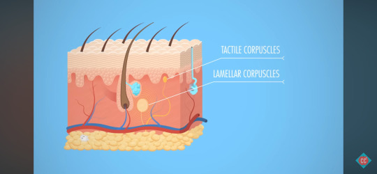
Tactile Corpuscles
transmits sensations of light touch and low frequency vibrations from periphery > central nervous system
E.g. Makes you aware of a tag scratching the back of your neck.
Lamellar Corpuscles
register the sense of pressure
E.g. Someone puts their hand on your shoulder.
Hair Follicle Receptors
feeling the breeze on skin/through your hair
More Functions: Removing Waste, Storing Blood & Regulating Body Temperature
"The integumentary system also plays a role in the excretion of waste."
Waste Removal
The integumentary system excretes small amounts of waste elimanted through the skin via sweat.
Urea, uric acid and ammonia, nitrogen-containing waste, are disposed via urine.
There's no evidence that heavy sweating rids the body of extra toxins, just water loss.
Blood Storage Unit
"About 5 percent of the entire blood volume is retained in your skin at any given time."
When more bloody supply is needed, like exercising, the nervous syste constricts dermal blood vessels to squeeze extra blood into circulation.
Body Temperature Regulation
During exertion, both blood and sweat glands regulate body temperature.
Insensible Perspiration
unnoticable sweat
The body excretes about 1.5L of sweat daily to maintain a comfortable body temperature.
Sensible Perspiration
noticeable sweat
Heavy exertion can cause 12L of sweat per day.
Vasoconstriction
To regulate heat loss, dermal blood vessels constrict, causing causing blood to head deeper into tissues to keep vital organs warm
Vasodilation
Once heat returns, blood vessels relax, allowing blood to return to the surface.
Skin Dyscolouration: Cyanosis, Jaundice or Erythema
Skin colour can indicate homeostatic imbalances.
As human skin colour spans, some conditions are easier to diagnose by discolouration of other tissues - mucous membranes, finger and toenail beds.
Cyanosis (Blue Skin)
In Caucasian people:
may indicate heart failure
poor circulation
severe respiratory issues
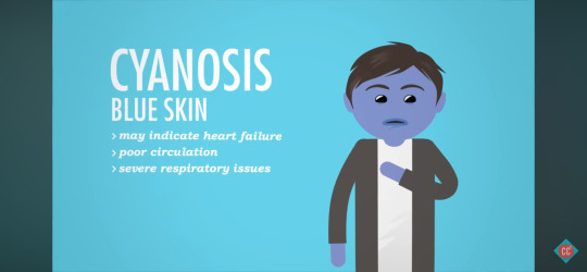
Blood depleted of oxygen turns darker in colour, which seen through the tissue of lips or skin, can look blue-ish.
Jaundice (Yellow Skin)
may indicate liver disorder
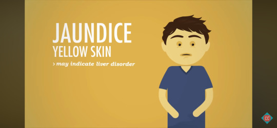
Yellow bile accumulates in the blood stream.
Erythema (Red Skin)
may indicate fever, inflammation, allergy
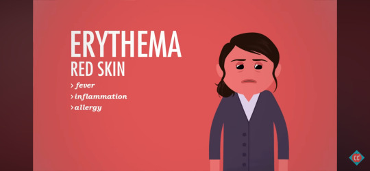
Fever, inflammation and allergy cause blood vessels expand and cause more blood flow to the skin's surface.
Melanin, Vitamin D & Skin Tone
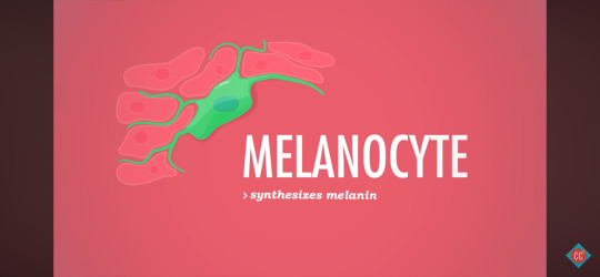
Melanocyte
synthesizes melanin
Melanin is a pigment produced by melanocyte cells in the epidermis.
Melanin
produces reddish yellow pigments
produces brownish black pigments
It's main job is to protect against sun's ultraviolet rays.
Vitamin D
Bones require Vitamin D to produce new bone cells, which is the only vitamin the body can produce on its own.
Skill cells contain a molecule that converts to Vitamin D when in contact with UV light.
Vitamin D Path:
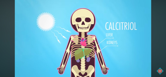
Skin > Bloodstream > Liver & Kidneys
In the liver and kidneys, Vitamin D becomes Activated D (Calcitriol).
Calcitriol circulated to all bones of the body.
How Does Hair Conditioner Work?
Skin Appendages
Hair
Nails
Sweat
Sebaceous Glands
Oil Glands
Hair
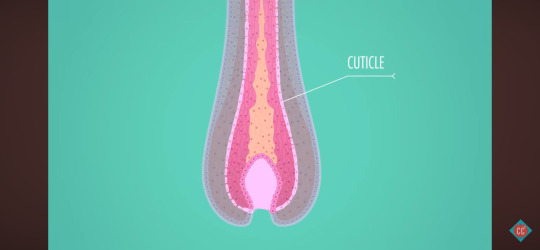
Hair, or pili, are flexible strands of dead keratin protein - like fingernails.
The outermost layer of the dead cells, the cuticle, look like overlapping roof shingles.
Shaft
keratinisation is complete
Root
keratinisation is still happening
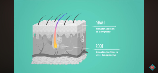
Each follicle is a tube of epidermal cells.
Like the epidermis, cells at the bottom of each follicle are young, continually dividing and pushing older cells up through the skin's surface.
Finger & Toe Nails
root of nail
nail body
nail bed
At the back of the nail bed, new cells divide at the root and are pushed forward, creating scaly-hard keratin.
Types of Sweat Glands: Eccrine, Apocrine, Mammary and Ceruminious
Sweat & Oil Glands
"There are up to three million toiny sudoriferous, or sweat glands, distributed throughout the body."
secrete salty, watery sweat
Eccrine & Apocrine Glands
Eccrine Glands
palms
forehead
soles of feet
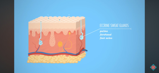
Eccrine glands are abundant.
They're simple coiled tubes that start in the dermis, extend through a duct and open into a pore on the skin's surface.
Apocrine Glands
armpit hair follicles
groin hair follicles
"deluxe" sweat - with fats and proteins
viscous
yellow-ish colour
odor
"You only have about 2000 of these, and they start cookin' around puberty, emptying into the hair follicles around the armpits and groin."
When the bacteria on the skin gets a hold of this sweat, it gets odorific, creating body odor.
Deodorants don't affect how much you sweat, but they reduce the smell by attacking odor-making bacteria.
Antiperspirants, using aluminum, block the sweat glands and prevent perspiration.
Modified Apocrine Sweat Glands
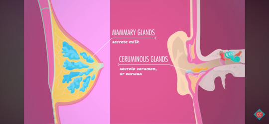
Mammary Glands
secrete milk
Ceruminious Glands
cerumen (earwax)
Sebaceous (Oil) Glands
Sebaceous Glands
oil glands
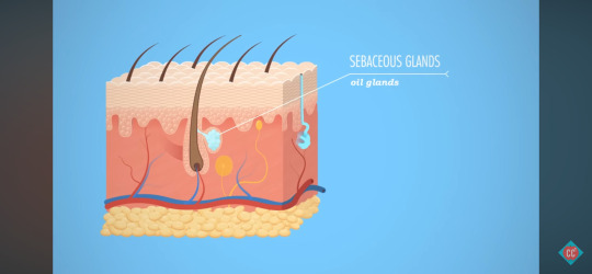
Sebaceous glands are everywhere but in thick skin on palms and foot soles.
Ducts are smaller on limbs but bigger on face, neck and upper chest.
Most sebaceous glands secrete sebum, an oily substance, into hair follicles that can travel to the skin's surface.
Although they cause pimples, their primary function is to soften and lubricate skin and hair, and slow skin's water loss in dry environments.
0 notes
Text
18th May 2024
Crash Course: Anatomy & Physiology - The Integumentary System, Skin Deep
youtube
Summary:
All About Skin
Epidermis, Dermis and Hypodermis
Melanin and Keratin Cells
Ensure You Get A Good Tattoo
Skin
Skin protects your body against infection and extreme temperatures, maintains your balance of fluids, and even synthesizes Vitamin D for personal use.
The nerve endings allow you to sense the outside world.
Sweat glands and blood vessels help maintain proper temperature and communicate - from health to emotions - through things like blushing, flushing and sweating
Integumentary System
Hair
Nails
Sweat
Oil Glands
Skin
Skin Layers: Epidermis, Dermis & Hypodermis
Epidermis
stratified squamous epithelial tissue
Dermis
sweating, blood circulation, feeling
Hypodermis (Subcutis)
adipose tissue
Types of Epidermal Cells: Keratinocytes, Melanocytes, Langerhan Cells and Merkel Cells
Keratinocytes
building blocks of the tough, fibrous protein keratin
make up the bulk of the epidermis

Provide structure, durability and waterproofing to hair, nails and outer skin.
Kertatinocytes are constantly dying and being replaced, losing milling of them daily, enough to completely replace the epidermis every 4 - 6 weeks.
Melanocyte
spider-shaped cell that synthesizes melanin
Skin colour isn't about the number of melanocytes but instead about the breath of their spidery cellular extensions, which in turn affect the amount of melanin that they contain.
Langherhan Cells (Denritic)
star-shaped
originate in bone marrow
defend the epidermis

Merkel Cells (Tactile)
combine with nerve endings to create a sensory receptor for touch

Found deep down at the boundary between the epidermis and dermis.
Thick Skin
Thick skin, found on the palms of hands and sole of feet, consists of five epidermal layers.
Thin Skin
Thin skin, found everywhere else, contains four epidermal layers.
Layers of Skin: Stratum Corneum, Stratum Lucidum, Straum Granulosum, Stratum Spinosum, and Stratum Basale
The deeper the epidermal layer, the younger the cells are.
Regeneration happens in the lower layers, with new cells moving up, maturing along the way, until they die on the surface.
This is because the epidermal is epithelial, so it's avascular, with all oxygen and nutrients coming from the dermis, so as they mature and move towards the surface, they lose their oxygen and nutrient supply and die.
Stratum Corneum
"horny layer"
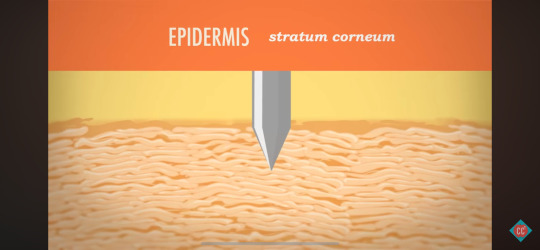
Outermost, roughest layer.
20 - 30 sheets of dead keratinocyte cells.
Stratum Lucidum
"clear layer"
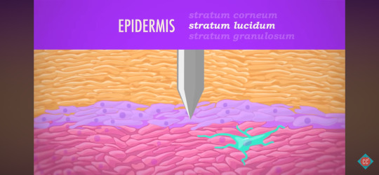
2 - 3 rows of clear, flat, dead keratinocytes that are only found in the thick skin of palms and soles.
Thin skin is only missing the stratum lucidum.
Stratum Granulosum
"granular layer"
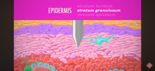
Layer contains living keratinocytes that form keratin like "crazy".
Looks grainy as cells are compressed and flattened as they move up through the epidermal layers, maturing as they go.
Stratum Spinosum
"spiny layer"
Prickly appearance under a microscope.
Closer to the point where regeneration, or mitosis, is active.
Stratum Basale
"basal layer"
A single layer of columnar cells.
Cell factory - where most cell production happens.
It connects the epidermis to the layer of skin below it, the dermis.
Pneumonic - Come, Let's Get Sun Burned!
C - Stratum Corneum
L - Stratum Lucidum
G - Stratum Granulosum
S - Stratum Spinosum
B - Stratum Basale
Sunburn
The ultraviolet radiation in sunlight can damage the epidermis, cause elastic fibers to clump up to form a leathery appearance.
Sunburn temporarily depresses the immune system, with immune cells in the epidermis - and radiation can alter skin cell's DNA, leading to skin cancer.
Layers of the Dermis: Papillary, Reticular and Hypodermis
Dermis
collagen fibers, elastin fibers
capillaries
blood vessels
nerve fibers
parts of hair follicles, oil glands/sweat glands with ducts that lead to the skin's surface
Papillary Layer
areolar connective tissues
dermal pippilae
In thick skin, papillae are tiny protrusions that form unique friction ridges that press up through the epidermis to help grip - fingerprints.
Reticular Layer
dense irregular connective tissue
The reticular layer, deeper and thicker, make up 80% of the dermis.
All the dynamic parts contained within the dermis, like nerve fibers and capillaries, are distributed between both its layers.
Any time you're cut deep enough to bleed or feel pain, you've broken through the epidermis layer and lacerated the dermis.
Tattoos must reach the dermis for permanency.
Hypodermis (Subcutis)
adipose connective tissue
Basically a seam of fat, it provides insulation, energy storage, shock absorption and anchors the skin.
0 notes
Text
Calendula: Calendula officinalis

• Family: Asteraceae (daisy/”first aid kit” family)
• Preparations/dose:
Tincture:
Fresh Flowers - weight:vol - 1:2, 100% alcohol in menstruum, dose: 1-2mL, 3-4x a day //
Dried Flowers weight:vol - 1:5 60-80% alcohol in menstruum, dose: 1-2mL 3-4x a day //
Infusion (Folk Method): Grind the dried herb into a fine powder. Place the herb into a jar and add a fixed oil of choice; olive, sesame, almond, etc. Leave an additional ¼ inch so the herb may absorb the extra oil. Cap jar and leave it out in the sun for 7-10d. Shake or stir every couple hours or at several times a day. When it is ready, strain out the leaves and sediment. Allow the infusion to sit in a glass jar in a cool dark place with proper labelling.
Salve: Measure out the dried powdered herb, add equal parts by volume of 190 proof ethyl alcohol (about 120ml). Mix herb with alcohol and stir until moist then place the sealed container safe overnight. Pour the oil into blender and add moist herbs, strain with a cheesecloth. Pour liquid into a double boiler and heat over low flame for a few hours until the alcohol is evaporated. You can check to see if there is any alcohol present by lighting the match to the surface of the mixture. Add beeswax to create the salve. Bring to boil and tightly cap (Green, 2000)
• Herbal actions: vulnerary, anti-inflammatory, lymphatic, hemostatic, antimicrobial, antifungal, antispasmodic, astringent, emmenagogue, cholagogue
• Indications: Calendula may be used externally to treat inflammation of the skin, bruising, strains, bleeding, minor burns and skin ulcers. Internally, calendula is also anti-inflammatory and can help treat gastric and duodenal ulcers, relieve gallbladder problems, treat swollen lymph glands, and ease indigestion (soothes digestive and mucous membranes). Calendula can also be used internally and externally to combat fungal infections. Calendula may stimulate the immune system (via bone marrow) and may help normalize menses and ease painful periods.
• Contraindications: Known allergy to Calendula or other members of the Asteraceae family.
• System affinities: Integumentary, lymph/immune, and hepatic
• Other notes:
Calendula could be beneficial for a mother wishing to reduce localized swelling and inflammation at the perineum after birth, as well as helping to heal wounds such as a perineal laceration or cesarean incision site. Since calendula reduces bruising, it could be used on a newborn’s skin for birth injuries. Calendula could also be helpful in taming yeast flare-ups on a mom’s nipples, and internally for the newborn’s mouth (thrush).
Energetics: warm and dry
No known contraindications at this time for Calendula. Avoid oral consumption during pregnancy due to potential to cause contractions. Calendula has been recognized as safe while breastfeeding.
Calendula is famous for its vulnerary properties, it’s been known to treat minor inflammations of the skin and mucosa and helps with healing minor wounds. It can be used in oil extracts or salve for treatments for inflamed, irritated, sore, or cracked nipples; an excellent resource as an anti inflammatory. There are also antimicrobial and antifungal benefits from the hydroethanolic extracts that may be used topically for treatment of infection (Romm, 2010). Commission E (2000) reports that it also possesses pain reduction properties, antifungal, antiviral, antiparasitic, and antibacterial activity with the presence of ethanol in addition to the increase of glycoprotein, nucleoprotein and collagen metabolism at the wound site.
//
Organoleptic notes: Such a lovely plant - the color is so striking. Gives me a new appreciation for marigolds. Distinctive, voluminous dried flower heads. Smells sweet, like caramelized honey.
Used the calendula salve regularly to soothe itchy razor bumps on my arm pits with great success. Applied a few times to belly stretch marks with less obvious success.
Plan is to infuse calendula oil to have on hand for postpartum healing.
0 notes
Last Seen Blogs

imtoohotmilk
very normal things happening here

seldomfollowed-blog
CURIOUSER.

inaaguirre20570-blog
A Few review Hoaxes And How You Can Defend Against

hudaalmazyed
Untitled

karuaiaerion
Kar's magic trunk of junk