#Bioelectronic
Text
Drug Discovery was the Leading Category in the Healthcare Bioconvergence Industry
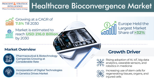
The healthcare bioconvergence market will power at a rate of 7.5% rate by the end of this decade, to touch USD 216 billion by 2030.The industry is growing due to the growing count of elderly people and the growing usage of stem cells for regenerating organs, tissues, and injured cells.
Moreover, the preference of users for combinations of cutting-edge technologies, including ML, ergonomics,…
View On WordPress
#Bioelectronic#Diagnostic and Biological Sensors#Drug Discovery#Healthcare Bioconvergence#Nanorobotics for Drug Delivery#Regenerative Medicine
0 notes
Text
Brain Stimulation study shows high electrical frequency stimulation can activate specific nerve fibers in the body

Brain Stimulation study shows high electrical frequency stimulation can activate specific nerve fibers in the body.
Feinstein Institutes Bioelectronic Medicine Researchers Target Specific Vagus Nerve Fibers for More Precise, Effective Stimulation.
MANHASSET, N.Y, November 04, 2022 - Electrically stimulating the vagus nerve has shown potential to treat inflammatory diseases, including rheumatoid arthritis, lupus and diabetes. Now, a team of bioelectronic medicine scientists at The Feinstein Institutes for Medical Research is one step closer to developing more precise, selective neuromodulation techniques. A new paper published in Brain Stimulation showed that delivering high-frequency electrical stimulation activates specific sensory nerve fibers of the vagus nerve and could be the key to better targeting and treating inflammation and disease.

Dr. Stavros Zanos recently led the study, which targets specific fibers in the vagus nerve for more precise electrical stimulation. (Credit: Feinstein Institutes)
The research study, led by Stavros Zanos, MD, PhD, associate professor at the Feinstein Institutes Institute of Bioelectronic Medicine, measured how vagus nerve stimulation (VNS) using kHz stimulation – an electrical stimulus that is commonly used to block nerve conduction at over 1,000 pulses per second affects the activity of different types of vagal fibers. Previous VNS therapies would stimulate the whole nerve bluntly, without targeting specific fibers, which could cause adverse effects and less effective therapy. For example, in past clinical trials, side effects from the vocal cords and from coughing reflex prevented clinicians from delivering the full dose of VNS, and thus limited the therapeutic effect.
“The vagus nerve is one of the most important nerves in our body, which helps us maintain our daily functions like breathing and our heart rate. Being able to target certain nerve fibers is a huge step for precision stimulation,” said Dr. Zanos, senior author of the paper. “This selective stimulation in the vagus nerve has not been achieved until now and our results will help researchers find new therapies to treat diseases.”

The vagus nerve is made up of 100,000 individual fibers. It acts as an information pipeline in the body, stretching from the brain to the organs helping to maintain bodily functions and immune response. If the vagus nerve is overactive or underactive inflammatory diseases can arise. The field of bioelectronic medicine combines molecular medicine, neuroscience and biomedical engineering to develop innovative therapies using computer chips and devices instead of drugs to treat those diseases.
By stimulating the vagus nerve with high frequency, Dr. Zanos and his team were able to consistently activate the small unmyelinated nerve fibers while at the same time bypassing the activation of larger fibers in the nerve – which are usually the first to be activated by VNS and are responsible for most unwanted side-effects. The study was done in two animal models (mice and rats) and if validated in humans, could be leveraged in novel bioelectronic therapies.
“Dr. Zanos discovered how to selectively activate nerve fibers traveling in the vagus nerve by manipulating the electric frequency,” said Kevin J. Tracey, MD, president and CEO of the Feinstein Institutes and Karches Family Distinguished Chair in Medical Research. “This work significantly advances the field of bioelectronic medicine and opens new pathways to explore experimental therapeutics.”
The Feinstein Institutes for Medical Research was recently awarded $6.7 million from the National Institutes of Health (NIH). The funding will help Dr. Zanos and his lab create a detailed map of the anatomy of the human vagus nerve. The funding is part of the NIH Common Fund’s Stimulating Peripheral Activity to Relieve Conditions (SPARC) program.
Read the full article
0 notes
Text
The scientific community has long been enamored of the potential for soft bioelectronic devices. But they’ve faced hurdles in identifying materials that are biocompatible and have all of the necessary characteristics to operate effectively.
The new research is a step in the right direction.
Researchers have modified an existing biocompatible material so that it conducts electricity efficiently in wet environments and can send and receive ionic signals from biological media.
Continue Reading.
49 notes
·
View notes
Text

New, more biocompatible materials for bioelectronic applications
Bioelectronics is a field of research in which biology and electronics converge. In medicine, for example, an external electric current is used to cure or monitor diseases of the nervous system, and also to monitor biomarkers in situ. Devices made of conductive materials are used for these applications.
The most widely used conductive polymer so far in energy and biomedical applications is PEDOT doped with PSS, known as PEDOT:PSS. Despite its exceptional properties, new conductive materials that can improve some of its limitations, such as biocompatibility, still need to be developed.
A study conducted by CIC biomaGUNE's Biomolecular Nanotechnology group is proposing a mechanism for doping PEDOT using a robust engineered protein (PEDOT:Protein); the outcome is a hybrid material with ionic and electronic conductivity, which is quite similar to PEDOT:PSS in some cases. The paper is published in the journal Small.
Read more.
#Materials Science#Science#Biocompatible#Biomaterials#Electronics#Bioelectronics#Polymers#Electrical conductivity#Proteins
12 notes
·
View notes
Text

Electrical Orchestration
How hollow organoids – lab-grown 3D tissue models – can be shaped and inflated by external electrical stimulation highlights the role of life's natural bioelectrical systems (as in water regulation) and has potential for applications in fields such as bioengineering and developmental biology
Read the published research article here
Image from work by Gawoon Shim and Isaac B. Breinyn, and colleagues
Department of Mechanical and Aerospace Engineering, Princeton University, Princeton, NJ, USA
Image originally published with a Creative Commons Attribution 4.0 International (CC BY 4.0)
Published in Nature Communications, April 2024
You can also follow BPoD on Instagram, Twitter and Facebook
#science#biomedicine#immunofluorescence#biology#organoids#electrical stimulation#bioelectronics#developmental biology
8 notes
·
View notes
Text


100 Years of Solitude || Treasure Planet || 2002
#lmao i was serious about becoming a variety blog#i wanna have a healthy mix of content on here because itll be fun#im starting with treasure planet#the url is staying the same tho because it is way too special to me to ever change LOL#anyways enjoy this everyone!!#jim hawkins#morph#ben#bioelectronic navigator#treasure planet#disney#disney movies#disney movie#r.t. makes gifs#r.t. posts gifs#disney gifs#disney movie gifs#movie gifs#r.t. talks#not sports#not mash#movies#movie#treasure planet gifs
180 notes
·
View notes
Text
Implantable sensor could lead to timelier Crohn’s treatment - Technology Org
New Post has been published on https://thedigitalinsider.com/implantable-sensor-could-lead-to-timelier-crohns-treatment-technology-org/
Implantable sensor could lead to timelier Crohn’s treatment - Technology Org
A team of Northwestern University scientists has developed the first wireless, implantable temperature sensor to detect inflammatory flareups in patients with Crohn’s disease. The approach offers long-term, real-time monitoring and could enable clinicians to act earlier to prevent or limit the permanent damage caused by inflammatory episodes.
The miniaturized implantable temperature sensor is small enough to fit on top of a U.S. dime.
More than 1 million Americans have Crohn’s disease, a chronic inflammatory bowel disease that affects the intestines, causes digestive issues and can lead to weight loss, malnutrition and other complications. People with mild cases are treated with oral medications, but these drugs typically fail over time, requiring approximately 70 percent of Crohn’s patients to undergo at least one surgery to remove portions of damaged intestines.
Because heat is indicative of inflammation, the Northwestern scientists tested whether a temperature sensor resting gently against the intestines of mice with Crohn’s disease could provide real-time insight into the disease’s progression and detect episodic flareups. Sure enough, they accomplished both goals in their research, published in the Nature Biomedical Engineering journal.
Arun Sharma, whose group led the animal testing, said clinicians currently do not have a way to quickly detect inflammatory events. Some go unnoticed by patients until the problem becomes so severe that it requires invasive surgery.
“The magnitude of the flareup can be measured regarding the heat signature,” said Sharma, co-corresponding author on the paper and a research associate professor of urology at Northwestern University Feinberg School of Medicine and of biomedical engineering at McCormick School of Engineering. “Is it so extensive that it will cause tissue damage over time?
“This could be potentially prevented if a clinician has this information readily at hand and can determine what type of therapy can be given to that person at that moment in time, rather than waiting weeks to get a blood analysis, tissue biopsy or fecal analysis. In the meantime, you’re losing valuable minutes regarding tissue damage with this inflammatory event.”
Sharma said this strategy of measuring temperature fluctuations could also be useful for patients with ulcerative colitis, another inflammatory bowel disease, or any condition with a prolonged inflammatory response. In their study, the researchers used the wireless sensors to track temperature fluctuations for nearly four months continuously.
Bioelectronics pioneer John Rogers, whose group led the device development, recently published another paper describing an ultrathin, soft implant that measures temperature and perfusion changes as a way to monitor the health of transplanted organs. Once again, the relationship between heat and inflammation was key, as excess inflammation around the transplanted organ can offer an early warning sign that the patient’s immune system is rejecting the new organ.
“To address Crohn’s disease, we developed an ultraminiaturized, precision temperature sensor with wireless communication capability,” said Rogers, co-corresponding author on the paper. “This tiny, soft device takes the form of a smooth, round capsule that rests within the GI system, without affecting natural physiological processes for long-term recordings. The data show some very unique signatures, in the form of perturbations to natural circadian cycles, known as ultradian rhythms, as early indications of inflammatory responses.”
Surabhi Madhvapathy, co-first author from the Rogers group who led the sensor engineering, said the scientists discovered that the ultradian temperature rhythms correlate to cyclic variations in stress levels and inflammatory markers in the blood.
“In addition to the short-term variations, we learned over the span of weeks to months, that the average temperature of the intestines decreases,” Madhvapathy said. “This decrease was indicative of the worsening tissue quality over time.”
Following these successful results in mice, the researchers plan to assess the sensor capabilities in human tissues that recreate the inflammatory gut conditions found in Inflammatory Bowel Disease.
Source: Northwestern University
You can offer your link to a page which is relevant to the topic of this post.
#Analysis#approach#bioelectronics#biopsy#Biotechnology news#blood#colitis#communication#Crohn's disease#data#development#Disease#drugs#engineering#Events#Featured life sciences news#form#gut#hand#Health#Heat#human#immune system#inflammation#inflammatory bowel disease (IBD)#InSight#issues#it#LED#Link
0 notes
Text
Bioelectronics Market Is Estimated To Witness High Growth Owing To Rising Applications In Healthcare
Bioelectronics is an interdisciplinary field that involves the merging of biology and electronics to develop devices and systems that combine biological and synthetic materials. Bioelectronics has applications in healthcare for developing implantable and wearable medical devices for monitoring health parameters such as electrocardiograms (ECG), electromyography (EMG), and electroencephalograms (EEG). Biosensors integrated into these devices can analyze blood, sweat or tissue samples to detect biomarkers for diseases. The global bioelectronics market is estimated to be valued at US$ 23530 Mn in 2023 and is expected to exhibit a CAGR of 11% over the forecast period 2023 to 2030, as highlighted in a new report published by Coherent Market Insights.
Market Dynamics
The rising applications of bioelectronics in healthcare is driving the growth of the global bioelectronics market. Bioelectronic devices aid in monitoring health parameters remotely and continuously, thereby improving disease diagnosis and management. This is expected to support market growth over the forecast period. Further, ongoing research focused on the development of advanced bioelectronic medicines is also fueling demand. For instance, researchers are working on developing electronic drug delivery systems coupled with biological components to treat diseases including diabetes, cancer, or inflammatory conditions with tailored drug release profiles. However, high costs associated with developing advanced bioelectronic systems and obtaining regulatory approvals pose a challenge to market players.
SWOT Analysis
Strength: Bioelectronics technology has capabilities that could positively impact and advance healthcare. It has the potential to improve disease diagnosis and monitoring through more precise and personalized detection methods. Devices incorporating bioelectronics may help enhance treatment through remote monitoring and administration of therapies. This represents a strength as patients could benefit significantly from more proactive and tailored care approaches.
Weakness: The bioelectronics field is still developing and refinements are ongoing to further strengthen performance of devices and integrate biocompatible materials. This presents some uncertainties that may discourage adoption until technologies achieve more proven results at commercial scales. Obtaining regulatory approval for new classes of medical devices can be lengthy and costly as well.
Opportunity: An aging global population is driving demand for advanced healthcare solutions. The medical technology industry is actively investing in bioelectronics as a means to deliver personalized care for chronic diseases and conditions. This growing preventive medicine trend presents commercial opportunities as new types of diagnostic tools and therapeutic devices are brought to markets. Biosensors also show promise for applications in food safety, environmental monitoring and national security.
Threats: Competing drug and device manufacturers may attempt to delay market entry of disruptive new bioelectronics through regulatory and legal challenges. Strict rules govern biosensing technologies and any adverse events involving new products could damage brands. Rapid changes occurring as the field evolves also present technical threats if standards and interfaces are not adequately stabilized before broad adoption.
Key Takeaways
The global bioelectronics market forecast is expected to witness high growth over the forecast period of 2023 to 2030 supported by a growing aging population with chronic conditions seeking more advanced healthcare solutions. The global bioelectronics market is estimated to be valued at US$ 23530 Mn in 2023 and is expected to exhibit a CAGR of 11% over the forecast period 2023 to 2030.
Regional analysis: The Asia Pacific region is dominating the global bioelectronics market with the highest growth rate over the forecast period. This can be attributed to factors such as rising medical expenditures, expanding biotechnology industries, and government initiatives and investments promoting development of indigenous bioelectronic technologies particularly in India, China, and South Korea. The large patient pools and growing middle-class populations seeking access to advanced healthcare in Asia Pacific economies also contribute to high market potential.
Key players operating in the bioelectronics market are BBI-Biotech GmbH Bioengineering AG, Danaher Corporation, Eppendorf AG, Getinge, Infors HT, Merck KGAA, Sartorius AG, Solaris Biotech Solutions, and Thermo Fisher Scientific, Inc. Leading players are focused on new product approvals, partnerships and acquisitions to enhance their product portfolio and geographic presence in this rapidly evolving industry.
Get more insights on this topic: https://www.newsstatix.com/bioelectronics-market-industry-insights-trends-bioelectronics-market/
Explore more information on this topic, Please visit: https://www.urdughr.com/2023/12/recycled-construction-aggregates-a-sustainable-solution-for-growing-construction-needs.html
#Bioelectronics#Bioelectronics Market#Bioelectronics Market size#Bioelectronics Market share#Bioelectronics Market demand#Bioelectronics Market analysis#Bioelectronics Market scope
0 notes
Text
#bioelectronicsandbiosensors#biosensors#bioelectronics#sensortechnology#bioelectronicsandbiosensorsmarketreport#bioelectronicsandbiosensorsmarketsize#bioelectronicsandbiosensorsmarketshare#bioelectronicsandbiosensorsmarketgrowth#bioelectronicsandbiosensorsmarketanalysis#grandresearchstore
0 notes
Link
Bioelectronics is a branch of medical science that deals with the application of ideologies of biological sciences in electrical/electronic engineering. In simpler terms, bioelectronics is the combination of biology and electronics. This is an essential emerging field in medicine. Bioelectronics has led to the expansion of various devices such as the pacemaker and a wide variety of therapeutic imaging devices that are easily accessible. The nervous system transmits an endless number of signals to carry out various functions of the human body. This creates a highly lucrative growth prospect for the study of bioelectronics and the related market is projected to flourish over the next few years.
0 notes
Link
REVA University has one of the best B Tech in Bioelectronics Engineering colleges. Bioelectronics is the discipline resulting from the convergence of biology and electronics and it has the potential to significantly impact many areas important to the nation’s economy and well-being, including healthcare and medicine, homeland security, forensics, and protecting the environment and the food supply.
0 notes
Text

New study challenges conventional understanding of charging process in electrochemical devices
A new study by researchers at the University of Cambridge reveals a surprising discovery that could transform the future of electrochemical devices. The findings offer new opportunities for the development of advanced materials and improved performance in fields such as energy storage, brain-like computing, and bioelectronics.
Electrochemical devices rely on the movement of charged particles, both ions and electrons, to function properly. However, understanding how these charged particles move together has presented a significant challenge, hindering progress in creating new materials for these devices.
In the rapidly evolving field of bioelectronics, soft conductive materials known as conjugated polymers are used for developing medical devices that can be used outside of traditional clinical settings. For example, this type of materials can be used to make wearable sensors that monitor patients' health remotely or implantable devices that actively treat disease.
The greatest benefit of using conjugated polymer electrodes for this kind of devices is their ability to seamlessly couple ions, responsible for electrical signals in the brain and body, with electrons, the carriers of electrical signals in electronic devices. This synergy improves the connection between the brain and medical devices, effectively translating between these two types of signals.
Read more.
#Materials Science#Science#Electrochemistry#Bioelectronics#Conjugated polymers#Polymers#University of Cambridge
11 notes
·
View notes
Text
Researchers have made a significant breakthrough in the development of an on-skin bioelectronic wearable sensor: the addition of wireless charging—without batteries—through a magnetic connection.
The team’s innovation, an important component to its existing ultrasoft, breathable, and stretchable material, provides the foundation for gathering precise vital sign measurements like blood pressure, electrical heart activity, and skin hydration.
The discovery could one day lead to early detection and timely interventions for chronic diseases such as heart disease, cancer, and diabetes.
Continue Reading.
71 notes
·
View notes
Text
Genetically Modified Bacteria Produce Energy From Wastewater

E. Coli is one of the most widely studied bacteria studied in academic research. Though most people probably associate it with food/water borne illness, most strains of E. Coli are completely harmless. They even occur naturally within your intestines. Now, scientists at EPFL have engineered a strain of E. Coli that can generate electricity.
The survival of bacteria depends on redox reactions. Bacteria use these reactions to interconvert chemicals in order to grow and metabolize. Since bacteria are an inexhaustible natural resource, many bacterial reactions have been industrially implemented, both for creating or consuming chemical substrates. For instance, you may have heard about researchers discovering bacteria that can break down and metabolize plastic, the benefits of which are obvious. Some of these bacterial reactions are anabolic, which means that they need to be provided external energy in order to carry it out, but others are catabolic, which means that the reactions actually create energy.
Some bacteria, such as Shewanella oneidensis, can create electricity as they metabolize. This could be useful to a number of green applications, such as bioelectricity generation from organic substrates, reductive extracellular synthesis of valuable products such as nanoparticles and polymers, degradation of pollutants for bioremediation, and bioelectronic sensing. However, electricity producing bacteria such as Shewanella oneidensis tend to be very specific. They need strict conditions in order to survive, and they only produce electricity in the presence of certain chemicals.
The method that Shewanella oneidensis uses to generate electricity is called extracellular electron transfer (EET). This means that the cell uses a pathway of proteins and iron compounds called hemes to transfer an electron out of the cell. Bacteria have an inner and outer cell membrane, so this pathway spans both of them, along with the periplasmic space between. In the past, scientists have tried to engineer hardier bacteria such as E. Coli with this electron-generating ability. It worked… a little bit. They were only able to create a partial EET pathway, so the amount of electricity generated was fairly small.
Now, the EPFL researchers have managed to create a full pathway and triple the amount of electricity that E. Coli can produce. "Instead of putting energy into the system to process organic waste, we are producing electricity while processing organic waste at the same time -- hitting two birds with one stone!" says Boghossian, a professor at EPFL. "We even tested our technology directly on wastewater that we collected from Les Brasseurs, a local brewery in Lausanne. The exotic electric microbes weren't even able to survive, whereas our bioengineered electric bacteria were able to flourish exponentially by feeding off this waste."
This development is still in the early stages, but it could have exciting implications both in wastewater processing and beyond.
"Our work is quite timely, as engineered bioelectric microbes are pushing the boundaries in more and more real-world applications" says Mouhib, the lead author of the manuscript. "We have set a new record compared to the previous state-of-the-art, which relied only on a partial pathway, and compared to the microbe that was used in one of the biggest papers recently published in the field. With all the current research efforts in the field, we are excited about the future of bioelectric bacteria, and can't wait for us and others to push this technology into new scales."
146 notes
·
View notes
Text
Shape-shifting ultrasound stickers detect post-surgical complications - Technology Org
New Post has been published on https://thedigitalinsider.com/shape-shifting-ultrasound-stickers-detect-post-surgical-complications-technology-org/
Shape-shifting ultrasound stickers detect post-surgical complications - Technology Org
Researchers led by Northwestern University and Washington University School of Medicine in St. Louis have developed a new, first-of-its-kind sticker that enables clinicians to monitor the health of patients’ organs and deep tissues with a simple ultrasound device.
Three variations of the soft, flexible ultrasound sticker device displayed on a finger. Image credit: Jiaqi Liu
When attached to an organ, the soft, tiny sticker changes in shape in response to the body’s changing pH levels, which can serve as an early warning sign for post-surgery complications such as anastomotic leaks. Clinicians can then view these shape changes in real-time through ultrasound imaging.
Currently, no existing methods can reliably and non-invasively detect anastomotic leaks — a life-threatening condition that occurs when gastrointestinal fluids escape the digestive system. By revealing the leakage of these fluids with high sensitivity and high specificity, the non-invasive sticker can enable earlier interventions than previously possible. Then, when the patient has fully recovered, the biocompatible, bioresorbable sticker simply dissolves away — bypassing the need for surgical extraction.
The study will be published March 8 in the journal Science. The paper outlines evaluations across small and large animal models to validate three different types of stickers made of hydrogel materials tailored for the ability to detect anastomotic leaks from the stomach, the small intestine and the pancreas.
“These leaks can arise from subtle perforations in the tissue, often as imperceptible gaps between two sides of a surgical incision,” said Northwestern’s John A. Rogers, who led device development with postdoctoral fellow Jiaqi Liu. “These types of defects cannot be seen directly with ultrasound imaging tools. They also escape detection by even the most sophisticated CT and MRI scans. We developed an engineering approach and a set of advanced materials to address this unmet need in patient monitoring. The technology has the potential to eliminate risks, reduce costs and expand accessibility to rapid, non-invasive assessments for improved patient outcomes.”
“Right now, there is no good way whatsoever to detect these kinds of leaks,” said gastrointestinal surgeon Dr. Chet Hammill, who led the clinical evaluation and animal model studies at Washington University with collaborator Dr. Matthew MacEwan, an assistant professor of neurosurgery. “The majority of operations in the abdomen — when you have to remove something and sew it back together — carry a risk of leaking. We can’t fully prevent those complications, but maybe we can catch them earlier to minimize harm. Even if we could detect a leak 24- or 48-hours earlier, we could catch complications before the patient becomes really sick. This new technology has potential to completely change the way we monitor patients after surgery.”
A bioelectronics pioneer, Rogers is the Louis Simpson and Kimberly Querrey Professor of Materials Science and Engineering, Biomedical Engineering and Neurological Surgery, with appointments at the McCormick School of Engineering and Northwestern University Feinberg School of Medicine. He also directs the Querrey Simpson Institute for Bioelectronics. At the time of the research, Hammill was an associate professor of surgery at Washington University. Rogers, Hammill and MacEwan co-led the research with Heling Wang, an associate professor at Tsinghua University in Beijing.
The importance of being early
All gastrointestinal surgeries carry the risk of anastomotic leaks. If the leak is not detected early enough, the patient has a 30% chance of spending up to six months in the hospital and a 20% chance of dying, according to Hammill. For patients recovering from pancreatic surgery, the risks are even higher. Hammill says a staggering 40-60% of patients suffer complications after pancreas-related surgeries.
The biggest problem is there’s no way to predict who will develop such complications. And, by the time the patient is experiencing symptoms, they already are incredibly ill.
“Patients might have some vague symptoms associated with the leak,” Hammill said. “But they have just gone through big surgery, so it’s hard to know if the symptoms are abnormal. If we can catch it early, then we can drain the fluid. If we catch it later, the patient can get sepsis and end up in the ICU. For patients with pancreatic cancer, they might only have six months to live as it is. Now, they are spending half that time in the hospital.”
In search of improved outcomes for his patients, Hammill contacted Rogers, whose laboratory specializes in developing engineering solutions to address health challenges. Rogers’ team had already developed a suite of bioresorbable electronic devices to serve as temporary implants, including dissolving pacemakers, nerve stimulators and implantable painkillers.
The bioresorbable systems piqued Hammill’s interest. The greatest odds of developing an anastomotic leak occur either three days or two weeks after surgery.
“We like to monitor patients for complications for about 30 days,” Hammill said. “Having a device that lasts a month and then disappears sounded ideal.”
Enhancing ultrasound
Instead of developing new imaging systems, Rogers speculated that his team might be able to enhance current imaging methods — allowing them to “see” features that otherwise would be invisible. Ultrasound technology already has many advantages: it’s inexpensive, readily available, does not require cumbersome equipment and does not expose patients to radiation or other risks.
But, of course, there is a major drawback. Ultrasound technology — which uses sound waves to determine the position, shape and structure of organs — cannot reliably differentiate between various bodily fluids. Blood and gastric fluid, for example, appear the same.
“The acoustic properties of the leaking fluids are very similar to those of naturally occurring biofluids and surrounding tissues,” Rogers said. “The clinical need, however, demands chemical specificity, beyond the scope of fundamental mechanisms that create contrast in ultrasound images.”
Ultimately, Rogers’ team devised an approach to overcome this limitation by using tiny sensor devices designed to be readable by ultrasound imaging. Specifically, they created a small, tissue-adhesive sticker out of a flexible, chemically responsive, soft hydrogel material. Then, they embedded tiny, paper-thin metal disks into the thin layers of this hydrogel. When the sticker encounters leaked fluids, it swells.
Making the invisible visible
The metal disks move apart as the hydrogel swells in response to changing pH. The ultrasound can then view these subtle changes in placement.
Watch the device expand in response to pH
“Because the acoustic properties of the metal disks are much different than those of the surrounding tissue, they provide very strong contrast in ultrasound images,” Rogers said. “In this way, we can essentially ‘tag’ an organ for monitoring.”
Because the need for monitoring extends only during a postsurgical recovery, Rogers team designed these stickers with bioresorbable materials. They simply disappear naturally and harmlessly in the body after they are no longer needed.
Computational collaborator Yonggang Huang, the Jan and Marcia Achenbach Professorship in Mechanical Engineering and professor of civil and environmental engineering at McCormick, used acoustic and mechanical simulation techniques to help guide optimized choices in materials and device layouts to ensure high visibility in ultrasound images, even for stickers located at deep positions within the body.
“CT and MRI scans just take a picture,” Hammill added. “The fluid might show up in a CT image, but there’s always fluid collections after surgery. We don’t know if it’s actually a leak or normal abdominal fluid. The information that we get from the new patch is much, much more valuable. If we can see that the pH is altered, then we know that something isn’t right.”
Rogers team constructed stickers of varying sizes. The largest measures 12 millimeters in diameter, while the smallest is just 4 millimeters in diameter. Considering that the metal disks are each 1 millimeter or smaller, Rogers realized that it might be difficult for radiologists to assess the images manually. To overcome this challenge, his team also developed software that can automatically analyze the images to detect with high accuracy any relative movement of the disks.
Improving quality of life
To evaluate the efficacy of the new sticker, Hammill’s team tested it in both small and large animal models. In the studies, ultrasound imaging consistently detected changes in the shape-shifting sticker — even when it was 10 centimeters deep inside of tissues. When exposed to fluids with abnormally high or low pH levels, the sticker altered its shape within minutes.
Rogers and Hammill imagine that the device could be implanted at the end of a surgical procedure. Or, because it’s small and flexible, the device also fits (rolled up) inside a syringe, which clinicians can use to inject the tag into the body.
“These tags are so small and thin and soft that surgeons can easily place collections of them at different locations,” Rogers said. “For example, if an incision extends by a few centimeters in length, an array of these tags can be placed along the length of the site to develop a map of pH for precisely locating the position of the leak.”
“It’s obviously an early prototype, but I can envision the final product where, at the end of surgery, you just place these little patches for monitoring,” Hammill said. “It does its job and then completely disappears. This could greatly impact patients, their recovery time and, ultimately, their quality of life.”
Next, Rogers and his team are exploring similar tags that could detect internal bleeding or temperature changes. “Detecting changes in pH is a good starting point,” Rogers said. “But this platform can extend to other types of applications by use of hydrogels that respond to other changes in local chemistry, or to temperature or other properties of clinical relevance.”
Source: Northwestern University
You can offer your link to a page which is relevant to the topic of this post.
#Accessibility#advanced materials#applications#approach#bioelectronics#bleeding#blood#Cancer#challenge#change#chemical#chemistry#Civil and environmental engineering#Collections#computed tomography#course#detection#development#devices#digestive system#electronic#electronic devices#engineering#Environmental#equipment#Features#fluids#Fundamental#Health#Health & medicine news
0 notes
Last Seen Blogs

minervabarbaras
URL UFT

sssseeekkkk
제목 없음

goldalify
Golda

touchtranscendslanguage
Touch Transcends Language

gviral
: // PERFECT SYSTEM.