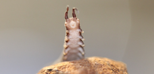
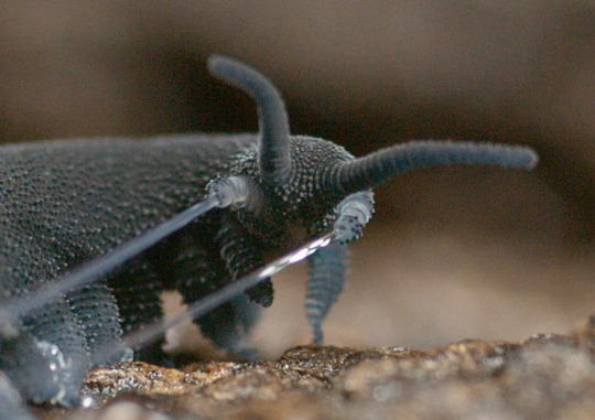
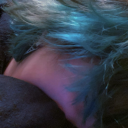
#Euperipatoides rowelli
Note
Do you have cool velvet worm facts? They're my favourite bug-creatures :3
I love velvet worms! Absolutely bizarre creatures. They don't have an exoskeleton and they move by changing the fluid pressure in their body. ALSO there's a species (Euperipatoides rowelli) that lives in social groups lead by a dominant female.
Let's admire that species.


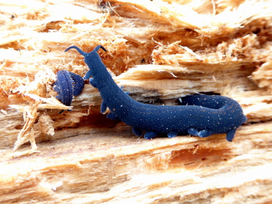
EXQUISITE. Photos by squiresk
5K notes
·
View notes
Photo

Velvet worms (Euperipatoides rowelli)
by Jackson Nugent
#velvet worms#Euperipatoides rowelli#Euperipatoides#Peripatopsidae#Euonychophora#Udeonychophora#Onychophora#wildlife: australia
18 notes
·
View notes
Text
Phylum #16: Onychophora, the velvet worms!

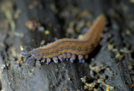
"Lobopods", unarmored relatives of arthropods, have a long and rich history. While early forms like Hallucigenia remain unclear as to their affinities, another lineage had already split from the first arthropods. Velvet worms still thrive today, having left the seas for rainforests and humid crevices.
Instead of the arthropods' articulated exoskeletons, velvet worms kept a softer, unarticulated cuticle, with tiny round "papillae" covered in bristles and scales providing their velvet-like appearance. They still moult their cuticle regularily, but do not gain new legs doing so - the babies already look like miniature adults!
Adapted to many terrains thanks to their flexible body and clawed legs, velvet worms are true ambush predators. They come with a special weapon: a glue-like slime, immobilizing prey and repelling predators, shot from appendages on each side of their mouth.
Velvet worms are also intelligent, social creatures. At least one species, Euperipatoides rowelli, is known for hunting together, living in groups of up to 15 individuals with complex interactions establishing their social hierarchy. Individuals within a group are often closely related, with most species giving live birth and taking care of their young!


374 notes
·
View notes
Text
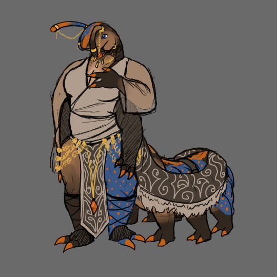
Still a wip, but I anthropomorphised my velvet worm sona... Still thinking of a name for her. She's a cross between a Euperipatoides rowelli and an Ooperipatus silvanus!
#anthro#anthro bug#bugsona#velvet worm#velvet worm character#fursona#bug furry#evanesce art#I have barely drawn the last few months because of uni sorry everyone#velvet worms are my fave invert :)
37 notes
·
View notes
Photo

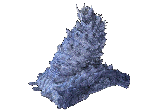
Velvet Worm (Tallaganda) as animal teacher:
Keywords: The ancient hunter. Entanglement. Dominance and submission. Drying out. The wanderer. A complex relationship with water. Fluid and flow. Steady as she goes. Nothing needs to change. Unchanging. Perfect as you are. Secretive. Hiding from the light. Shadows have great yields. The benefits of shadow work. Look to the dark. Avid and hungry for more. Complex social interactions. Where do you stand among your friends? Establishing rank. Night secrets. Not everything needs to be revealed right now. Using what works well for you. Do you really need anything new?
*
original art, water/colour pencil and ink on illustration board.
42 notes
·
View notes
Text
everyone shut the FUCK up im thinking about euperipatoides rowelli


and how much i want to pet them gently like god’s littlest dog
2 notes
·
View notes
Text
We detected a strict female-dominated feeding hierarchy: the first individual to feed was always a female, and she remained the sole feeder for 45–60 min (Fig. 2).
This female fed in bouts of up to 10 min, interrupted by short intervals during which she moved around the Petri dish. During her solitary feeding time, males and young of the group remained distant, often in physical contact with one another.
The other females assumed a ‘waiting’ position close to the prey, circled it, and occasionally tried to feed. If noticed by the first female, they were attacked by her, bitten, kicked and pushed away.
After 45–60 min, the first female tolerated other individuals at the food. Now, the remaining females started feeding in groups, soon followed by the males and young (Fig. 2). The number of feeding males reached its maximum once the females started leaving the food.
All individuals eventually fed, but rarely all simultaneously. After 3 h, feeding was over, and the group reaggregated once the individuals had cleaned their mouthparts and antennae.
from: Social behaviour in an Australian velvet worm, Euperipatoides rowelli (Onychophora: Peripatopsidae) by Judith Reinhard and David M. Rowell (2005)
reading more about Euperipatoides rowelli, the velvet worm with surprisingly complex social and hunting/feeding behavior and i LOVE these guys
3 notes
·
View notes
Photo

“At our Chair of Biomedical Physics, a new nano computer tomography device was developed, that creates three-dimensional x-ray images at resolutions up to 100 nanometers. The first test object was the locomotor system of the velvet worm – to be seen in the video.“
#transparent#cursing the gif tree#velvet worm#claws#CT#fruits edit#sloppy editing#TUMuenchen1#youtube#2017#Technical University of Munich#xray#euperipatoides rowelli#nano CT
129 notes
·
View notes
Text
Nanometer-Scale 3D Images Show Velvet Worm Up Close
A new type of computed tomography lets scientists take a closer look at biological structures
Photo: Müller, Pfeiffer /TUM, courtesy PNAS
The surface of a velvet worm leg is shown next to an image highlighting the muscular fibers within. Both images were produced using a new computed tomography system. Here is it is showing a resolution of 400 nanometers.
Euperipatoides rowelli is ready for its closeup.
Biomedical physicists have used a new computed tomography (CT) machine to produce 400-nanometer-scale 3D images of a tiny limb of a newborn velvet worm to test the system’s ability to produce hyper-detailed scans of biological samples.
Such images could help scientists probe evolutionary linkages among fundamental and ancient animal species. Its inventors also hope it can illuminate fleshy features for medical applications such as tumor analysis.
The tabletop tomography setup was assembled by researchers at the Technical University of Munich, who discuss its pros and cons in a recent paper in the Proceedings of the National Academy of Sciences.
The system’s key advantage is that it employs a new type of X-ray tube produced in Sweden by manufacturer Excillum. It features a very thin water-cooled tungsten target sitting on a diamond layer. The thinner the target, the smaller can be the X-ray focal spot can be (up to a point).
Mark Müller, a physicist at the Technical University of Munich, says the X-ray tube’s ability to project a very small focal spot—and by small he means on the order of 200 to 300 nanometers—makes it possible for the machine to generate images at extremely high resolutions.
“The idea was to build an in-house system that can reach higher resolutions than commercially available devices,” he says.
With it, Müller and a group of fellow researchers in Franz Pfeiffer’s biomedical physics group created images of Euperipatoides rowelli showing singular muscle fibers. They built a 3D picture of the outside and inside of a worm limb with a resolution of 400 nanometers. The width of a human hair is 40 times that size.
These images have captured the attention of a velvet worm expert just a few hours away—Georg Mayer, who heads up the zoology department at the University of Kassel, also in Germany. He’s interested in arthropods, or animals with jointed legs, including spiders, crabs, and mantises, and how their limbs got to be that way.
Arthropods are ancient animals, but not as ancient as the velvet worm. With a lineage that dates back 500,000 years, it is also unique in being a worm that has legs. Mayer is exploring the evolutionary link between worms and arthropods, which may be reflected in the musculature of the velvet worm leg.
“The idea was to build an in-house system that can reach higher resolutions than commercially available devices.”
—Mark Muller
The images from Munich, Mayer says, can show Euperipatoides leg muscles at the level of single strands. “We want to compare the musculature arrangement in the limbs with that of arthropods,” he says. “From that, we hope to say this muscle evolved from that one, and this other one got lost.”
In order to produce a scan in their new machine, researchers place an object (in this case, the velvet worm) between the X-ray source, contained within the tube, and a detector. The sample under observation is placed very close to the focal spot, with maybe a millimeter between them. The source focuses a stream of electrons from the back of the tube onto the tungsten target. The resulting collision between the electrons and target scatters the X-rays, which then pass through the sample. The detector on the other side then picks up the image produced by the X-rays.
Because X-rays pass through most objects, they can create an image of almost anything placed between the source and the detector. By rotating their sample, the Munich crew constructed a 3D image of the velvet worm’s leg.
Mayer had already been examining worm images produced by the Deutsches Elektronen-Synchrotron, a particle accelerator outside of Hamburg. But the resolution of those images is not high enough to resolve single muscle fibers, and using a synchrotron is a rather expensive way to do worm research.
Müller says the advantage of the Munich system over other micro-imaging techniques is that it can quickly create high-resolution images without using more complex X-ray optics, which saves money. And, because it doesn’t require samples to be cut into small sections, as does confocal laser-scanning microscopy, the tissue is preserved.
Euperipatoides is more than just legs (although its jaws, slime papillae, and antenna are, technically speaking, modified forms of legs). Researchers at the Max Planck Institute of Colloids and Interfaces in Potsdam are also interested in how these other fibers form and reform, hoping they might point the way to making new kinds of recyclable materials.
All told, this is enough to take a closer look at the velvet worm. A much, much closer look.
Nanometer-Scale 3D Images Show Velvet Worm Up Close syndicated from http://ift.tt/2Bq2FuP
0 notes
Photo

“VELVET WORM - Euperipatoides rowelli”
#transparent#cursing the gif tree#velvet worm#euperipatoides rowelli#measurement#youtube#fruits edit#sloppy editing#TUMuenchen1#2017#CT#Technical University of Munich#xray#nano CT
114 notes
·
View notes
Photo
Novel Nano-CT Device creates high-resolution X-Ray Images
#transparent#cursing the gif tree#claws#velvet worm#CT#xray#fruits edit#youtube#2017#Technical University of Munich#segmentation#TUMuenchen1#euperipatoides rowelli#nano CT
38 notes
·
View notes
Text
Nanometer-Scale 3D Images Show Velvet Worm Up Close
A new type of computed tomography lets scientists take a closer look at biological structures
Photo: Müller, Pfeiffer /TUM, courtesy PNAS
The surface of a velvet worm leg is shown next to an image highlighting the muscular fibers within. Both images were produced using a new computed tomography system. Here is it is showing a resolution of 400 nanometers.
Euperipatoides rowelli is ready for its closeup.
Biomedical physicists have used a new computed tomography (CT) machine to produce 400-nanometer-scale 3D images of a tiny limb of a newborn velvet worm to test the system’s ability to produce hyper-detailed scans of biological samples.
Such images could help scientists probe evolutionary linkages among fundamental and ancient animal species. Its inventors also hope it can illuminate fleshy features for medical applications such as tumor analysis.
The tabletop tomography setup was assembled by researchers at the Technical University of Munich, who discuss its pros and cons in a recent paper in the Proceedings of the National Academy of Sciences.
The system’s key advantage is that it employs a new type of X-ray tube produced in Sweden by manufacturer Excillum. It features a very thin water-cooled tungsten target sitting on a diamond layer. The thinner the target, the smaller can be the X-ray focal spot can be (up to a point).
Mark Müller, a physicist at the Technical University of Munich, says the X-ray tube’s ability to project a very small focal spot—and by small he means on the order of 200 to 300 nanometers—makes it possible for the machine to generate images at extremely high resolutions.
“The idea was to build an in-house system that can reach higher resolutions than commercially available devices,” he says.
With it, Müller and a group of fellow researchers in Franz Pfeiffer’s biomedical physics group created images of Euperipatoides rowelli showing singular muscle fibers. They built a 3D picture of the outside and inside of a worm limb with a resolution of 400 nanometers. The width of a human hair is 40 times that size.
These images have captured the attention of a velvet worm expert just a few hours away—Georg Mayer, who heads up the zoology department at the University of Kassel, also in Germany. He’s interested in arthropods, or animals with jointed legs, including spiders, crabs, and mantises, and how their limbs got to be that way.
Arthropods are ancient animals, but not as ancient as the velvet worm. With a lineage that dates back 500,000 years, it is also unique in being a worm that has legs. Mayer is exploring the evolutionary link between worms and arthropods, which may be reflected in the musculature of the velvet worm leg.
“The idea was to build an in-house system that can reach higher resolutions than commercially available devices.”
—Mark Muller
The images from Munich, Mayer says, can show Euperipatoides leg muscles at the level of single strands. “We want to compare the musculature arrangement in the limbs with that of arthropods,” he says. “From that, we hope to say this muscle evolved from that one, and this other one got lost.”
In order to produce a scan in their new machine, researchers place an object (in this case, the velvet worm) between the X-ray source, contained within the tube, and a detector. The sample under observation is placed very close to the focal spot, with maybe a millimeter between them. The source focuses a stream of electrons from the back of the tube onto the tungsten target. The resulting collision between the electrons and target scatters the X-rays, which then pass through the sample. The detector on the other side then picks up the image produced by the X-rays.
Because X-rays pass through most objects, they can create an image of almost anything placed between the source and the detector. By rotating their sample, the Munich crew constructed a 3D image of the velvet worm’s leg.
Mayer had already been examining worm images produced by the Deutsches Elektronen-Synchrotron, a particle accelerator outside of Hamburg. But the resolution of those images is not high enough to resolve single muscle fibers, and using a synchrotron is a rather expensive way to do worm research.
Müller says the advantage of the Munich system over other micro-imaging techniques is that it can quickly create high-resolution images without using more complex X-ray optics, which saves money. And, because it doesn’t require samples to be cut into small sections, as does confocal laser-scanning microscopy, the tissue is preserved.
Euperipatoides is more than just legs (although its jaws, slime papillae, and antenna are, technically speaking, modified forms of legs). Researchers at the Max Planck Institute of Colloids and Interfaces in Potsdam are also interested in how these other fibers form and reform, hoping they might point the way to making new kinds of recyclable materials.
All told, this is enough to take a closer look at the velvet worm. A much, much closer look.
Nanometer-Scale 3D Images Show Velvet Worm Up Close syndicated from http://ift.tt/2Bq2FuP
0 notes
Last Seen Blogs
deezybotpress
She Ate Her Tale
cuntorcut
cece

sunsetsinframes
maría

jxmieoleksiaks
oh you and your hockey.