
jmdhistology
JMD Histology Inc.
JMD Histology, Inc. is a premier provider of high-quality, cost-efficient, technically superior histology slides and histopathology services
25 posts
Don't wanna be here? Send us removal request.
Last Seen Blogs

wechat-mi700108
女性婚姻爱情守护指导WECHAT》MI700108

dcdiario15
Metanóia

bigfatbimbo
totally radical dude

reformistcall
عقيدة الدولة السعودية

apuntes-cristianos
Gracia Sobre Gracia
Photo
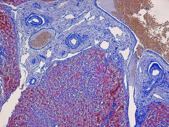
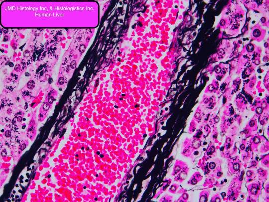
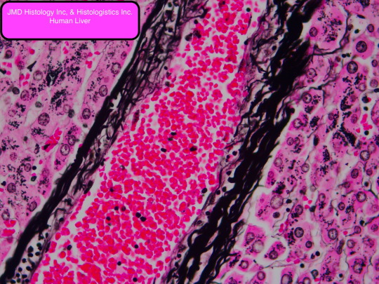
JMD Histology Inc. & Histologistics Incorporated Human Liver Rhodanine stain for copper. Excess copper is found in the cytoplasm of cells bound to copper-associated protein. DMAB-rhodanine is a bidentate chelating agent, that has a strong affinity for proteinaceous copper deposits. Increased amounts of copper are found in the liver in several disease states including Wilson’s disease, the excess copper has a toxic effect causing liver damage ranging from acute hepatitis and advanced cirrhosis. copper deposits brick red, nuclei: pale blue, bile: green
#Histology#histologyclass#histologylab#histology_lab#histopathology#pathology#pathologyapparel#copper#anatomy#science#sciences#scientist
9 notes
·
View notes
Photo
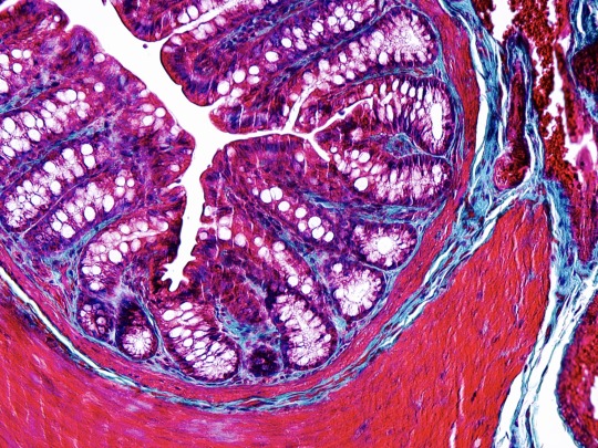
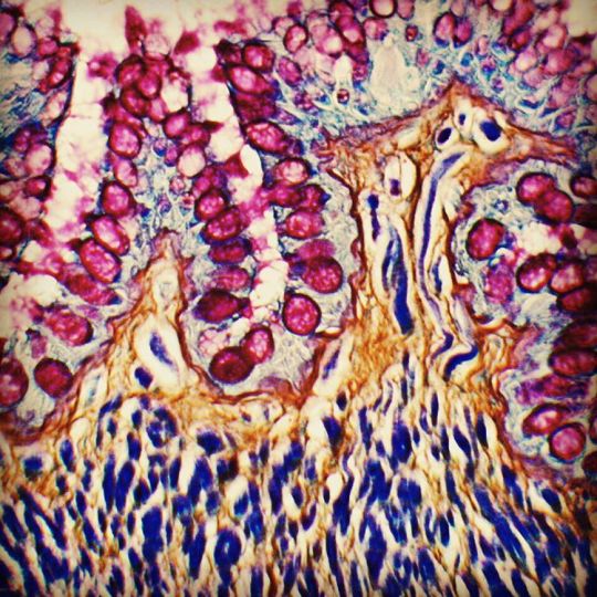
JMD Histology Inc. & Histologistics Incorporated Masson's trichrome: Is a three-colour staining protocol. It was evolved from Claude L. Pierre Masson's (1880-1959) original formulation have different specific applications. All are suited for distinguishing cells from surrounding connective tissue. Red keratin and muscle fibers, blue or green collagen and bone, light red or pink cytoplasm, and dark brown to black cell nuclei. show less
14 notes
·
View notes
Photo
JMD Histology Inc. & Histologistics Incorporated
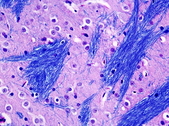
Rat Brain LFB H&E,
Luxol fast blue stain: Is commonly used to observe myelin under light microscopy, created by Heinrich Klüver and Barrera in 1953. LFB is commonly used to detect demyelination in the central nervous system (CNS), but cannot discern myelination in the peripheral nervous system. Luxol fast blue is a copper phthalocyanine dye that is soluble in alcohol and is attracted to bases found in the lipoproteins of the myelin sheath.
3 notes
·
View notes
Photo
JMD Histology Inc. & Histologistics Incorporated PERIODIC ACID SCHIFF: This stain is used for the demonstration of glycogen. Tissue sections are first oxidized by periodic acid. The oxidative process results in the formation of aldehyde groupings through carbon-to-carbon bond cleavage. Oxidation is completed when it reaches the aldehyde stage. The aldehyde groups are detected by the Schiff reagent. A colorless, unstable dialdehyde compound is formed and then transformed to the colored final product by restoration of the quinoid chromophoric grouping.
4 notes
·
View notes
Photo
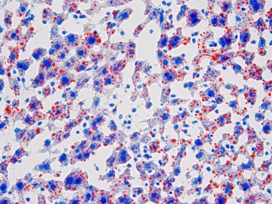
JMD Histology JMD Histology Inc., & Histologistics Incorporated What Does Oil Red O Stain? Oil Red O (‘ORO’) is used to demonstrate the presence of fat or lipids in fresh, frozen tissue sections. ORO is a fat-soluble diazo dye, it is classified as one of the Sudan dyes which have been in use since the late 1800s. Like most stains used to detect lipids, ORO is not a true special stain, since it can’t form bonds with lipid components. It is actually a pigment that functions as an oil-soluble colorant, and the technique represents a physical method of staining
#histologyslide#histologylab#Histology#lipids#science#research#researcher#histopathology#pathology_lab
2 notes
·
View notes
Photo
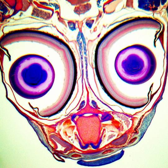
Mallorys PAS trichrome staining of a zebrafish JMDHistology.com #pathologyproject #histopathology #pathology #funnyhistology #histologytime
7 notes
·
View notes
Photo
Pas Maoris trichrome staining. JMDHistology.com #histologytime #funnyhistology #pathology #histopathology #pathologyproject #pathologist #pathologylab #science #biology
#histopathology#biology#pathologyproject#science#histologytime#pathologist#pathologylab#pathology#funnyhistology
8 notes
·
View notes
Photo
PGP 9.5 Immunostaining#histologyslides #histologylove #histology101 #histopathology #pathology #pathologylab #immunopathologyig #clinicalpathology #pathologyapparel #
#histology101#histologylove#pathologyapparel#clinicalpathology#immunopathologyig#pathologylab#histologyslides#histopathology#pathology
1 note
·
View note
Photo
Sirius Red Fast Green stain : Sirius Red binds to all types of collagen, whereas fast green stains non-collagenous proteins. This method has been applied to the measurement of collagen contents in various tissues. Please visit JMDHistology.com for all of your pathology histology and research needs #pathologylab #pathologyapparel #pathology #immunopathologyig #histology #histology101 #histology_lab #histologylove
#histology101#histologylove#pathologyapparel#immunopathologyig#pathologylab#histology#pathology#histology_lab
2 notes
·
View notes
Photo
Modified Gomori Methenamine-Silver (GMS) Nitrate Stain (Microorganism Stain) The Modified Gomori Methenamine-Silver Nitrate Stain (GMS Stain) is intended for use in the histologic visualization of fungi, basement membrane and some opportunistic organisms such as Pneumocystis carinii. Pneumocystis carinii is an opportunistic pathogen that causes severe pulmonary disease in humans, dogs, rats, mice and other vertebrate species with acquired, induced, or inherited immune deficiency syndromes. In addition, this procedure will reveal Actinomyces and related species, Nocardia asteroids, and certain encapsulated #histopathology #pathologylab #pathologyapparel #histopathology #clinicalpathology #histology #histology101 #histopathology #histologylab #histologylove #science #sciencefair #scientist #sciencemuseum #histologyart #histologylove #researcher
#histopathology#histology101#sciencemuseum#pathologyapparel#clinicalpathology#researcher#science#sciencefair#scientist#pathologylab#histology#histologyart#histologylove#histologylab
2 notes
·
View notes
Photo
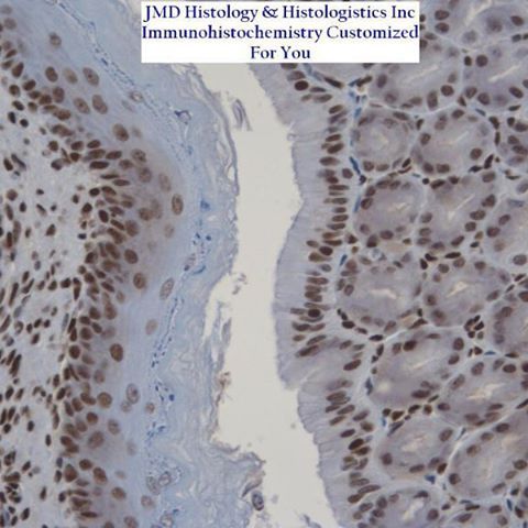
JMDHistology.com #researcher #histologylove #histologyart #sciencemuseum #scientist #histologylab #histology #histologyclass #pathology_lab #histopathology
#histopathology#histologylove#sciencemuseum#researcher#histologyclass#histology#scientist#histologylab#pathology_lab#histologyart
1 note
·
View note
Photo
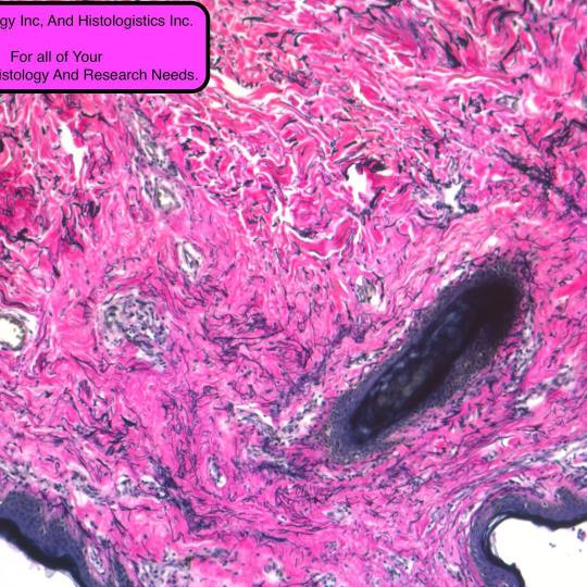
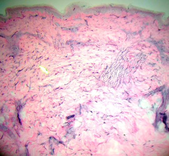
Human Skin: VVG staining #histopathology #pathology_lab #histologyclass #histology #histologylab #histologyart #histologyslide #pathologyproject
#histopathology#pathologyproject#histologyclass#histologyslide#histology#histologylab#pathology_lab#histologyart
1 note
·
View note
Photo
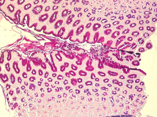
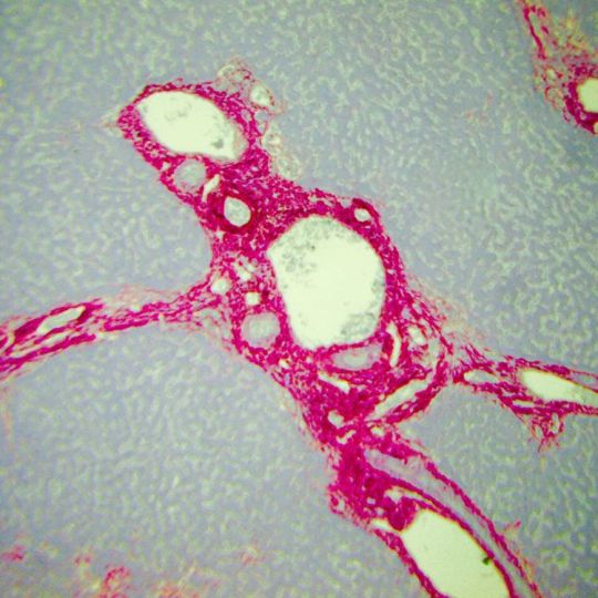
Human Liver, reticulum staining#pathologyproject #histologyslide #histologyart #histologylab #histology #histology101 #histology_lab #pathology #science #scientist #sciences #sciencecenter
#histology101#pathologyproject#sciencecenter#science#histologyslide#sciences#scientist#histology#pathology#histologyart#histology_lab#histologylab
1 note
·
View note
Photo
JMDHistology.com #sciences #sciencecenter #scientist #pathology #histology #histology101 #histologyart #histologylab #histology_lab
#histology101#sciencecenter#sciences#scientist#histology#histologylab#pathology#histology_lab#histologyart
1 note
·
View note
Photo
JMD Histology Inc. & Histologistics Incorporated Superior Quality, Unbeatable Turnaround time! Please Visit Us For All Of Your Histology, Pathology, and Research Needs. We Specialize In All Aspects Of Routine Histology, IHC, In Situ Hybridization (ISH), Immunofluorescence Techniques, Tissue Micro Arrays. Plastic, Embedding, Sectioning and Staining
1 note
·
View note
Photo
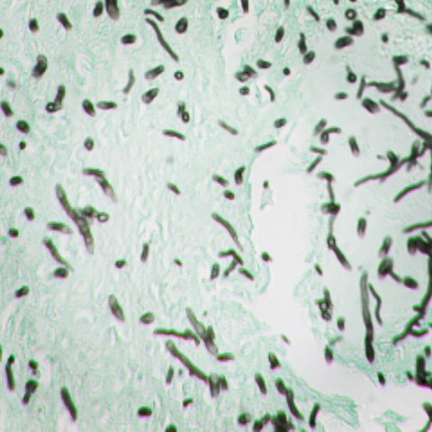
JMDHistology.com & Histologistics.com
The Reticulum Stain is used in the histological demonstration of reticular fibers. The main function of reticular fibers is to provide support. They are normally found throughout the body, particularly in liver, lymph node, spleen and kidney. Ammoniacal silver stains are the most commonly used methods for demonstration of reticular fibers.
3 notes
·
View notes
Photo

Human Skin: VVG staining #histopathology #pathology_lab #histologyclass #histology #histologylab #histologyart #histologyslide #pathologyproject
#histopathology#pathologyproject#histologyclass#histologyslide#histology#histologylab#pathology_lab#histologyart
1 note
·
View note