#utricular
Text
Teacher fucks student
Latina sucking the black off his Dick
Jordan thot ass
Hardcore gang bang with naughty babes with loads of goo
I live ass fuck
rough double anal gangbang orgy
Bbc bust huge load on latina ass
Spex british teen gets facialized by old man
Busty asian slut Jade Kush
Gay sex movietures of boy and his grandmother More Bukkake with
#wallpapers#corticating#concumbency#vagas#oilskinned#utricular#semipatriotic#coproduced#parker#stingrays#twinjets#p0rnhub#antilogarithm#moralioralist#pyrometrical#brass-colored#calcitonin#Hazlitt#pirrie#wallwort
0 notes
Text
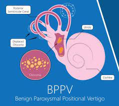
BPPV o VPPB, como guste
Post infográfico para darle salida a lo que he investigado estos días sobre el vértigo paroxístico.
Antes que nada, un lexicón:
Oto: oído.
Otoconia: literalmente, polvo del oído.
Otolito: piedra del oído; "piedras" o estructuras microscópicas que habitan en el oído interno, cuya función es regular la percepción de gravedad y aceleración.
(Por favor alguien corríjame si estoy equivocada, pero hasta ahora no he logrado encontrar diferencia sustancial entre otolito, otolitos, otoconio, otoconios y otoconia, parecen intercambiables, a veces nomás pluralizables).
Vértigo (movimiento, mareo) paroxístico (depende del movimiento) postural (y de la postura) benigno (no es peligroso, no empeora, no es contagioso).
Ahora, un bloque informativo, cortesía de este maravilloso estudio:
En el oído interno hay dos órganos (esdrújulos), el utrículo y el sáculo, que detectan los movimientos de desplazamiento y aceleración de la cabeza. Están recubiertos por un tejido epitelial (piel, digamos, o escamas o folículos) sensorial llamado mácula utricular, a su vez formada por células ciliadas y de soporte. Encima de todo esto hay una capa gelatinosa y, luego, una membrana con cristales de carbonato de calcio incrustados. Estos son los otoconios.
Los otoconios, como buenas piedras, pesan, lo que ocasiona que la membrana sea más pesada que las demás estructuras circundantes. Entonces, cuando la cabeza se inclina o se desplaza, la membrana se desplaza igualmente a causa de la gravedad en un movimiento que podemos visualizar como "inclinación" (shear) o incluso como un desfase o rezago. Los filamentos de la parte inferior de la membrana se activan y surge (brota, aparece, emerge, nace) la "recepción" / percepción.
A veces, algunos de los otoconios se desprenden del utrículo (siempre del utrículo, aunque el sáculo también contiene otoconios, los suyos no pueden viajar, son flojos) y migran hasta el sistema de canales semicirculares, donde comienzan a enviar señales equivocadas que se convierten en vértigo, dado que el mundo real no se corresponde con la percepción.
El desplazamiento, entonces, se vuelve no sólo más lento, sino completamente absurdo. Te acuestas y la habitación gira, pues tu oído insiste en que estás parada. Te pones de pie y te caes al suelo, el oído empeñado en que así tenían que ser las cosas.
Los cristales desaparecen por sí solos, aunque este proceso puede llegar a tardar años. Por fortuna, algunos genios idearon una serie de maniobras que ayuda a corregir el camino de los cristales (poesía) y devolverlos a donde pertenecen.
Esta operación, como dije antes, me recuerda a los juguetes noventeros en los que había que mover una especie de pecerita de plástico en varias direcciones hasta que un arito encajara en un palo o hasta que una bolita abandonara un laberinto.
Así se siente el vértigo, diría, como andar a ciegas por un laberinto.
Esta es una de las infinitas metáforas que me vienen a la mente cuando intento explicarme. Más: explicarme en otro idioma. Las demás, que no resultan tan informativas, las omito. En su lugar, dejo una imagen.
1 note
·
View note
Link
div class=\'at-above-post addthis_tool\' data-url=\'http://samakbahar.com/vemp-test/\'/divThe vibration stimuli as well as sound stimuli that are used to elicit oVEMPs excite many structures — the cochlea, the otoliths, the semicircular canals, proprioceptive input, so there are many choices for input. There is some evidence that these are utricular in origin. Curthoys et al (2016) reported in guinea pigs that both bone...!-- AddThis Advanced Settings above via filter on get_the_excerpt --!-- AddThis Advanced Settings below via filter on get_the_excerpt --!-- AddThis Advanced Settings generic via filter on get_the_excerpt --!-- AddThis Share Buttons above via filter on get_the_excerpt --!-- AddThis Share Buttons below via filter on get_the_excerpt --div class=\'at-below-post addthis_tool\' data-url=\'http://samakbahar.com/vemp-test/\'/div!-- AddThis Share Buttons generic via filter on get_the_excerpt --
0 notes
Text
In vivo recording of the vestibular microphonic in mammals
Publication date: Available online 26 July 2017
Source:Hearing Research
Author(s): C.J. Pastras, I.S. Curthoys, D.J. Brown
BackgroundThe Vestibular Microphonic (VM) has only featured in a handful of publications, mostly involving non-mammalian and ex vivo models. The VM is the extracellular analogue of the vestibular hair cell receptor current, and offers a tool to monitor vestibular hair cell activity in vivo.ObjectiveTo characterise features of the VM measured in vivo in guinea pigs, using a relatively simple experimental setup.MethodsThe VM, evoked by bone-conducted vibration (BCV), was recorded from the basal surface of either the utricular or saccular macula after surgical removal of the cochlea, in 27 guinea pigs.ResultsThe VM remained after vestibular nerve blockade, but was abolished following end-organ destruction or death. The VM reversed polarity as the recording electrode tracked across the utricular or saccular macula surface, or through the utricular macula. The VM could be evoked by BCV stimuli of frequencies between 100 Hz and 5 kHz, and was largest to vibrations between 600 Hz and 800 Hz. Experimental manipulations demonstrated a reduction in the VM amplitude with maculae displacement, or rupture of the utricular membrane.ConclusionsResults mirror those obtained in previous ex vivo studies, and further demonstrate that vestibular hair cells are sensitive to vibrations of several kilohertz. Changes in the VM with maculae displacement or rupture suggest utricular hydrops may alter vestibular hair cell sensitivity due to either mechanical or ionic changes.
from #Head and Neck by Sfakianakis via simeraentaxei on Inoreader http://ift.tt/2vb36cn
via IFTTT
from OtoRhinoLaryngology - Alexandros G. Sfakianakis via Alexandros G.Sfakianakis on Inoreader http://ift.tt/2uBeuwH
0 notes
Text
In vivo recording of the vestibular microphonic in mammals
Publication date: Available online 26 July 2017
Source:Hearing Research
Author(s): C.J. Pastras, I.S. Curthoys, D.J. Brown
BackgroundThe Vestibular Microphonic (VM) has only featured in a handful of publications, mostly involving non-mammalian and ex vivo models. The VM is the extracellular analogue of the vestibular hair cell receptor current, and offers a tool to monitor vestibular hair cell activity in vivo.ObjectiveTo characterise features of the VM measured in vivo in guinea pigs, using a relatively simple experimental setup.MethodsThe VM, evoked by bone-conducted vibration (BCV), was recorded from the basal surface of either the utricular or saccular macula after surgical removal of the cochlea, in 27 guinea pigs.ResultsThe VM remained after vestibular nerve blockade, but was abolished following end-organ destruction or death. The VM reversed polarity as the recording electrode tracked across the utricular or saccular macula surface, or through the utricular macula. The VM could be evoked by BCV stimuli of frequencies between 100 Hz and 5 kHz, and was largest to vibrations between 600 Hz and 800 Hz. Experimental manipulations demonstrated a reduction in the VM amplitude with maculae displacement, or rupture of the utricular membrane.ConclusionsResults mirror those obtained in previous ex vivo studies, and further demonstrate that vestibular hair cells are sensitive to vibrations of several kilohertz. Changes in the VM with maculae displacement or rupture suggest utricular hydrops may alter vestibular hair cell sensitivity due to either mechanical or ionic changes.
from #ENT via xlomafota13 on Inoreader http://ift.tt/2vb36cn
from OtoRhinoLaryngology - Alexandros G. Sfakianakis via Alexandros G.Sfakianakis on Inoreader http://ift.tt/2uBrwdS
0 notes
Text
In vivo recording of the vestibular microphonic in mammals
Publication date: Available online 26 July 2017
Source:Hearing Research
Author(s): C.J. Pastras, I.S. Curthoys, D.J. Brown
BackgroundThe Vestibular Microphonic (VM) has only featured in a handful of publications, mostly involving non-mammalian and ex vivo models. The VM is the extracellular analogue of the vestibular hair cell receptor current, and offers a tool to monitor vestibular hair cell activity in vivo.ObjectiveTo characterise features of the VM measured in vivo in guinea pigs, using a relatively simple experimental setup.MethodsThe VM, evoked by bone-conducted vibration (BCV), was recorded from the basal surface of either the utricular or saccular macula after surgical removal of the cochlea, in 27 guinea pigs.ResultsThe VM remained after vestibular nerve blockade, but was abolished following end-organ destruction or death. The VM reversed polarity as the recording electrode tracked across the utricular or saccular macula surface, or through the utricular macula. The VM could be evoked by BCV stimuli of frequencies between 100 Hz and 5 kHz, and was largest to vibrations between 600 Hz and 800 Hz. Experimental manipulations demonstrated a reduction in the VM amplitude with maculae displacement, or rupture of the utricular membrane.ConclusionsResults mirror those obtained in previous ex vivo studies, and further demonstrate that vestibular hair cells are sensitive to vibrations of several kilohertz. Changes in the VM with maculae displacement or rupture suggest utricular hydrops may alter vestibular hair cell sensitivity due to either mechanical or ionic changes.
from #Audiology via xlomafota13 on Inoreader http://ift.tt/2vb36cn
from OtoRhinoLaryngology - Alexandros G. Sfakianakis via Alexandros G.Sfakianakis on Inoreader http://ift.tt/2vMufzD
0 notes
Last Seen Blogs

llxca-blog
don't threaten me with a good time

khaozic
ZAKARIYA

moneyheist
Untitled
moneyheist
Untitled

thatguywiththehairomg
JD’s Left Glove