#raretumor
Photo

Different types of brain tumors from Miami Neuroscience Center. Not all tumors are cancerous. Ependymoma (the kind I had) are. These tumors (and others) can spread to the spinal column. It is not known what causes them. Follow me on my journey and you can also donate for brain tumor research. #braintumorresearch #brain #brainhealth #braincancer #braintumors #braintumorsurvivor #ependymoma #subependymoma #raretumor #raretumorawareness https://www.instagram.com/p/ChcSFZzOF7d/?igshid=NGJjMDIxMWI=
#braintumorresearch#brain#brainhealth#braincancer#braintumors#braintumorsurvivor#ependymoma#subependymoma#raretumor#raretumorawareness
0 notes
Text

I write this on my iPhone, sitting next to my dad, who is currently getting his 4th Chemo Therapy Treatment of Carboplatin and Taxol. The drugs are chemical bombs and each week the accumulative damage grows. They pre-treat him with histamine blocking meds so he doesn’t have reactions, but he has reactions during the infusion, like he can’t breath. The nurses are well aware and calmly manage the reactions with more meds. These meds cause him to become very drowsy, so the remainder of the day becomes about keeping him from falling.
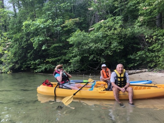
I still am trying to process all that has occurred since early August 2018. I look back on these pictures of our last outing at Lake Jocassee and never would have guessed how things would change just a week later. I’ve often wondered how cancer strikes people so quickly, now I know. I am writing this so I will never forget each minute that will forever live with me. I am also using this as a way to cope and understand something that is unfamiliar and terrifying.
My parents have always taken care of themselves and one another. They have been very lucky to have good health and I have been lucky to have them as energetic as they are in their eighties. When they moved up here from Florida, I was delighted I was going to finally be able to spend more time with them - like daily and weekly vs. just twice a year. They moved 15 minutes away or a lovely 60 min bike ride through rolling countryside and mountains. I was giddy and felt the universe shift a bit. I felt pulled to them. They are in fact two of the coolest, funniest, and open-minded people I know.
Shortly after this kayak trip (photos above) they decided to make a pact to live to 100 and created a “bucket list”. They were thankful for their health and never took it for granted. Perhaps the bucket list idea was a way to for them to celebrate how young they felt or perhaps they recognized they were chronologically getting up there.
Paddling on Jocassee was relaxing, calm, and beautiful; Certainly an experience they would have loved to have recreated again and I am hopeful they will. It may look different in the future, but I suspect the beauty and calmness of the lake will bath their brains in peace.
A week after snapping these pics, I got a call from my mom, she was on her way to the ER with my Dad. I was working one floor up and met them in the ER. While we waited, I learned my Dad had been feeling fatigued for several months and had developed shortness of breath over last few weeks. It wasn’t evident on the kayak trip that he was struggling, but it was obvious in the ER. My mom said they had been to their primary care several times and their primary care doc reassured him it was natural aging, as tests did not reveal anything to be concerned about.
As we sat for 6 hours in the waiting area, I was certain it was nothing serious. Afterall, my dad had no other health issues other than a little hypertension. His meds consisted of an 81 mg baby aspirin and amlodipine 2.5 mg each day - what a lucky guy. I was thinking maybe he had pleurisy or walking pneumonia.
We finally were shown to a room and labs were drawn. We were relieved to finally get things moving. By this time my sister, Lori, and I were getting silly from the fatigue of waiting. We were thoroughly entertained by a belligerent drunk guy on a stretcher in the halllway who seemed to draw all the attention of the medical staff while we well-behaved folks waited for answers.
I noticed my dad’s HR would easily jump to a sinus tach in the 130s with just a little bit of movement. Something didn’t seem right, but I was not going to speculate or think the worst. I was just his daughter, at his side, keeping the mood light.
We were informed by the physician assistant caring for us that his left diaphragm was elevated and was probably the cause of his shortness of breath. I was a little taken back as this was an unusual finding that left me with a knot in my stomach. Not too long after this finding he was whisked away for a CT of his chest.
He returned to the room and we waited for results. The PA came in with a sticky note and said she read off it: “You have a very large anterior mediastinal mass...No one here will operate because of your age...We are discharging you and you will need to see an oncologist.”
Our mouths dropped. My stomach bottomed-out as she said “mass” and my face flushed. We all just blankly looked at one another. Go home?
I spoke to a good nurse friend in recovery and she called the thoracic resident. I spoke to the PA who delivered the news and said, “We can’t go home. He is short of breath. He and my mom live alone. His Heart rate is bouncing up to 130s. He is weak. Please admit him and consult thoracic surgery.” My dad chimes in, “I’m not a throw away!” Meaning he doesn’t want to be dismissed because of his chronological age. He was far healthier than most half his age and this deserved a second look. The radiologist who read the report never actually saw my dad, but he did see a birthdate.
The next day, the interventional radiologist who read his CT and gave us the crappy news also did a needle biopsy of this baseball size mass.
We went home on a Wednesday after 2 days and waited. We were waiting for results and waiting for an appointment with a thoracic surgeon. Waiting is tough and if you are sick you will learn the meaning of patience.
We made it to Sunday when I thought something wasn’t right with my dad. He continued to have episodes of shortness of breath, but something was still off. I knew he had anxiety, but this was different. He said he felt fine and I almost left it at that. As a nurse you learn to listen to your 6th sense.
My parents live in a remote part of the county where everything is 30 min away. I left there house and an hour later returned with a pulse oximeter that I purchased from a CVS drug store. His oxygenation was 95% not bad for a guy now breathing 40 times a minute with 1.25 lung capacity. However, his pulse read 155 and I was baffled. No way?! I palpated his radial artery and it was a match. Off we went to the ER...
ER visit number II was faster as we went to a smaller satellite hospital 30 min from their home. The rhythm was too fast on the monitor to establish what it was so the ER MD attempted to chemically cardiovert him with adenosine. Adenosine is pushed quickly through an IV. It stops and restarts the heart. I can not lie, I was nervous. It’s so diffferent when this is your own family member. My mom tearfully excused herself and I stayed by his bedside. The ER doc informed my dad it would suck, and we proceeded. It sucked. He felt his heart stop and I watched his eyes bulge and panic come across his face for 3 of the longest seconds of my life. We were able to see he had an underlying atrial flutter. We were started on a verapamil drip and were transported to the main hospital for management by a cardiologist. His heart converted back to a normal rhythm on the verapamil drip before we left the ER in transport to Main hospital at 1 am. We were under the impression it was stress related to the new shitty diagnosis and having to wait on results.
The next day he had an echocardiogram to look at the structure and function of his heart. He was started on a Metoprolol a drug that blocks adrenaline and keeps heart rate lower and it was doing its’ job.
He spent 2 nights in hospital and outside of naps, lacked solid hours of good sleep. We finally got word that his ECHO results were good. No one said a word about metastatic disease to his pericardium. We were told he had a small ring of fluid within the pericardial sack, but it wasn’t a lot and certainly not something they felt needed draining. The atrial flutter responded well to the metoprolol and we were discharged home to once again wait for our thoracic surgery appointment.
We finally made it to the thoracic surgeon to learn of what was growing in my dad’s mediastinum. I was hoping for a thymoma, but instead we drew the really short stick with a highly aggressive, highly invasive cancer called: Squamos Cell Thymic Carcinoma.
WTF? Come on! Can we not catch a break here?
I had never heard of this type of cancer and neither have many in the medical field cause in addition to being aggressive and invasive, it is also a rare tumor. A rare tumor that hasn’t impacted enough lives that researchers devote a lot of time, money and effort into understanding it. Not only that, but sadly, most people die before any data can be collected. Once you get short of breath, dry cough and fatigue it is usually advanced.
PET Scan had some questionable lymph nodes light up, but no other disease was noted distal to the mediastinal cavity.
We hoped it could be removed. Excising the tumor was first choice in the management of this cancer and had the best outcomes, but to do this the surgeon would need to get clean margins. The thoracic surgeon wanted a cardiac MRI to examine if this tumor had invaded any of his great vessels. CT scans had only shown that the tumor was abutting the ascending aorta, but we needed to be certain cause the surgery involved opening his sternum with a saw and recovery would be 5-6 weeks. The surgeon emphasized that he didn’t want to operate and create trauma without being able to get the entire tumor. He didn’t want to delay care in a time-is-of-the-essence scenario.
It was 6pm on a Monday evening just days out from last hospitalization, when I returned to their house to check on him. Earlier that morning, my mom and I took his mini Pomeranian back to the vet and learned it was dying. The vet apologized and said it was time. We put my dad’s 18 y/o Pom, Ben, to sleep at 10:30. My mom held him and he passed. We were a mess. We told my dad and his response seemed flat. Distant.Something else was on his mind.
I stayed close and felt something was amiss, something was unfolding, progressing. I was thinking is he getting an infection? His temp was 100.2, slightly more SOB, and his pulse was 95-110 at rest, on a beta blocker. Nowhere near his norm and I could not ignore this or excuse it. My dad is precious to me. I looked at my mom and dad, apologized as I informed them we needed to go back to the ER. They were agreeable. I think he was relieved I recognized something was wrong.
Shortly after arrival at the satellite ER labs were drawn and ultrasound of his heart was done by ER doc. He said there appeared to be a large fluid collection around my dad’s heart. We were again admitted to ICU for a condition called Cardiac Tamponade. Early the next morning he had the fluid drained 600 ml from around his heart. The fluid build up which is inside the pericardial sac squeezes the heart. The heart can be stunned and go into failure. The fluid that was drawn off was sent for cytology. It was suspicious. It was likely metastatic disease.
In fact after annoying the cardiologist with repeated questions in the hallway, he motioned me over to his computer screen. He showed me the ECHO and pointed out the thickening of the pericardium and showed me a mass dangling from his ventricle. I didn’t need to wait for cytology. This was confirmation for me that we were very far into a disease process. My face flushed, my heart sank, and my stomach dropped as I comprehended the situation. I thanked the MD and my mom asked what he was showing me. I told her. I saw the color leave her face.
The thoracic surgeon was still hoping to remove the mass as the CT didn’t show it had invaded the great vessels, but he did want a Cardiac MRI which was on the back burner. We were still in ICU cause the Cardiac Tamponade and procedure to drain the fluid triggered a lot of Atrial Flutter and Atrial Fibrillation. We waited for the Cardiac MRI for 3 days. There is only one machine and his was repeated twice before they got quality images. The thoracic surgeon finally met with us and after consulting his partners, radiologist, and oncologist, it was decided surgery was just too risky and he wasn’t certain he could get clear margins. He stressed how he didn’t want to create more problems or delay my dad in getting treatment if there were complications. We very much appreciated the thoughtfulness of his answer. We really didn’t have a minute to spare. The surgeon decided to cut a window in my dad’s heart so the cancer did not build up more fluid and compress this vital organ again. The cancer cells would drain into his belly instead of filling the pericardial sack.
We were discharged home in a questionable state: weak. At first we were told he would stay until he was walking well, but the hospital was full and we were off-loaded unexpectedly. Home is a place with stairs. Stairs to to get in and stairs to get out and the most movement he had done in a week was walking 25 ft with a walker and that was exhausting for him. I was concerned about falls. How were me and my mom going to get 170 lb man up 5 steps safely? He was too weak. He hadn’t eaten, he had not slept in 10 days. We were behind the eight ball and chemo had not even started.
Chemo is rough. To survive chemo, one needs some level of fitness, meaning able to perform ADLs independently and move often. We were overwhelmed. The next week was labor intensive and emotionally draining. Here we were home and we were struggling. He still wasn’t eating, still not sleeping, and my radar was on constant alert. I spent my days observing and looking for subtle changes. Oh and there were changes that needed immediate attention as he flipped in and out of rapid atrial fibrillation and got urinary tract infection.
I was scared and my dad was terrified. In times when we were alone, he would ask me: “How did this happen?” He would shake his head as if disappointed in his body. Disbelief. He was unable to comprehend it and he too was terrified.
To be continued...

1 note
·
View note
Text
Dedifferentiated Chondrosarcoma of the Larynx: A Case Report and Review of the Literature- Juniper Publishers

Abstract
Background: Laryngeal chondrosarcomas are rare, slow-growing, cartilaginous tumors. Dedifferentiated chondrosarcomas, a rare entity of chondrosarcoma, are more aggressive and associated with a more ominous prognosis. Definite diagnosis can be established by incisional biopsy and histopathologic examination. Histopathologic examination reveals a cartilaginous tumor with a malignant spindle cell component. Definitive reatment of dedifferentiated chondrosarcomas of the larynx is total laryngectomy. There have been 14 case reports of laryngeal dedifferentiated chondrosarcoma reported since 1988. The average life expectancy reported is 6 months, and a 5-year survival rate of 10.5%.
Case presentation: We present a case of dedifferentiated chondrosarcoma arising in the cricoids cartilage of a male patient, who presented with 3-week history of dyspnea, stridor, dysphonia and intermittent aphonia. As a result, he underwent a total laryngectomy, and received adjuvant radiation therapy.
Conclusion: Laryngeal dedifferentiated chondrosarcoma is a rare entity. Symptoms include dyspnea, hoarseness, dysphagia, and a painless neck mass. Due to the aggressiveness of the tumor, it is essential to include it as a differential diagnosis among the other laryngeal tumors.
Keywords: Chondrosarcoma; Dedifferentiated; Larynx; Malignancy
Abbreviations: DC: Dedifferentiated Chondrosarcoma; PL: Partial Laryngectomy; TL: Total Laryngectomy; BND: Bilateral Neck Dissection; TT: Total Thyroidectomy; WE: Wide Excision; ART: Adjuvant Radiotherapy; ARC: Adjuvant Radiochemotherapy; DRMD: Died Of Regional Metastatic Disease; DDMD: Died Of Distant Metastatic Disease; NED: Alive With No Evidence Of Malignancy; AMD: Alive With Metastatic Disease; DOD: Died Of Other Cause
Introduction
Chondrosarcoma (CS) is a slow-growing malignant mesenchymal tumor with cartilaginous differentiation. CS, as a whole, is relatively common [1-4]. These tumors are most commonly located in the pelvis, femur, ribs, humerus, scapula, fibula, sacrum, or sternum; accounting for about 10-20% of malignant primary bone tumors [1-3,5-8]. Paranasal sinuses, nasal cavity, temporal bone, mandible and larynx were the most common sites of head and neck chondrosarcomas origin [9]. Laryngeal chondrosarcomas are a raretumor, and account for only <0.2% of all head and neck malignancies [1,2,4,6,10-12]. Among the subtypes of CS, dedifferentiated CS (DC) is a rare, sinister variant associated with a poor prognosis of an average life-expectancy of 6 months, and a 5-year survival rate of 10.5%.
There have been numerous reports of laryngeal CS, however, there are only 14 reports of laryngeal DC. Here we present a case study of DC of the cricoid cartilage, including the clinical presentation, investigations, and management.
Case Presentation
A 56-year-old Caucasian male was referred to the ENT clinic with a 3-week history of dyspnea, stridor, dysphonia and intermittent aphonia. Associated symptoms included frequent throat clearing, globus pharyngeus, hoarseness and dysphagia. The patient lost 40 pounds over a 4- month period, and progressively became more fatigued. He quit smoking 15 years ago. Prior to that, he smoked 10 cigarettes daily for 20 years. He denied the use of alcohol or recreational drugs. Past medical history was significant for hypertension, prediabetes, sleep apnea, COPD and GERD. The senior author felt the later three diagnoses were secondary to the laryngeal chondrosarcoma as they resolved post-laryngectomy. His head and neck examinations were normal.
Flexible endoscopic laryngoscopy revealed left vocal cord immobility, with the vocal cords in themedian position, and what appeared endoscopically to be subglottic stenosis. He underwent a head, neck and chest CT scan to rule out any sinister lesions along the course of the recurrent laryngeal nerve. Imaging revealed a 4 x 4cm left cricoid chondrosarcoma (Figure 1).
The CT scan revealed a chondrosarcoma, a poorly defined mixed soft tissue and calcified mass measuring 3.0 x 2.0cm in the axial dimension, and 3.5cm in the superior-inferior dimension, centered on theposterior left lateral aspect of the cricoid cartilage. The mass extended superiorly to the false vocal cords, and inferiorly, just below the true vocal cords. Just below the true vocal cords, there was significant narrowing of the airway. The lumen measured 2.5cm in the anterior-posterior dimension, and only 4mm in the transverse dimension. Destruction of most of the cricoid cartilage was identified. There was no evidence of cervical lymphadenopathy. The lungs were clear and there was no mediastinal lymphadenopathy. Diffuse fatty filtration of the liver was present with a poorly defined 1.2cm hypodense mass in the right lobe.
Shortly after the CT scan was done, the patient was admitted to hospital with severe respiratory distress and complete aphasia, he subsequently underwent a microlaryngoscopy, biopsy and tracheostomy. The patient's dyspnea resolved following the procedure. Due to the histological findings concluding laryngeal chondrosarcoma with a non-functional larynx, he underwent a narrow field total laryngectomy, cricopharyngeal myotomy, and tracheoesophageal puncture with placement of voice prosthesis. Postoperatively, he recovered well and his speech was 100% intelligible.
Within the total laryngectomy specimen, a firm tumor located in the left larynx was identified (Figure 2).The tumor extended proximally from the false vocal cords, and distally, into the subglottis. The tumor invaded both, the thyroid and cricoid cartilages. The resection superior and inferior margins were negative. The anterior margins were 4mm, and the posterolateral and posterior margins appeared to potentially involve the tumor. The histological findings, shown in Figure 3, revealed areas of well-differentiated cartilaginous regionsjuxtaposed with poorly- differentiated spindle cell sarcoma, arising from the cricoid cartilage. This finding was consistent with dedifferentiated chondrosarcoma.
A PET scan was completed to rule out persistent or recurrent disease in the pharyngeal region. There was no evidence of hypermetabolic adenopathy, pulmonary nodules, or hypermetabolic activity in the neckintraabdominal or pelvic organs. However, focal hypermetabolism was present in the superior aspect of thesternal body, either associated with a fracture or alternatively, a metastatic lesion. Further, imaging with theMRI revealed a healing non-displaced sternal fracture. There was no evidence of metastatic bone disease.
Radiation treatment to the laryngeal bed is recommended for intermediate and poorly dedifferentiated chondrosarcoma due to an ominous prognosis, high risk of local recurrence and distant metastases. Furthermore, in the patient's case, radiation is recommended due to the close margins revealed in the total laryngectomy specimen. In this case, a total dose of 66Gy in 33 fractions of external beam radiation therapy was completed. The patient will continue to receive ongoing assessment, including 3-monthly appointments for the first two years, 4-monthly appointments in the third year, and following with 6-monthly appointments. Moreover, a chest x-ray twice annually, and further CT scans as indicated.
Discussion
Laryngeal chondrosarcoma is the most common non- epithelial laryngeal neoplasm [7,13-15]. The incidence of chondrosarcoma increases with age, with the majority of cases arising in the sixth and seventh decade of life [3,6,7,10,11,13,1517]. Unlike other head and neck chondrosarcomas, laryngeal chondrosarcomas are relatively slow growing, low- grade neoplasms. Local excision is curative in most cases. Laryngeal chondrosarcomas typically originate from hyaline cartilage. Approximately 75% of cases arise from the posterior lamina of the cricoid cartilage [3,4,6-8,10-12,15,16,18-22]. The rest originate from the thyroid, arytenoid, epiglottis and accessory cartilages [3,6,12,15,16,18,20-22]. The exact pathogenesis of laryngeal chondrosarcomas remains unknown [2,3,6,7,11,1517,19].
Dedifferentiated chondrosarcomas of the larynx are extremely rare. Currently there are 14 cases reported in the literature, including this report (Table 1 & Figure 4). The term dedifferentiated chondrosarcoma was first termed by Dahlin and Beabout in 1971 [9,18,22,23]. DC, a rare entity of chondrosarcoma, has an estimated incidence of 8-14% of all laryngeal chondrosarcomas [9]. DC are aggressive and present with a poor prognosis [13,20]. DC commonly arise among the older adult population, with a male predilection [10]. Compared to the cases of DC reported in the literature, the clinical presentation, investigations and treatments were all similar.
Diagnosis of DC includes a combination of pertinent information on history, physical examination, imaging, and histological and immunocytochemical analyses. Definite diagnosis can be established by incisional biopsy and histopathological examination. The utility of FNA has only been described in 1 DC case. FNA can be performed in an outpatient setting with minimal complications; it is also efficient and cost- effective. Despite the advantages, the senior author does not recommend FNA in the case of chondrosarcoma; there is a risk of collecting non- representative tissue, it is difficult distinguishing benign and malignant tumors, and grading and classifying lesions [9].
Symptomology
Presenting symptoms vary depending on the anatomic location of the tumor. In the literature, common symptoms reported include hoarseness due to narrowing of the glottis and compression of the inferior laryngeal nerves, dyspnea and airway obstruction as a result of endolaryngeal and subglottic growth, dysphagia due to extralaryngeal growth of the tumor originating in the posterior cricoid, and a painless neck mass from tumor the involving the thyroid cartilage [7,10,11,12,13,18]. Rapid progression of symptoms over days or weeks should raise the suspicion of the lesion may be more aggressive than the standard laryngeal well-differentiated chondrosarcoma.
Histopathology
Histopathologic grading of chondrosarcomas is based on the criteria first described by Lichtenstein and Jaffe in 1943. The grading system stratifies the tumor into three different grades, from low to high grade according to cellularity, nuclear size and pleomorphism, necrosis and mitotic activity. This classification system assists with delineating the tumor's aggressiveness and prognosis, and subsequent treatment. Higher-grade neoplasms are associated with higher aggressiveness and a poorer prognosis [6,7,15,16,20,23]. Histological analysis remains the gold- standard for diagnostic purposes [3,6,10]. The histopathologic hallmark of DC concluded in case studies is the presence of an abrupt transition between low grade cartilaginous component juxtaposed on a high- grade, noncartilaginous, spindle cell sarcoma with pleomorphism, vesicular nuclei, giant cells, and numerous mitoses[4,5,9,10,13,18,23]. The cartilaginous portion of the tumor can contain some cells of DC, and thus the entire tumor must be examined.
Morphologically, the features of DC are similar to undifferentiated sarcoma, osteosarcoma, rhabdomyosarcoma, leiomyosarcoma or angiosarcoma [6,10,13,18].
Immunohistochemistry
Immunochemical findings were not described in this case, however other studies concluded that the malignant spindle cell component of DC is strongly positive for vimentin, focally positive for alpha-1-anti-chymotrypsin, and negative for cytokeratins, S-100 protein, desmin and muscle-specific act in [9,13,18,22].
Imaging
According to the literature, CT scan is the imaging modality of choice. Stippled to coarse calcifications within the tumor is a patho gnomonic finding for laryngeal chondrosarcoma. This feature can be seen in tumors of any grade. Thus, imaging alone is not sufficient to make the diagnosis of dedifferentiated chondrosarcoma [2, 4,6,7,10,12,15,16,18]. This finding cannot be appreciated on MRI. MRI is a useful complementary image modality for determining the extent of the tumor, as well as treatment planning and prognosis.
Destructive invasion of the cartilage, bone or soft tissue may suggest a high-grade tumor [10]. One study outlined the feasibility of multimodality imaging with PET and MRI, including DW-MRI. PET and MRI provide additional information regarding the hyper metabolic, aggressive dedifferentiated component, and the hyper metabolic, low-grade component [4].
Treatment
The standard treatment for laryngeal cartilaginous tumors is surgical excision, with preservation of the function of the larynx, if feasible [2-4,8,11,14,16,17,20]. However, due to the aggressiveness of the DC, the definitive treatment remains total laryngectomy [4,21]. Nonetheless, partial laryngectomy has been reported in a few cases. It is also recommended that patients with laryngeal DC receive adjuvant radiotherapy [2]. Although in this case, the patient received adjuvant radiation therapy, there have been previously reported cases in the literature in which the patient did not receive adjuvant therapy following surgical excision [5,17]. The role of radiation therapy remains undetermined [4]. Follow-up reveals a variable clinical course among the reported cases. Some patients remain disease free, whilst others develop metastatic disease [10,20].
Prognosis
Compared to low-grade chondrosarcomas, DC has been shown to have a poorer prognosis with a high rate of recurrence and a predisposition of distant metastases [3,4,8,13,18,23]. The average life-expectancy reported is 6 months, and a 5-year survival rate of 10.5%. Approximately 70% of patients develop pulmonarymetastatic disease some time during their disease course [18,23].
Conclusion
DC is a rare entity in the larynx. DC typically arises in the cricoid cartilage, presenting with rapidly Progressive dyspnea, hoarseness, dysphagia, and a painless neck mass. Due to the aggressiveness of thetumor, it is essential to include DC as a differential diagnosis among the other laryngeal tumors [24,25].
Acknowledgement
Thank you to Dr. Brent Wilde for providing photographs of the histology slide and of the gross specimen of the dedifferentiated chondrosarcoma of the larynx.
For more Open access journals please visit our site: Juniper Publishers
For more articles please click on Journal of Cell Science & Molecular Biology
0 notes
Photo

Finally got my dad out into the sunshine on such a beautiful day. Spending time in the 🌞 does a soul good. Chemo ✔️ Radiation✔️ and we are managing some severe weakness and profound fatigue. Rescan this week and 🙏 no new lesions. Despite how much this sucks and how many dismal days we have had, we are thankful for each day and we have tried very hard to stay in the moment. Alternatives do not serve us well. This was a good day. A good moment. https://gailkattouf.tumblr.com/post/179379161312/liters-of-iv-fluids-2-units-of-blood-and-we-made #raretumor #thymiccarcinoma #justthisday #sun #onedayatatime #thismoment #thisday #sunshine #cancersucks https://www.instagram.com/p/BsREPrfAJxC/?utm_source=ig_tumblr_share&igshid=lkznzx4t7yih
0 notes
Last Seen Blogs

summer-blues-stuff
Summer Blues

ratbastardthebastardking
Bow to the Bastard King

philosophythoughts
~speechless ~

jujutsugojou
Not bloody optimal

ut-assassintale
Assassintale