#eyeground
Text
Topher Phoenix Bareback Fucks A Fan
Bound teen rides intruder
Mati y las mellis
Summer blowjob and german blonde homemade xxx Hot Sneaky Yoga
novinha levando enrabada no pelo
Big brother fingering his real sister pussy while she sleep
Masturbacion en el coche
Milf Thing MILF sex pro Julia never lost her taste for jizz
Hairy ebony MILF shaving her pussy Pt. 3
Busty Tranny Eating Her Own Jizz
#scewing#notebooks#predeceased#Thera#advocatess#semi-intelligently#ungazing#smooth-tined#exterritorial#throwing-in#insecticidal#rochester#well-consenting#argol#snappingly#taivert#undefectiveness#tgurl#eyeground#Ctenostomata
1 note
·
View note
Text
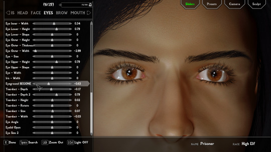
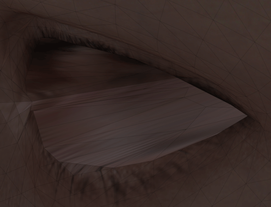
Eyeground BEGONE!
I officially made the decision today that I refuse further bullying from the Skyrim Eyeground. BEGONE! x'D
I made a slider today. It's going to be added to the CBVE.
Fed up with it so much. I accept some air between eyeball and tearline. I can live with that. What I can't live anymore is with this space invading eyeground, that behaves like "Leg Spreader Steve" in a train. EFM gives you already some editing. But it's done with Low Poly Head, which doesn't have the vertices to make this as detailed as I did. That's why my eyeground edit can only be added to HPH, sadly. ;_; There are more vertices to move around. So I did just that. Gonna see if I add more definition, like maybe outer, middle and inside eyeground sliders.
#modding#skyrim se#skyrim mods#skyrim#skyrim modding#mod author#the elder scrolls#pretty eyes#eyes#wip#CustomBetterVanillaEyes
4 notes
·
View notes
Text
Jingluo Tong with the same origin of medicine and food ---- Clove Poria Pill and Bulao Pill, manual eye patch, and secret Chinese medicine ointment in water Medical Case I药食同源之经络通----丁香茯苓丸与不老丸及手工眼贴、秘制中药水中药膏健康三宝医学案例一
Hand made traditional Chinese medicine eye patch
1、 Main functions of manual eye patch
Activates the meridians and blood vessels. The first pulse is the meridians, and the second is the blood. Manual eye patch can prevent and assist in the treatment of symptoms caused by myopia and visual fatigue (decreased vision, blurred vision, soreness and pain, photophobia and squinting, foreign body sensation, visual fatigue, eye dryness, etc.), and assist in the treatment of senile degenerative eye diseases such as cataracts, floaters, presbyopia, glaucoma, etc. Juvenile pseudomyopia below 200 degrees can completely restore normal vision.
2、 Recommended usage time of eye patch
1. It takes 15-20 minutes! Through acupoint massage and penetration, eye fatigue, itching, photophobia, squinting and other symptoms can be effectively applied, and you can enjoy a cool, moist and comfortable private massage.
2. In a 7-day period, it can obviously relieve the symptoms of dry and itchy eyes, dazzling eyes, pain, frequent blinking, etc. caused by myopia; Love eye care and enjoy a healthy life
3. After 3 months of continuous use, pseudomyopia below 200 degrees can be restored to the level of ordinary people.
4. Many presbyopians insist on using it for about a month and will remove the presbyopia.
Give yourself 1 hour, form good habits, and accompany you healthily!
3、 Instructions for using eye patches
Uncover the white paper on the eye patch to reveal the black plaster eye patch.
Close the eyes, apply the eye patch to the eyes, gently smooth the eye patch, and let the eye patch fully contact the eye skin.
Keep your eyes closed for 20 minutes to 2 hours, relax and enjoy the cool feeling of the good eye patch.
Put the white paper that has been uncovered back to its original place. After the next reuse (only for myself), take off the eye patch and continue to close your eyes for 10 minutes. When you open your eyes, you can see things much clearer than before.
4、 It is recommended to use the eye patch for conditioning cycle (individual differences exist)
1. Overuse of eyes due to asthenopia: 1-2 conditioning cycles
2. Myopia within 300 degrees: 1-2 conditioning cycles; Within 500 degrees: 2-3 conditioning cycles; Above 600 degrees; 3-5 conditioning cycles
3. Presbyopia: 2-5 conditioning cycles
5、 Take the middle-aged and elderly patients with ophthalmopathy as an example:
1. Eyeground disease, presbyopia, floater disease, vitreous opacity, macular degeneration, optic nerve atrophy and other middle-aged and elderly eye diseases.
2. Always use it three times a day. It is generally recommended to take about three courses of treatment (depending on the actual situation of eye disease)
Moisten eyes, remove turbidity, repair damage, supplement nutrition, reverse eye aging, care for eye health, and give the elderly a pair of bright eyes.
6、 Taboos for use of eye patch:
People with eye wounds, ulcerative inflammation, and eyeground bleeding. Red eye disease (infectious conjunctivitis), acute keratitis and other inflammatory eye patients.
Although the eye patch is a pure traditional Chinese medicine patch, and there are few cases of allergy, it still reminds users of allergic constitution to use it after testing.
Friends, this hospital is really
It's good. I ate it
Less than one bottle, I have to
The disc is protruding, and the waist and legs are painful
After all, please feel free to eat!
08:56
Master Zhu
I ate Jingluo Tonghe No
Less than one bottle of the old pill, the blood sugar and blood pressure drop straightly, which is very effective
0909
Aunt Ma made a mistake. It should be that the meridians are not connected
Wrong, Aunt Ma is me
It was said that the aunt who spent more than 10000 yuan in March of the year before and failed to cure her back and leg pain may be nearly three months old now. Yesterday afternoon, she took a bottle of ageless pills and insisted on using the handmade eye patch. The aunt told me that her daughter asked her about her eyes
What's the matter with the eyes? She didn't know what C 'meant. Her daughter finally said that her eyes did not fall off, but also improved. The heavy eyelids of her youth reappeared. Very beautiful
An aunt, but she is a little overweight, and seems to have passed 70kg
Auntie has lost three kilograms by eating the meridians, and she is still worried! Auntie will tell you again, good things. Don't worry about it. How many people want to lose weight and can't lose it! The aunt carefully calculated the amount of money to be no more than 200 yuan at most: she also agreed happily
Thanks, Aunt Ma. I'm waiting
09:17
The meridian unblocking, namely the clove poria pill, can unblock all the blood stasis in the whole body, and the five viscera and six fu organs. The spleen and stomach are well conditioned and well transported
Excess things that the body does not need will disappear and be removed naturally,,. Fat, thin naturally thin. Jingluo Tong will master our body's balance. Don't worry too much.
The people with rheumatoid arthritis in our group are too thin. After eating for two months, they obviously know that they are fat and their legs are painful. They must be getting better and better.
It was said that in March
手工中药眼贴
一、手工眼贴的主要作用
通经络、活血脉。通脉活脉,第一个脉是经络,第二个脉是血脉。手工眼贴可预防和辅助调理近视和视疲劳引起的症状(视力下降、视力模糊、酸胀疼痛、畏光眯眼,眼异物感,视物疲劳、眼涩、眼干等),以及辅助调理老年退行性眼部疾病如白内障、飞蚊症、老花眼、青光眼等。青少年200度以下的假性近视可以完全恢复正常视力。
二、眼贴推荐使用时间
1、15-20分钟既可见效!通过穴位按摩和渗透,眼疲劳、瘙痒、畏光眯眼等症一贴见效,享受清凉、滋润、舒适的私人按摩。
2、7天一个阶段,明显缓解近视带来的眼睛干痒、花眼、疼痛、频繁眨眼等症;爱上眼部保健,享受健康生活
3、坚持使用3个月,200度以下的假性近视,可以恢复到普通人水平。
4、不少老花眼者坚持使用一个月左右,会把老花镜摘除。
给自己1小时,养成好习惯,健康陪伴您!
三、眼贴使用步骤说明
揭开眼贴上的白纸,露出黑膏药眼贴。
闭上眼睛,将眼贴敷于眼部,轻轻抚平眼贴,让眼贴与眼部肌肤充分接触。
闭目养神二十分钟至两小时,放松身心,享受好眼贴带给您的清凉感受。
把揭开下来的白纸继续放回原处,待下次重复使用(只限本人)取下眼贴后继续闭目养神10分钟,睁开眼睛,看东西感觉比以前清晰了很多。
四、眼贴建议使用调理周期 (存在个别差异)
1、视疲劳用眼过度者:1-2个调理周期
2、近视眼300度以内:1-2个调理周期;500度以内:2-3个调理周期;600度以上;3-5个调理周期
3、老花眼:2-5个调理周期
五、以中老年眼病患者为例:
1、眼底病变、老花眼、飞蚊症、玻璃体混浊、黄斑变性、视神经萎缩等中老年眼病。
2、一定要坚持使用,一日3次,一般建议3个疗程左右(应根据眼疾实际情况而定)
润眼、拔浊、修复损伤、补充营养,逆转眼部老化,呵护眼部健康,给老人一双明亮的双眼。
六、眼贴使用禁忌:
眼睛有伤口,破溃性炎症,眼底出血的人。红眼病(传染性结膜炎)、急性角膜炎等炎症性眼部患者。
虽然眼贴是纯中药贴剂,过敏的案例极少,但仍然提醒过敏体质的用户测试后再用。
群友们,这医院真
的不错啊,我吃了
不到一瓶,我得椎
盘突出,腰腿疼已
经好了,你们请放心的吃吧!
08:56
朱师傅
我吃了经络通和不
老丸不到一瓶,血糖,血压直线下降,这效果真牛
0909
马阿姨给写错了,应该是经络通不
错,马阿姨就是我
曾经说过年前三月花了一万多没有治好腰腿疼的阿姨,吃到现在可能快三个月了,昨天下午有拿了一瓶不老丸,手工眼贴也在坚持用,阿姨告诉我,她女儿问她眼
睛怎么了,她不知 c‘ 啥意思,女儿最后才说,眼睛不掉皮下来,还提升了,年轻时的重眼皮又再现了。很漂亮的
位阿姨,不过阿姨稍微胖了点,好像也70公斤过了
阿姨吃经络通瘦了有三公斤,还在担心呢!阿姨再给您说一次,好事情,真不要担心,有多少人想瘦还瘦不下來呢!这位阿姨细算钱最多不超过二百:也高兴的答应
要谢我,马阿姨我可在等着呢
09:17
经络通即丁香茯苓丸,能通调全身上下一切瘀堵,及五脏六腑,脾胃调理好了,运化好了
身体不需要的多余东西自然消失除去,该胖的自然会 ,,。 胖,该瘦的自然会瘦。经络通会掌握咱们身体的平衡,都放心,不要有太多担心。
咱群里的类风湿关节炎的人,原来就是太瘦了,吃了两个月经络通,明显知道自己胖了,腿疼再就不说了,肯定是越来越好了。
曾经说过年前三月
0 notes
Text
Proliferative Diabetic Retinopathy
Diabetes often leads to damage of the blood vessels, especially if the blood sugar levels are not well controlled. Swelling, leaking or blockage of the blood vessels in the eye causes a serious condition, proliferative diabetic retinopathy.
CAUSES OF DIABETIC RETINOPATHY
The retina is a light-sensitive tissue lining the back of the eye. In your eye, the light rays that pass through the pupil, are focused in the lens, and penetrate to the retina, where they are transformed into signals that are transmitted by the optic nerve to the brain and interpreted as images. Therefore, it is as essential for your vision, as a processor for a computer. Your retina has multiple delicate blood vessels that deliver oxygen and nutrients to sustain its function.
Elevated blood sugar leads to thinning of the blood vessel walls and clumping of the red blood cells. These two processes lead to blood leakage and vessel obstruction. Retina becomes swollen when the blood leaks from the vessels damaged by diabetes. When the blood vessels get blocked, retinal cells become deprived of oxygen and nourishment in the area of the blood vessel obstruction. A small specialized area in the center of the retina, called macula, is especially sensitive to the consequences of the blood vessel damage. Macula is essential for clearly seeing the details of the objects located in front of you. If macula becomes swollen (a condition called macular edema), it causes blurry vision, and an impairment of the ability to recognize faces or read.
THE FOUR STAGES OF DIABETIC RETINOPATHY
The National Eye Institute (NEI) defines four distinct stages, through which the diabetic retinopathy may progress. These are mild, moderate, and severe non-proliferative diabetic retinopathy, and proliferative retinopathy. The first three diabetic retinopathy stages differ by the number of the swollen, distorted, and blocked blood vessels in the retina. Macular edema may develop even at the second stage of the disease progression. During the third stage, oxygen and nutrient deprivation of the retina caused by the vessel blockage triggers secretion of a special molecule that stimulates the new blood vessel growth. This molecule is called vascular endothelial growth factor, VEGF in short. VEGF secretion promotes diabetic retinopathy to its most advanced, proliferative stage. It is characterized by the growth of the new blood vessels in the retina, a process called neovascularization.
Proliferative retinopathy is especially harmful for vision. The newly formed blood vessels are extra fragile, and the blood leaking from them is spilled from the retina to vitreous gel, a jelly-like transparent substance inside the eye, through which the light passes on its way from the lens to the retina. The blood accumulation in vitreous gel blocks vision partially, causing appearance of black “floaters” in your field of vision, or completely, causing blindness. Neovascularization of the retina can scar this delicate tissue, causing retinal detachment from the back of the eye. Detached retina cannot convert the light rays to nerve signals, leading to partial or complete blindness.
SYMPTOMS OF RETINOPATHY
In summary, the diabetic retinopathy may cause macular edema even at the initial stages of development. In its advanced, proliferative stage, it causes the blood accumulation in the vitreous (vitreous hemorrhage), and retinal scarring and detachment. The retinopathy usually develops simultaneously in both eyes, causing the following symptoms:
• Blurry vision, or changes from clear to blurry vision and back
• Floaters and dark or black spots appearing in the field of vision
• Poor night vision
• Changes in color perception, with colors appearing faded or washed off
DETECTION AND DIAGNOSIS OF DIABETIC RETINOPATHY
Diabetic retinopathy is detected during a comprehensive eye exam. It may include the following tests, exams and procedures:
• Visual activity test. The eye chart reading measures your ability to see at various distances.
• Tonometry. A test that measures pressure inside the eye.
• Funduscopy. Its a fancy name of an eye exam performed using a magnifying glass. The drops placed on the eye surface widen (dilate) the pupil, so the physician can visually examine the “eyegrounds”, including the retina, retinal blood vessels, and the optic nerve. Fundoscopy detects changes in blood vessels (aneurisms), leaky blood vessels and fatty deposits, macular edema, and changes in the lens and abnormalities in the optic nerve.
• Optical coherence tomography (OCT). This test resembles an ultrasound exam but uses light instead of sound waves. OCT provides detailed images of eye tissues and complements the visual inspection of the retina.
• Fluorescein angiogram. In this procedure a fluorescent dye is injected into an arm vein. When the dye reaches the eye, multiple detailed pictures of the retinal blood vessels can be taken, revealing blood leaks and blood vessel changes that otherwise escaped detection.
The last two procedures are used if macular edema or progressive diabetic retinopathy are suspected.
DIABETIC RETINOPATHY TREATMENT
Many treatment methods for the diabetic retinopathy are focused on repairing or removing the damaged blood vessels and restoring the blood flow in the retina. For the best effect, two or more therapies may be combined
ANTI-VEGF INJECTION THERAPY
. VEGF, a vascular endothelial growth factor, is a key molecule that induces neovascularization, promoting advancement of diabetic retinopathy to its fourth stage, the proliferative retinopathy. Drugs that counteract VEGF action are injected in the vitreous gel of the eye every month for half a year. After that the frequency of the injections is gradually decreased, and the treatment is completed within five years. Anti-VEGF drugs include Avastin (bevacizumab), Lucentis (ranibizumab), and Eylea (aflibercept). Avastin is approved by the U.S. Food and Drug Administration (FDA) as an anti-cancer medication, but it is also used to treat eye conditions, including macular edema. Lucentis and Eylea are approved for treating macular edema and diabetic retinopathy. Anti-VEGF therapy is showing a great promise for treatment of macular edema and proliferative diabetic retinopathy.
PANRETINAL LASER SURGERY
. This treatment is also called scatter laser surgery, or photocoagulation. It involves making several thousand microscopic laser burns to shrink the abnormal blood vessels. The burns are aimed in the areas away from macula to preserve the central vision. The side effects of panretinal laser surgery include some loss of peripheral vision, and defects in night and color vision, caused by laser-induce damage of parts of the retina.
Vitrectomy. If proliferative retinopathy results in accumulation of blood in the center of the eye, which blocks vision, the vitreous gel is surgically removed. This procedure includes removing the vitreous gel by suction and replacing it with sterile saline solution to preserve the pressure in the eye and maintain the eye shape. Vitrectomy can be done under local or general anesthesia; it often requires a hospital stay, and recovery takes several weeks. When both eyes require the surgery, the second vitrectomy is performed after the first eye is completely recovered.
If proliferative diabetic retinopathy is combined with macular edema, specific treatments for the latter can be combined with the panretinal laser surgery and anti-VEGF therapy.
Injection or implantation of corticosteroids. Corticosteroids have anti-angiogenic, anti-permeability, and anti-fibrotic properties. It means that they prevent the new blood vessel growth, decrease leakage of fluid into the retina, and prevent retinal scarring. Injections of steroids, commonly triamcinolone, into the vitreous gel are performed in the same way as in the anti-VEGF therapy. Implants are also placed in the vitreous and deliver sustained amount of medication for a defined time. Some implants are designed for a short-term treatment, like Ozurdex (dexamethasone). Iluvien (fluocinolone acetonide) is used for longer treatment. The flexibility of choice of the steroid treatment regimen is essential, steroids are known to increase the pressure in the eye, promoting glaucoma development. Glaucoma is more common in diabetics than in general population, therefore consider discussing the risks and benefits of the steroid treatment with your physician.
Focal/Grid macular laser surgery. Opposite to the panretinal surgery, this treatment specifically targets the macula. 10-100 laser burns are inflicted to remove and seal the damage blood vessels to prevent blood leakage and minimize macular swelling. This treatment can be combined with anti-VEGF therapy or used as a second line of defense if the anti-VEGF therapy turned out ineffective.
OTHER DIABETES-RELATED OPHTHALMIC COMPLICATIONS
Although diabetic retinopathy is the most common and the most serious eye disease related to diabetes, other complications, such as glaucoma and cataracts, are known to affect vision in diabetic patients.
Glaucoma is a condition when the optic nerve is damaged and fails to transmit signals from the retina to the brain. In most cases, glaucoma is caused by the increased pressure in the eye. In diabetes, the growth of the new blood vessels in the iris can block the fluid flow in the eye. The pressure inside the eye increases, and a condition called a neovascular glaucoma may develop. It is also known that people with diabetes develop a more common type of glaucoma, an open-angle glaucoma, twice as frequently as non-diabetics. However, the opposite is also true: glaucoma patients have an increased chance of developing diabetes. Therefore, it is not clear if a high blood sugar causes the open-angle glaucoma, or the two diseases share some common risk factors.
Cataracts is a common cause of blindness caused by clouding of the lens. The incidence of cataracts in diabetics is twice as high as in non-diabetics. It might be caused by a chronic lens swelling caused by the constantly elevated blood sugar. Also, sudden sharp changes of the blood sugar concentration may cause distortion of the lens shape.
IN CONCLUSION
Diabetes contributes to development of serious eye diseases, such as proliferative diabetic retinopathy, macular edema, glaucoma and cataracts. Your best way to prevent the diabetes-related blindness is to keep your blood sugar in check by combination of diet, exercise, and medications. The incidence of diabetic retinopathy development in those diabetic patients who maintain their hemoglobin A1c levels below 7.0, is the same as in non-diabetics.
Diseases of the eye are easier to treat when they are detected at early stages. Therefore, all diabetic patients should get a comprehensive eye exam at least once a year. Regular ophthalmologist visits ensure the timely detection and treatment of the diabetic retinopathy, glaucoma and cataracts.
Knowledge is power. If you learned something new, or know some people who would benefit from reading this article, please share it on social media of your choice.
0 notes
Last Seen Blogs


dekakirigiri-blog
Super High School Level Detective

junkshopblog
Rie !!

robolvr
#ASFR

hb6466
İsimsiz