ubidimaging054-blog
UBid Imaging Chicago Naperville Blog Page
17 posts
Don't wanna be here? Send us removal request.
Last Seen Blogs

triumphengineersassociates
Triumph Engineers & Associates Pvt Ltd

aaannchan
Aaannchan

fuckyeahthatsart
Fuck Yeah That's Art

looking4funsblog
For fun
looking4funsblog
For fun
Text
About CT Scan Of The Abdomen
Computed Tomography (CAT or CT) Scan Of The Brain
What is a brain CT scan?
Computed tomography (CAT or CT scan) is a procedure that involves noninvasive diagnostic imaging using a combination of computer technology and X-rays to produce axial or horizontal images (commonly known as slices) of a patient’s body. CAT scan images are well-defined and provide detailed images of any body part, including the muscles, organs, bones, and fat. These scans are much finer and more detailed than typical X-ray images.
In normal X-rays, energy beams are aimed at the part of the body being studied. A plate placed behind the part of the body being examined then capture the energy beam variations after they have passed through the muscle, bone, skin, and any other tissue. While this information can be acquired using a standard X-ray, many internal structure and organ details are not available.
In CAT or computed tomography scans, the energy beam moves in circular motions around the body. By doing this, it becomes easier for the scan to capture different views of the same structure or organ. The information is then sent to a computer, which then interprets the data before displaying it in 2D or two-dimensional form on a computer monitor.
CT/CAT scans can be performed with or without contrast – which is the substance, either injected through an IV (intravenous) line or taken by mouth, that causes particular tissues or organs being studied to be seen or viewed more clearly. Contrast examinations could require a patient to fast for a specified period before they undergo the procedure. Your technician or physician will advise you about this before proceeding with the procedure.
Brain CT/CAT scans can provide more comprehensive information about brain structures and tissues than a standard head X-ray, therefore, providing more detailed brain injury or disease information than the standard X-ray.
Other brain scan related procedures used to diagnose disorders of the brain include brain magnetic resonance imaging or MRI scanning, cerebral arteriogram, and brain positron emission tomography or PET scanning.
The Anatomy of the Brain
Our central nervous system is comprised of the brain and the spinal cord. The brain is the most important organ in the body and controls motor skills, emotions, memory, touch, thought, temperature, vision, hunger, respiration and all other processes that regulate our body.
A Look At The Different Parts of Our Brain
The brain is divided into three parts. These are the cerebellum, brainstem, and cerebrum.
Cerebrum. This part, also known as the front of the brain or supratentorial, is made up of the left and right brain hemispheres. Its functions include coordination of movement, hearing, judgement, vision, temperature, initiation of movement, reasoning, learning, problem solving, and emotions.
Brainstem. Also known as the middle of the brain or midline, the brainstem is made up of the pons, medulla, and midbrain. It is responsibilities include relaying of sensory messages (loud, hot, pain, etc.), movement of the mouth and eyes, regulating body temperature, respiration, hunger, involuntary muscle movements, consciousness, swallowing, vomiting, coughing, and sneezing.
Cerebellum. Also known as the back of the brain or infratentorial, the cerebellum is located at the back of our head. Its main responsibility is to maintain posture, equilibrium, and balance, and to coordinate voluntary muscle movements.
More precisely, other parts of our brain include:
Pons. Pons are quite deep in the brain and are located within the brainstem. They contain many of the areas responsible for controlling face and eye movements, equilibrium, hearing, and facial sensation.
Medulla. The medulla is the lowest of part of our brainstem and is the most important part of the entire organ. It is where important lungs and heart control centers are located.
The spinal cord. An extensive bundle of nervous fibers located in our back, the spinal cord, extends from the brain’s base down to the lower back. It is responsible for carrying messages to the brain, from the brain, and the rest of our body.
Frontal Lobe. The frontal lobe is the largest section of our brain and is located at the front of our head. It is involved in movement and personality characteristics.
Parietal Lobe. The parietal lobe, which is the middle part of our brain, plays a crucial role as it is what helps us understand spatial relationships (this is where the body is compared to different objects around it) and to identify objects. This part is also involved in the interpretation of touch and pain.
Occipital Lobe. The back part of our brain, the occipital lobe is involved in matters related to vision.
Temporal Lobe. The temporal lobes, which are the sides of our brain, are involved in speech, sense of smell, and memory.
When is a CT/CAT scan of the brain https://ubidmri.com required?
A CT/CAT scan of the brain can be performed to evaluate the brain for injuries, structural anomalies like hydrocephalus, intracranial bleeding, tumors and other lesions, brain function and other conditions, and especially if other types of examinations like a physical examination or X-rays are inconclusive.
CAT/CT scans of the brain could also be used to evaluate the effects of brain tumor treatment and to detect any clots within the organ that could be responsible for strokes. Another use of a brain CAT scan is to provide clarity and guidance for brain tissue biopsies or surgery.
There are other reasons why doctors could recommend a CT/CAT scan of the brain.
Risks Of Brain CT/CAT scans
You may need to ask your health care provider about the amount and levels of radiation used during a CAT procedure and the related risks in relation to your particular condition. It’s a good idea to maintain a record of your radiation exposure history if you have had other types of CT or X-rays scans performed on you in the past. Some of the known radiation exposure risks are related to the total number of times a patient has undergone X-ray examinations and treatments.
If you suspect that you are pregnant or are pregnant, then it is important that you let your doctor know well in advance. Exposure to radiation during pregnancy could affect the fetus, resulting in birth defects. If it is really necessary that you have a CAT scan of the brain, then special precautionary measures will have to be employed to ensure that the least amount of radiation is exposed to the fetus.
For nursing mothers, it is advisable that they wait for at least 24 hours before resuming breastfeeding. This way, the contrast material will have exited their systems.
If a contrast material is used, there are chances you’ll probably react to the mater. Patients who are sensitive to medications or are allergic should notify their physician beforehand. Also, it is important to notify your doctor if you have any kidney problems, or have ever reacted to any contrast media before. A documented allergic reaction to seafood isn’t considered an iodinated contrast contraindication.
Patients with kidney problems or who have experienced kidney failure before should notify their physicians beforehand as the media is known to cause kidney failure. At the same time, patients on diabetes medication Glucophage (metformin) are advised to notify they physician before having an IV contrast as it could cause a rare condition known as metabolic acidosis. If you take this drug (Metformin), you are advised to stop using it for some time during the procedure and wait for forty-eight hours after the procedure before taking it. A blood test may be required to see how well your kidney is functioning before you can start using Metformin again.
There could be other risks, but it all depends on your particular medical condition. Make sure that you discuss any concerns you have with your doctor beforehand.
How to prepare for a brain scan
You should ideally wear loose fitting, comfortable clothing as you go to your exam. Before the procedure starts, you may be given a gown to wear.
Any metal objects including things like dentures, jewelry, hair pins, eyeglasses, etc. may affect the CT images and should therefore be removed before your exam or left at home altogether. You might also be asked to remove any removable dental work and hearing aids. If possible, you may be asked to remove any piercings. Women might also be asked to remove bras that contain metal underwire.
You will also be asked in advance not to drink or eat anything several hours beforehand, particularly if your exam involves the use of a contrast material. Make sure that you inform your doctor of any medications that you’re taking, and any allergies you might have. In case you have any known allergies to contrast dyes or material, your physician might prescribe some medications (mostly a steroid) to minimize the risk of developing an allergic reaction. You should take the medications as instructed, typically 12 hours before the administration of the contrast material. To prevent unnecessary delays, be sure to contact your physician before the exact time of the exam.
Don’t forget to let your doctor know any recent medical conditions or illnesses and whether you have a history of asthma, heart disease, kidney disease, diabetes, or thyroid problems. Any of these conditions will increase the risk of developing an unusual adverse effect. Your radiologists should also know if you have multiple myeloma, asthma, or any other disorder of the heart, thyroid gland, or kidneys, or if you have diabetes; especially if you’re taking Glucophage.
For women, you should always inform your CT technologist and your physician if there is any possibility that you may be pregnant.
What to expect
What does the equipment to look like?
The CT scanner is a distinctly shaped, large, boxlike machine short tunnel or hole at its center. You’ll lie down on a special, narrow examination table which slides in and out of the tunnel. The x-ray tube and an electronic x-ray detector will be rotating around you, and are located on opposite sides of each other in a ring-like structure called a Gantry.
In a separate room will be the computer workstation that processes the imaging information, and it’s where the technologist will be operating the scanner. The technologist will also be monitoring your examination in a direct visual contact and will in most cases be able to talk to you and hear you using a speaker and microphone.
How the procedure works
In a lot of ways, CT scanning works quite similarly to other x-ray examinations. Different parts of the body will absorb varying degrees of x-rays. This crucial difference in absorption rates allows body parts to be distinguished from each other on a CT electronic image or x-ray film.
Conventional x-ray exams have a small amount of radiation aimed at and passed through the target body part being examined, then an image is recorded on a special electronic image recording plate. On the x-ray, bones will appear white; soft tissue including organs like the liver or heart will show up in shades of grey; air appears black.
With CT scanning, a number of x-ray beams coupled with a set of x-ray detectors (electronic) rotate around you. In the process, they’ll measure the amount of radiation that’s absorbed in different parts of your body. At times, the examination table will move during the scan to ensure that the x-ray beam takes a spiral path. A specialized computer software is then used to process the large volume of data to create a 2-D cross-sectional images of your body, and then display them on a monitor.
You can think of CT imaging as observing a loaf of bread by first cutting it into many thin slices, such that when the image slices are reassembled through the computer software, the result into a very detailed multidimensional view of the loaf’s interior.
Recent advancements in refinements in detector technology allow almost all the modern CT scanners to get multiple image slices in just a single rotation. Such scanners are referred to as multi-detector CT or multi-slice CT, and allow for thinner image slices to be obtained in very short amount of time, which results into additional view capabilities and more detail.
The more modern CT scanners are so fast that they only need a few seconds to scan through large sections of your body. They’re even faster in small children. Such amounts of speeds are beneficial to all patients, but more so children, the critically ill, and the elderly, all of whom might have difficulty staying still, even for the brief amount of time required for images to be taken. The CT scanner technique can be adjusted for children to fit their size and area of interest, and minimize the dose of radiation.
Some CT exams may require the use of a contrast material to enhance the visibility of the area of interest.
CT scanning procedure
The technologist starts by positioning the patient on the CT examination table, often lying flat on their back. Pillows and straps might be used to help a patient maintain the correct posture and position as well as help them remain still during the exam.
Most scanners are fast enough to scan children without the need for sedation. In some special cases, the use of sedation may be required if the child cannot hold still. This is because motion leads to blurring of the image and degrades the quality of the examination, just as it affects conventional photographs.
Depending on the type of exam, if a contrast material is to be used, it’s either swallowed or injected via an intravenous line (IV). In some rare cases, it may also be administered by enema. Next, the table moves quickly through the scanner to find the correct position to start the scan. The table then moves slowly into the machine for the CT scanning to start. Based on the kind of CT scan required, the machine might take several passes.
You might be asked to momentarily hold your breath as the scan is performed. Any type of motion, whether body movements or breathing can cause artefacts on the final image. Such degradation of image quality resembles the blurring you will see on a photograph of a moving object.
Once the examination is complete, you’ll have to wait for a few minutes for the technologist to verify that the taken images are of high quality, enough for accurate interpretation. A head CT scan is usually completed within 10 minutes.
What can I expect to happen during and following the procedure?
In general, CT exams are easy, fast and painless. Using multidetector CT reduces how long a patient must lie still.
Although no pain is caused by the actual scanning process, needing to lie still for several minutes might result in some discomfort. The CT exam might be stressful for you, if you have chronic pain, are claustrophobic or have difficulty staying still. The nurse or technologist, under a physician’s direction, might offer some medication to you to help make the CT scanning procedure more tolerable.
If there is any intravenous contrast material utilized, you will feel a pinprick sensation when the needle gets inserted inside of your vein. Most likely you will have a flushed, warm sensation while the contrast materials are being injected and there will be a metallic taste inside of your mouth that will last for one to two minutes at most. You might have the feeling that you need to urinate; however, that will subside quickly and is a contrast effect.
After you have entered the CT scanner, there might be special light lines projected onto your body. These are used to make sure your body is positioned properly. With a modern CT scanner, all you will hear are slight whirring, clicking and buzzing noises as the internal parts of the CT scanner, which usually are not visible to you, are revolving around you throughout the imaging process.
During your CT scan, you will be alone inside of the exam room, unless it is a special situation. For example, at times, a parent who has a lead shield on might remain with their child in the room. However, the technologist can hear, see, and speak to use through the built-in intercom system at all times.
A parent might be allowed inside of the room with a pediatric patient but will have to wear a lead apron so that radiation exposure is minimized.
After the CT exam is complete, the technologist will remove the intravenous line that was used for injecting the contrast material, and a small dressing will be placed over the tiny hole that the needle made to cover it. You can then resume your regular activities.
Who interprets my exam results and how will I receive them?
A radiologist who has expertise in interpreting and supervising radiology exams will analyze your images and then an official report will be sent to your primary physician or the physician who referred you for a CT exam. Your physician will discuss your results with you.
It might be necessary to have follow-up exams. Your physician will explain exactly why another exam has been requested. At times a follow-up exam is conducted because there is a potential abnormality that needs to be evaluated further with a special imaging technique or additional views. A follow-up exam might also be necessary in order to monitor any changes in a known abnormality over time. Some the best way to know whether a treatment is working or not or a finding is stable or has changed over time is through follow-up exams.
What are a CT scan’s benefits and risks?
Benefits
– A CT scan is accurate, noninvasive and painless.
– One major advantage that a CT has is its capability to image blood vessels, soft tissue, and bone simultaneously.
– CT scanning, unlike conventional x-rays, provides images that are very detailed of many kinds of tissue in addition to the blood vessels, bones, and lungs.
– CT exams are simple and fast; in an emergency situation, they are able to reveal bleeding and internal injuries quickly enough to help save a life.
– It has been shown that CT is a cost-effective imaging tool for a broad array of various clinical issues.
– CT is less sensitive compared to MRI to patient movement.
– Unlike MRI, a CT scan may be performed even if you have any kind of implanted medical device.
– A diagnosis that is determined by a CT scan might eliminate the need for surgical biopsy and exploratory surgery.
– Following a CT exam, no radiation stays inside of the patient’s body.
– There should be no immediate side effects to the X-rays that are used in CT scans.
Risks
– There always is a slight chance of cancer due to radiation exposure. However, the benefit of receiving an accurate diagnosis from the scan far outweighs any risk involved.
– The effective dose of radiation for the procedure does vary.
– Women always should inform their physician and CT or x-ray technologist if there is a possibility they are pregnant.
– In general, CT scanning is not recommended for a woman who is pregnant, unless it is medically necessary due to the potential risk for the baby. However, with head CT scanning, this risk is minimal.
– Intravenous contrast manufacturers indicate that a baby should not be breastfed by the mother for 24-48 hours after being given contrast medium. However, both the European Society of Urogenital Radiology and American College
0 notes
Text
About CT Scan Of The Abdomen
Abdominal CAT Or CT Scans
What is an abdominal CT scan?
Computed tomography, also known as a CAT or CT scan, is a diagnostic imaging technique that is noninvasive and uses a combination of computer technology and x-ray to visualize the internal organs. The images, or slices, produced are of the axial or horizontal planes of the body. The images produced by a CT scan can include blood vessels, bones, organs, fat, and muscles. CT is used for much more detailed images than can be achieved with x-ray and for structures that cannot be visualized via x-ray.
During a traditional x-ray, the radiation energy is directed at the body part that requires imaging. A plate is positioned under or behind the part being assessed, which is used to capture the radiation beam as it passes through the body part. Although x-ray is useful for diagnosing conditions or injuries in some body parts, such as bone, conditions affecting the internal organs and soft tissues cannot be visualized with x-ray.
During a CT scan, the small amount of radioactive energy moves around the body in a circular pattern. This creates images of the organs from multiple angles. The resulting data is interpreted by a computer and creates a two dimensional form, which is presented on the computer monitor.
CT scans are performed with or without a contrast agent. A contrast is a substance that is either consumed by mouth or administered intravenously, which enhances the visualization of the organs being observed under CT. Depending on the reason for a CT scan or the exact organs being scanned, you may need to fast before the imaging process. If this is necessary, your doctor will give you specific pre-test instructions.
Abdominal CT scans are more informative than x-rays of the abdomen. They are frequently used to help diagnose conditions of the abdominal organs or determine the extent of injuries.
CT scans are also used during other procedures to help determine the placement of instruments, such as needles, during the course of biopsies or to remove (aspirate) fluid from the abdomen. Abdominal CT is also used to monitor various conditions, such as tumors, before, during, and after treatments.
Abdominal conditions can be diagnosed using various diagnostic imaging procedures. These can include standard x-ray, endoscopy, abdominal ultrasound, colonoscopy, abdominal angiogram, and CT scans of the kidney, liver, pancreas, and/or gallbladder.
Why are abdominal CT scans used?
There are numerous organs located within the abdomen and abdominopelvic region. These organs are components of the endocrine, gastrointestinal, urinary, and reproductive systems. When an abdominal CT scan is performed, it may be used to identify lesions, tumors, intra-abdominal bleeding, injuries, obstructions, infections, unexplained pain, or other problems, especially when other diagnostic imaging tests or physical examinations are inconclusive.
If an abdominal tumor is present, an abdominal CT may be used to help monitor the effectiveness of treatments. Abdominal CTs are also used to aid in guiding the needle during fluid aspiration or biopsies of abdominal organs.
Your doctor may recommend an abdominal CT for other reasons.
Are there risks associated with an abdominal CT scan?
You should discuss with your doctor any concerns you have about the use of radiation to perform a CT and how it affects your specific situation. You should consider keeping thorough records of any history of radiation exposure, such as other x-rays and CT scans, so you can discuss your past history of radiation exposure with your doctor. Any risks associated with radiation exposure from CT and x-ray are cumulative, meaning the risk increases with multiple exposures to diagnostic imaging radiation over time.
You should inform your health care professional if you are currently pregnant or believe you might be pregnant. Exposure to radiation during pregnancy is associated with birth defects.
When contrast is used during a CT, there is the risk of having an allergic reaction. If you are allergic or have sensitivities to any medications, you should always notify your physician. Inform them if you have any past or current kidney problems and any previous history of reactions to contrast agents. Having a seafood allergy is not contraindicated with iodinated contrasts. If you currently take metformin (Glucophage), or similar medications, you may need to stop your medications at least 48 hours before you are administered contrast. When contrast is combined with these medications, it may cause a dangerous change in blood pH, called metabolic acidosis.
Patients with a history of kidney failure or renal disease should inform their doctor immediately. Contrast agents may contribute to kidney failure in some people, particularly if they are experiencing dehydration or have a history of renal disease.
Depending on your specific medical needs or underlying conditions, there are other risks that need to be discussed with your doctor. Always discuss your concerns with your medical team before agreeing to have the procedure.
Some medical conditions or factors may impede the accuracy of an abdominal CT, such as:
The presence of metal objects inside the abdomen, such as rods, pins, or surgical clips.
Any residual barium from a recent barium enema.
Gas and/or stool within the bowel.
A complete hip replacement.
How to prepare for an abdominal scan
You should ideally wear loose fitting, comfortable clothing as you go to your exam. Before the procedure starts, you may be given a gown to wear.
Any metal objects including things like dentures, jewelry, hair pins, eyeglasses, etc. may affect the CT images and should therefore be removed before your exam or left at home altogether. You might also be asked to remove any removable dental work and hearing aids. If possible, you may be asked to remove any piercings. Women might also be asked to remove bras that contain metal underwire.
You will also be asked in advance not to drink or eat anything several hours beforehand, particularly if your exam involves the use of a contrast material. Make sure that you inform your doctor of any medications that you’re taking, and any allergies you might have. In case you have any known allergies to contrast dyes or material, your physician might prescribe some medications (mostly a steroid) to minimize the risk of developing an allergic reaction. You should take the medications as instructed, typically 12 hours before the administration of the contrast material. To prevent unnecessary delays, be sure to contact your physician before the exact time of the exam.
Don’t forget to let your doctor know any recent medical conditions or illnesses and whether you have a history of asthma, heart disease, kidney disease, diabetes, or thyroid problems. Any of these conditions will increase the risk of developing an unusual adverse effect. Your radiologists should also know if you have multiple myeloma, asthma, or any other disorder of the heart, thyroid gland, or kidneys, or if you have diabetes; especially if you’re taking Glucophage.
For women, you should always inform your CT technologist and your physician if there is any possibility that you may be pregnant.
What to expect
What does the equipment to look like?
The CT scanner is a distinctly shaped, large, boxlike machine short tunnel or hole at its center. You’ll lie down on a special, narrow examination table which slides in and out of the tunnel. The x-ray tube and an electronic x-ray detector will be rotating around you, and are located on opposite sides of each other in a ring-like structure called a Gantry.
In a separate room will be the computer workstation that processes the imaging information, and it’s where the technologist will be operating the scanner. The technologist will also be monitoring your examination in a direct visual contact and will in most cases be able to talk to you and hear you using a speaker and microphone.
How the procedure works
In a lot of ways, CT scanning works quite similarly to other x-ray examinations. Different parts of the body will absorb varying degrees of x-rays. This crucial difference in absorption rates allows body parts to be distinguished from each other on a CT electronic image or x-ray film.
Conventional x-ray exams have a small amount of radiation aimed at and passed through the target body part being examined, then an image is recorded on a special electronic image recording plate. On the x-ray, bones will appear white; soft tissue including organs like the liver or heart will show up in shades of grey; air appears black.
With CT scanning, a number of x-ray beams coupled with a set of x-ray detectors (electronic) rotate around you. In the process, they’ll measure the amount of radiation that’s absorbed in different parts of your body. At times, the examination table will move during the scan to ensure that the x-ray beam takes a spiral path. A specialized computer software is then used to process the large volume of data to create a 2-D cross-sectional images of your body, and then display them on a monitor.
You can think of CT imaging as observing a loaf of bread by first cutting it into many thin slices, such that when the image slices are reassembled through the computer software, the result into a very detailed multidimensional view of the loaf’s interior.
Recent advancements in refinements in detector technology allow almost all the modern CT scanners to get multiple image slices in just a single rotation. Such scanners are referred to as multi-detector CT or multi-slice CT, and allow for thinner image slices to be obtained in very short amount of time, which results into additional view capabilities and more detail.
The more modern CT scanners are so fast that they only need a few seconds to scan through large sections of your body. They’re even faster in small children. Such amounts of speeds are beneficial to all patients, but more so children, the critically ill, and the elderly, all of whom might have difficulty staying still, even for the brief amount of time required for images to be taken. The CT scanner technique can be adjusted for children to fit their size and area of interest, and minimize the dose of radiation.
Some CT exams may require the use of a contrast material to enhance the visibility of the area of interest.
CT scanning procedure
The technologist starts by positioning the patient on the CT examination table, often lying flat on their back. Pillows and straps might be used to help a patient maintain the correct posture and position as well as help them remain still during the exam.
Most scanners are fast enough to scan children without the need for sedation. In some special cases, the use of sedation may be required if the child cannot hold still. This is because motion leads to blurring of the image and degrades the quality of the examination, just as it affects conventional photographs.
Depending on the type of exam, if a contrast material is to be used, it’s either swallowed or injected via an intravenous line (IV). In some rare cases, it may also be administered by enema. Next, the table moves quickly through the scanner to find the correct position to start the scan. The table then moves slowly into the machine for the CT scanning to start. Based on the kind of CT scan required, the machine might take several passes.
You might be asked to momentarily hold your breath as the scan is performed. Any type of motion, whether body movements or breathing can cause artefacts on the final image. Such degradation of image quality resembles the blurring you will see on a photograph of a moving object.
Once the examination is complete, you’ll have to wait for a few minutes for the technologist to verify that the taken images are of high quality, enough for accurate interpretation. A head CT scan is usually completed within 10 minutes.
What can I expect to happen during and following the procedure?
In general, CT exams are easy, fast and painless. Using multidetector CT reduces how long a patient must lie still.
Although no pain is caused by the actual scanning process, needing to lie still for several minutes might result in some discomfort. The CT exam might be stressful for you, if you have chronic pain, are claustrophobic or have difficulty staying still. The nurse or technologist, under a physician’s direction, might offer some medication to you to help make the CT scanning procedure more tolerable.
If there is any intravenous contrast material utilized, you will feel a pinprick sensation when the needle gets inserted inside of your vein. Most likely you will have a flushed, warm sensation while the contrast materials are being injected and there will be a metallic taste inside of your mouth that will last for one to two minutes at most. You might have the feeling that you need to urinate; however, that will subside quickly and is a contrast effect.
After you have entered the CT scanner, there might be special light lines projected onto your body. These are used to make sure your body is positioned properly. With a modern CT scanner, all you will hear are slight whirring, clicking and buzzing noises as the internal parts of the CT scanner, which usually are not visible to you, are revolving around you throughout the imaging process.
During your CT scan, you will be alone inside of the exam room, unless it is a special situation. For example, at times, a parent who has a lead shield on might remain with their child in the room. However, the technologist can hear, see, and speak to use through the built-in intercom system at all times.
A parent might be allowed inside of the room with a pediatric patient but will have to wear a lead apron so that radiation exposure is minimized.
After the CT exam is complete, the technologist will remove the intravenous line that was used for injecting the contrast material, and a small dressing will be placed over the tiny hole that the needle made to cover it. You can then resume your regular activities.
Who interprets my exam results and how will I receive them?
A radiologist who has expertise in interpreting and supervising radiology exams will ubid-imaging-and-diagnostics.business.site analyze your images and then an official report will be sent to your primary physician or the physician who referred you for a CT exam. Your physician will discuss your results with you.
It might be necessary to have follow-up exams. Your physician will explain exactly why another exam has been requested. At times a follow-up exam is conducted because there is a potential abnormality that needs to be evaluated further with a special imaging technique or additional views. A follow-up exam might also be necessary in order to monitor any changes in a known abnormality over time. Some the best way to know whether a treatment is working or not or a finding is stable or has changed over time is through follow-up exams.
What are a CT scan’s benefits and risks?
Benefits
– A CT scan is accurate, noninvasive and painless.
– One major advantage that a CT has is its capability to image blood vessels, soft tissue, and bone simultaneously.
– CT scanning, unlike conventional x-rays, provides images that are very detailed of many kinds of tissue in addition to the blood vessels, bones, and lungs.
– CT exams are simple and fast; in an emergency situation, they are able to reveal bleeding and internal injuries quickly enough to help save a life.
– It has been shown that CT is a cost-effective imaging tool for a broad array of various clinical issues.
– CT is less sensitive compared to MRI to patient movement.
– Unlike MRI, a CT scan may be performed even if you have any kind of implanted medical device.
– A diagnosis that is determined by a CT scan might eliminate the need for surgical biopsy and exploratory surgery.
– Following a CT exam, no radiation stays inside of the patient’s body.
– There should be no immediate side effects to the X-rays that are used in CT scans.
Risks
– There always is a slight chance of cancer due to radiation exposure. However, the benefit of receiving an accurate diagnosis from the scan far outweighs any risk involved.
– The effective dose of radiation for the procedure does vary.
– Women always should inform their physician and CT or x-ray technologist if there is a possibility they are pregnant.
– In general, CT scanning is not recommended for a woman who is pregnant, unless it is medically necessary due to the potential risk for the baby. However, with head CT scanning, this risk is minimal.
– Intravenous contrast manufacturers indicate that a baby should not be breastfed by the mother for 24-48 hours after being given contrast medium. However, both the European Society of Urogenital Radiology and American College of Radiology (ACR) have noted that available data suggests it is safe to breastfeed after intravenous contrast has been received. Please consult ACR’s Manual on Contrast Media along with its references for further information.
– The risk is extremely rare o serious reaction to any contrast materials containing iodine, and radiology departments are very well-equipped to handle them.
– Since children are more sensitive to the effects of radiation, they only should have a CT exam if it is essential for a diagnosis to be made, and repeated CT exams should not be done unless they are absolutely necessary. The lose-dose technique should always be done in any CT scans performed on children.
– What limitations does CT Scanning of the Head have?
A very large person might not fit inside of the opening on a conventional CT scanner or might be over the moving table’s weight limit – which is usually 450 pounds.
When compared with MRI imaging, on CT scans the precise soft tissue details (especially of the brain and its disease processes) are not as visible. The CT scan is not very sensitive when it comes to detecting meninges inflammation – which is the membranes that cover the brain.
0 notes
Text
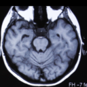
About CT Scans Of The Head
Head CT Scans
A CT Scan of the head utilizes special x-ray equipment to help in the assessment of head injuries, dizziness, severe headaches, and other symptoms of bleeding, aneurysm, brain tumors, and stroke. This can also help the physician to assess your sinuses, face, and skull or to develop a plan for radiation therapy in brain cancer treatment. In some emergency cases, the scan can reveal internal bleeding and injuries quickly enough to save lives.
If there’s a possibility that you’re pregnant, tell your doctor and be sure to discuss any recent medical conditions, illnesses, allergies, and medications you’re taking. Before the procedure, you will be instructed not to eat or drink anything for a few hours in advance. In case you have a known allergic reaction to contrast material, the doctor may prescribe some medications to reduce the impact of the allergy. These medications should be taken 12 hours before the exam. Leave all your jewelry at home, and wear comfortable loose clothing. You may be asked to wear a gown.
What’s Involved in the CT Scanning of the Head?
Computer tomography, commonly referred to as a CAT or CT scan is a diagnostic medical test that produces multiple pictures or images of the inside of the body just like the traditional x-rays.
The CT scan generates cross-sectional images which can be reformatted in multiple planes, and can even be used to generate 3-D images. The images can be printed on a film, viewed on a computer monitor, or transferred to a storage medium such as a DVD or CD.
In comparison to the traditional x-rays, CT images of various internal organs, blood vessels, soft tissue, and bones, have a much greater detail, especially of blood vessels and soft tissues. CT scanning also gives a much greater detail on head injuries, brain tumor, stroke, and other brain conditions than the regular radiographs (x-rays).
Common uses of CT scans
• Head CT scans are typically used to detect:
• Bleeding caused by a leaking or ruptured aneurysm in patients with a sudden severe headache
• Brain injury, bleeding, and skull fractures in patients with head injuries
• A stroke, particularly with the new technique referred to as Perfusion CT
• Bleeding or a blood clot within the brain shortly after symptoms of a stroke are recorded
• Enlarged ventricles (brain cavities) in patients with hydrocephalus
• Brain tumors
• Malfunctions or diseases of the skull
CT scanning is also used in:
• assessing the extent of soft tissue and bone damage in patients with facial trauma before surgical reconstruction.
• Determining whether inflammation of other changes have occurred in the paranasal sinuses
• Diagnosing diseases of the temporal bone located on the side of the skull, which might be the cause of hearing problems
• Planning radiation therapy for cancer of the brain and other tissues
• Assessing arteriovenous malfunctions or aneurysms through the CT angiography technique
How to prepare for a head scan
You should ideally wear loose fitting, comfortable clothing as you go to your exam. Before the procedure starts, you may be given a gown to wear.
Any metal objects including things like dentures, jewelry, hair pins, eyeglasses, etc. may affect the CT images and should therefore be removed before your exam or left at home altogether. You might also be asked to remove any removable dental work and hearing aids. If possible, you may be asked to remove any piercings. Women might also be asked to remove bras that contain metal underwire.
You will also be asked in advance not to drink or eat anything several hours beforehand, particularly if your exam involves the use of a contrast material. Make sure that you inform your doctor of any medications that you’re taking, and any allergies you might have. In case you have any known allergies to contrast dyes or material, your physician might prescribe some medications (mostly a steroid) to minimize the risk of developing an allergic reaction. You should take the medications as instructed, typically 12 hours before the administration of the contrast material. To prevent unnecessary delays, be sure to contact your physician before the exact time of the exam.
Don’t forget to let your doctor know any recent medical conditions or illnesses and whether you have a history of asthma, heart disease, kidney disease, diabetes, or thyroid problems. Any of these conditions will increase the risk of developing an unusual adverse effect. Your radiologists should also know if you have multiple myeloma, asthma, or any other disorder of the heart, thyroid gland, or kidneys, or if you have diabetes; especially if you’re taking Glucophage.
For women, you should always inform your CT technologist and your physician if there is any possibility that you may be pregnant.
What to expect
What does the equipment to look like?
The CT scanner is a distinctly shaped, large, boxlike machine short tunnel or hole at its center. You’ll lie down on a special, narrow examination table which slides in and out of the tunnel. The x-ray tube and an electronic x-ray detector will be rotating around you, and are located on opposite sides of each other in a ring-like structure called a Gantry.
In a separate room will be the computer workstation that processes the imaging information, and it’s where the technologist will be operating the scanner. The technologist will also be monitoring your examination in a direct visual contact and will in most cases be able to talk to you and hear you using a speaker and microphone.
How the procedure works
In a lot of ways, CT scanning works quite similarly to other x-ray examinations. Different parts of the body will absorb varying degrees of x-rays. This crucial difference in absorption rates allows body parts to be distinguished from each other on a CT electronic image or x-ray film.
Conventional x-ray exams have a small amount of radiation aimed at and passed through the target body part being examined, then an image is recorded on a special electronic image recording plate. On the x-ray, bones will appear white; soft tissue including organs like the liver or heart will show up in shades of grey; air appears black.
With CT scanning, a number of x-ray beams coupled with a set of x-ray detectors (electronic) rotate around you. In the process, they’ll measure the amount of radiation that’s absorbed in different parts of your body. At times, the examination table will move during the scan to ensure that the x-ray beam takes a spiral path. A specialized computer software is then used to process the large volume of data to create a 2-D cross-sectional images of your body, and then display them on a monitor.
You can think of CT imaging as observing a loaf of bread by first cutting it into many thin slices, such that when the image slices are reassembled through the computer software, the result into a very detailed multidimensional view of the loaf’s interior.
Recent advancements in refinements in detector technology allow almost all the modern CT scanners to get multiple image slices in just a single rotation. Such scanners are referred to as multi-detector CT or multi-slice CT, and allow for thinner image slices to be obtained in very short amount of time, which results into additional view capabilities and more detail.
The more modern CT scanners are so fast that they only need a few seconds to scan through large sections of your body. They’re even faster in small children. Such amounts of speeds are beneficial to all patients, but more so children, the critically ill, and the elderly, all of whom might have difficulty staying still, even for the brief amount of time required for images to be taken. The CT scanner technique can be adjusted for children to fit their size and area of interest, and minimize the dose of radiation.
Some CT exams may require the use of a contrast material to enhance the visibility of the area of interest.
CT scanning procedure
The technologist starts by positioning the patient on the CT examination table, often lying flat on their back. Pillows and straps might be used to help a patient maintain the correct posture and position as well as help them remain still during the exam.
Most scanners are fast enough to scan children without the need for sedation. In some special cases, the use of sedation may be required if the child cannot hold still. This is because motion leads to blurring of the image and degrades the quality of the examination, just as it affects conventional photographs.
Depending on the type of exam, if a contrast material is to be used, it’s either swallowed or injected via an intravenous line (IV). In some rare cases, it may also be administered by enema. Next, the table moves quickly through the scanner to find the correct position to start the scan. The table then moves slowly into the machine for the CT scanning to start. Based on the kind of CT scan required, the machine might take several passes.
You might be asked to momentarily hold your breath as the scan is performed. Any type of motion, whether body movements or breathing can cause artefacts on the final image. Such degradation of image quality resembles the blurring you will see on a photograph of a moving object.
Once the examination is complete, you’ll have to wait for a few minutes for the technologist to verify CT Scan Of The Head that the taken images are of high quality, enough for accurate interpretation. A head CT scan is usually completed within 10 minutes.
What can I expect to happen during and following the procedure?
In general, CT exams are easy, fast and painless. Using multidetector CT reduces how long a patient must lie still.
Although no pain is caused by the actual scanning process, needing to lie still for several minutes might result in some discomfort. The CT exam might be stressful for you, if you have chronic pain, are claustrophobic or have difficulty staying still. The nurse or technologist, under a physician’s direction, might offer some medication to you to help make the CT scanning procedure more tolerable.
If there is any intravenous contrast material utilized, you will feel a pinprick sensation when the needle gets inserted inside of your vein. Most likely you will have a flushed, warm sensation while the contrast materials are being injected and there will be a metallic taste inside of your mouth that will last for one to two minutes at most. You might have the feeling that you need to urinate; however, that will subside quickly and is a contrast effect.
After you have entered the CT scanner, there might be special light lines projected onto your body. These are used to make sure your body is positioned properly. With a modern CT scanner, all you will hear are slight whirring, clicking and buzzing noises as the internal parts of the CT scanner, which usually are not visible to you, are revolving around you throughout the imaging process.
During your CT scan, you will be alone inside of the exam room, unless it is a special situation. For example, at times, a parent who has a lead shield on might remain with their child in the room. However, the technologist can hear, see, and speak to use through the built-in intercom system at all times.
A parent might be allowed inside of the room with a pediatric patient but will have to wear a lead apron so that radiation exposure is minimized.
After the CT exam is complete, the technologist will remove the intravenous line that was used for injecting the contrast material, and a small dressing will be placed over the tiny hole that the needle made to cover it. You can then resume your regular activities.
Who interprets my exam results and how will I receive them?
A radiologist who has expertise in interpreting and supervising radiology exams will analyze your images and then an official report will be sent to your primary physician or the physician who referred you for a CT exam. Your physician will discuss your results with you.
It might be necessary to have follow-up exams. Your physician will explain exactly why another exam has been requested. At times a follow-up exam is conducted because there is a potential abnormality that needs to be evaluated further with a special imaging technique or additional views. A follow-up exam might also be necessary in order to monitor any changes in a known abnormality over time. Some the best way to know whether a treatment is working or not or a finding is stable or has changed over time is through follow-up exams.
What are a CT scan’s benefits and risks?
Benefits
– A CT scan is accurate, noninvasive and painless.
– One major advantage that a CT has is its capability to image blood vessels, soft tissue, and bone simultaneously.
– CT scanning, unlike conventional x-rays, provides images that are very detailed of many kinds of tissue in addition to the blood vessels, bones, and lungs.
– CT exams are simple and fast; in an emergency situation, they are able to reveal bleeding and internal injuries quickly enough to help save a life.
– It has been shown that CT is a cost-effective imaging tool for a broad array of various clinical issues.
– CT is less sensitive compared to MRI to patient movement.
– Unlike MRI, a CT scan may be performed even if you have any kind of implanted medical device.
– A diagnosis that is determined by a CT scan might eliminate the need for surgical biopsy and exploratory surgery.
– Following a CT exam, no radiation stays inside of the patient’s body.
– There should be no immediate side effects to the X-rays that are used in CT scans.
Risks
– There always is a slight chance of cancer due to radiation exposure. However, the benefit of receiving an accurate diagnosis from the scan far outweighs any risk involved.
– The effective dose of radiation for the procedure does vary.
– Women always should inform their physician and CT or x-ray technologist if there is a possibility they are pregnant.
– In general, CT scanning is not recommended for a woman who is pregnant, unless it is medically necessary due to the potential risk for the baby. However, with head CT scanning, this risk is minimal.
– Intravenous contrast manufacturers indicate that a baby should not be breastfed by the mother for 24-48 hours after being given contrast medium. However, both the European Society of Urogenital Radiology and American College of Radiology (ACR) have noted that available data suggests it is safe to breastfeed after intravenous contrast has been received. Please consult ACR’s Manual on Contrast Media along with its references for further information.
– The risk is extremely rare o serious reaction to any contrast materials containing iodine, and radiology departments are very well-equipped to handle them.
– Since children are more sensitive to the effects of radiation, they only should have a CT exam if it is essential for a diagnosis to be made, and repeated CT exams should not be done unless they are absolutely necessary. The lose-dose technique should always be done in any CT scans performed on children.
– What limitations does CT Scanning of the Head have?
A very large person might not fit inside of the opening on a conventional CT scanner or might be over the moving table’s weight limit – which is usually 450 pounds.
When compared with MRI imaging, on CT scans the precise soft tissue details (especially of the brain and its disease processes) are not as visible. The CT scan is not very sensitive when it comes to detecting meninges inflammation – which is the membranes that cover the brain.
0 notes
Text
About CT Scan Of The Chest
Computed Tomography (CT or CAT) Scan of the Chest
Computer tomography, also referred to as a CAT scan or CT scan is a non-invasive imaging diagnostic procedure, which uses computer technology and X-rays to produce axial or horizontal images 9also called slices) of the human body. The CAT scan essentially shows detailed images of the part of the body in question, including muscles, bones, organs, and fats. CT scans are in essence more detailed than the standard X-rays.
For standard x-rays, an energy beam is aimed to the part of the body being studies. A plate, usually placed behind the body, captures the variations of the beam as it passes through the skin, tissue, bone, or muscle. However, though you can get a lot of information about the internal organs and structures on a standard X-ray, a lot of detail is not availed.
The CT scan has the X-ray moving in a circle around the body. What this does is allow for many different views of the same structure or organ. Then, the information is sent to the computer, which will interpret the data and display the result on a 2-dimensional format on a computer monitor.
In essence, CT scans can be done with or without contrast – the substance taken orally or injected into an intravenous (IV) channel to help the particular part, tissue, or organ become more visible under the beams. In this case, contrast examinations often need you to keep away from taking food orally for a period of time before the procedure is done. Your doctor should notify you about this in due time.
Chest CT scans can offer a much-detailed information about the various structures and organs located inside the chest region, than standard X-rays would. Therefore, this is a more effective diagnostic procedure as it provides more information when it comes to injuries and diseases that are related to the thoracic (chest) organs.
CT scans on the chest can also be used to see the placement of needles in biopsy procedures for thoracic tumors or organs or during the withdrawal (aspiration) of fluid from the thoracic cavities. Such scans are essential for monitoring tumors as well as other conditions of the chest area before, during, and after treatment.
Reasons for CT scan of the Chest
The chest region contains vital organs of the cardiovascular and respiratory systems, plus the esophagus, which includes the hollow tube of muscle that extends from below the tongue to the stomach. For this reason, a chest CT scan can be used to assess the chest and the organs of the chest for tumors and other injuries, lesions, infections, bleeding, obstructions, unexplained chest pains, intrathoracic bleeding, as well as many other conditions, especially when the other forms of diagnosis and examination have proved ineffective (such as physical examinations and standard x-rays).
The CT scan performed of the chest can also be used for evaluating the effect of the various forms of treatment for thoracic tumors. Another use is offering guidance for biopsies or aspiration of the tissues located in the chest. Keep in mind that the doctor can recommend a CT scan for a variety of reasons, and it is best to trust his expertise.
Risk involved in CT scans
It is a good idea to ask your doctor amount of radiation that they use during the CT scan procedure as well as the risks involved when it comes to your particular situation. You may also want to keep an updated record of the previous history of exposure to radiation, including other previous CT scans, and other forms of x-rays so that you can inform your doctor before the procedure commences. The risks associated with radiation exposure are usually related to the cumulative amount of x-ray examinations and treatments over a period of time.
If you are actually pregnant, or you suspect that you are pregnant, it’s always good to inform your doctor. Exposing your body to radiation during pregnancy poses a risk for birth defects. When a contrast is used during the procedure, there is a risk for developing allergic reactions. In this case, let your doctor know if you have had an allergic reaction to any of the contrast media or any form of kidney problems. Reported seafood allergies are not considered contradictions to the iodinated contrast.
For patients with any form of kidney failure or kidney problems, be sure to notify the doctor. The reason for this is that sometimes the contrast media might cause kidney failure, especially for patients that are dehydrated or have underlying kidney problems. Patients who are currently under the diabetes prescription called metformin (Glucophage) or any of its derivatives, some forms of contrast poses a risk of a condition referred to as metabolic acidosis, which is a dangerous change to the blood pH.
There are several other risks that may arise depending on the particular state of your health. In this case, be sure to have a talk with your doctor and discuss all the concerns that you might have prior to the CT scan procedure.
Other conditions or factors might interfere with the accuracy of chest CT scans. Such factors include the following:
Body Piercings on the chest
Metallic objects placed within the chest like a pacemaker and surgical clips
Barium in the esophagus from a recent barium examination
Preparation for a CT scan
In John Hopkins radiology center, patients who are yet to undergo a computed tomography angiography (CTA) scan are given a basic set of instructions when they make the appointment.
Precautions
Should you be pregnant or suspect that you might be pregnant, please consult your doctor before you schedule your scan. Other options available for you will be discussed between you and your doctor.
Clothing
For clothing, keep in mind that you might be asked to change into an official patient gown. If this is the case, a gown will be provided to you. In addition, a locker will be assigned to ensure that your personal belongings are secured. Be sure to remove any form of piercings and leave all your valuables and jewelry at home.
Contrast Media
Most CT scans are usually done without a contrast media. The role of a contrast media is to improve the ability of the radiologist to view the images of the internal organs of the body.
Allergies
Make sure you inform the access center representatives if you have previously had any allergic reactions to contrast media, when you schedule the CT scan. Contrast IV will may not be administered to you if you have had an anaphylactic reaction in the past to any contrast media. If your allergy was mild and moderate during the previous use of contrast, then some medication will be administered prior to the chest CT scan. All of these plans will have to be discussed to you in detail in your appointment with our doctors for the exam. However, any known reactions to contrast media must be reported and discussed with your personal physician.
Food
You can eat and drink normally and even take your prescribed medications if your doctor decided to go with a CT scan without contrast. However, if your doctor ordered a CT scan with contrast, you are not supposed to eat and/or drink anything 3 hours prior to CT scan. Only clear liquids should be taken, and you may still take your prescribed medicine prior to the scan.
Diabetics
If you are a diabetic, you should eat a light meal three hours prior to the exam’s scheduled time. You might be asked to pause the use of medication for 48 hours after the scan, depending on the type of medication you use. At John Hopkins radiology center, you will be given detailed information on how to go about this, following your exam.
Medications
All patients may take the various forms of prescribed medicine as usual.
Don’t forget that based on your medical condition, the doctor might request a more specific preparation, which will be availed to you in detail.
CT scan Details: Procedure
CT scans can be performed on an outpatient basis and even as part of your stay in the hospital’s premises. The procedure will vary depending on the condition as well as the preferential aspects of your physician.
You might be asked to change into the patients’ gown, and this will be offered to you. In addition, a secure locker will be assigned to you where you can keep all https://en.wikipedia.org/wiki/?search=MRI Scans Of The Chest of your personal items. Again, be sure to remove any forms of piercings and leave any valuables such as jewelry at home.
If your procedure involves the use of contrast, an intravenous (IV) line I started in the arm or hand for injection of the media. For the orally administered contrast, you will essentially be given a specially prepared liquid contrast preparation that you need to swallow. In some cases, the contrast can be given rectally.
Next, you will lie on the scan table, which slides into the large circular opening of the CT scanning machine. For comfort and preventing movement during the procedure, pillows and straps might be used. A technologist will be in the other room where the controls for the scanner are located. Nonetheless, you will be in a constant sight of the technologist via a window. You can communicate with the technologist Looking for the best imaging? via the speakers located inside the scanner. You also have a call button at your comfort to let them know if you are experiencing any problems as the procedure occurs. The technologist should be in constant communication with you, as they watch you every step of the procedure.
While the scanner starts to rotate around you, x-ray beams will be passing through the body for some amount of time. There are clicking sounds, which are very normal. The rays absorbed by your body tissue can be detected by the machine, and will be transmitted to the computer. This data will be translated into an image, and the radiologist can interpret it. More importantly, be sure to remain still throughout the procedure. You may also be asked to hold your breath several times during the procedure.
Contrast media may make you feel some effects when it is injected into the (IV) line. Such effects include a salty and metallic taste in your mouth, a flushing sensation, nausea or even vomiting, and a brief headache. The effects usually last for a short amount of time. Alert the technologist should you feel any form of breathing difficulties, numbness, sweating, or heart palpitations during the procedure.
Although the CT scan is not painful, the need to lie still for the entire procedure can be uncomfortable and even cause some pain, especially if you previously had an invasive procedure such as surgery or even an injury. In this case, the technologist will use all comfort measures possible and complete the procedure as quick as possible to minimize pain or discomfort.
How to prepare for a chest scan
You should ideally wear loose fitting, comfortable clothing as you go to your exam. Before the procedure starts, you may be given a gown to wear.
Any metal objects including things like dentures, jewelry, hair pins, eyeglasses, etc. may affect the CT images and should therefore be removed before your exam or left at home altogether. You might also be asked to remove any removable dental work and hearing aids. If possible, you may be asked to remove any piercings. Women might also be asked to remove bras that contain metal underwire.
You will also be asked in advance not to drink or eat anything several hours beforehand, particularly if your exam involves the use of a contrast material. Make sure that you inform your doctor of any medications that you’re taking, and any allergies you might have. In case you have any known allergies to contrast dyes or material, your physician might prescribe some medications (mostly a steroid) to minimize the risk of developing an allergic reaction. You should take the medications as instructed, typically 12 hours before the administration of the contrast material. To prevent unnecessary delays, be sure to contact your physician before the exact time of the exam.
Don’t forget to let your doctor know any recent medical conditions or illnesses and whether you have a history of asthma, heart disease, kidney disease, diabetes, or thyroid problems. Any of these conditions will increase the risk of developing an unusual adverse effect. Your radiologists should also know if you have multiple myeloma, asthma, or any other disorder of the heart, thyroid gland, or kidneys, or if you have diabetes; especially if you’re taking Glucophage.
For women, you should always inform your CT technologist and your physician if there is any possibility that you may be pregnant.
What to expect
What does the equipment to look like?
The CT scanner is a distinctly shaped, large, boxlike machine short tunnel or hole at its center. You’ll lie down on a special, narrow examination table which slides in and out of the tunnel. The x-ray tube and an electronic x-ray detector will be rotating around you, and are located on opposite sides of each other in a ring-like structure called a Gantry.
In a separate room will be the computer workstation that processes the imaging information, and it’s where the technologist will be operating the scanner. The technologist will also be monitoring your examination in a direct visual contact and will in most cases be able to talk to you and hear you using a speaker and microphone.
How the procedure works
In a lot of ways, CT scanning works quite similarly to other x-ray examinations. Different parts of the body will absorb varying degrees of x-rays. This crucial difference in absorption rates allows body parts to be distinguished from each other on a CT electronic image or x-ray film.
Conventional x-ray exams have a small amount of radiation aimed at and passed through the target body part being examined, then an image is recorded on a special electronic image recording plate. On the x-ray, bones will appear white; soft tissue including organs like the liver or heart will show up in shades of grey; air appears black.
With CT scanning, a number of x-ray beams coupled with a set of x-ray detectors (electronic) rotate around you. In the process, they’ll measure the amount of radiation that’s absorbed in different parts of your body. At times, the examination table will move during the scan to ensure that the x-ray beam takes a spiral path. A specialized computer software is then used to process the large volume of data to create a 2-D cross-sectional images of your body, and then display them on a monitor.
You can think of CT imaging as observing a loaf of bread by first cutting it into many thin slices, such that when the image slices are reassembled through the computer software, the result into a very detailed multidimensional view of the loaf’s interior.
Recent advancements in refinements in detector technology allow almost all the modern CT scanners to get multiple image slices in just a single rotation. Such scanners are referred to as multi-detector CT or multi-slice CT, and allow for thinner image slices to be obtained in very short amount of time, which results into additional view capabilities and more detail.
The more modern CT scanners are so fast that they only need a few seconds to scan through large sections of your body. They’re even faster in small children. Such amounts of speeds are beneficial to all patients, but more so children, the critically ill, and the elderly, all of whom might have difficulty staying still, even for the brief amount of time required for images to be taken. The CT scanner technique can be adjusted for children to fit their size and area of interest, and minimize the dose of radiation.
Some CT exams may require the use of a contrast material to enhance the visibility of the area of interest.
CT scanning procedure
The technologist starts by positioning the patient on the CT examination table, often lying flat on their back. Pillows and straps might be used to help a patient maintain the correct posture and position as well as help them remain still during the exam.
Most scanners are fast enough to scan children without the need for sedation. In some special cases, the use of sedation may be required if the child cannot hold still. This is because motion leads to blurring of the image and degrades the quality of the examination, just as it affects conventional photographs.
Depending on the type of exam, if a contrast material is to be used, it’s either swallowed or injected via an intravenous line (IV). In some rare cases, it may also be administered by enema. Next, the table moves quickly through the scanner to find the correct position to start the scan. The table then moves slowly into the machine for the CT scanning to start. Based on the kind of CT scan required, the machine might take several passes.
You might be asked to momentarily hold your breath as the scan is performed. Any type of motion, whether body movements or breathing can cause artefacts on the final image. Such degradation of image quality resembles the blurring you will see on a photograph of a moving object.
Once the examination is complete, you’ll have to wait for a few minutes for the technologist to verify that the taken images are of high quality, enough for accurate interpretation. A head CT scan is usually completed within 10 minutes.
What can I expect to happen during and following the procedure?
In general, CT exams are easy, fast and painless. Using multidetector CT reduces how long a patient must lie still.
Although no pain is caused by the actual scanning process, needing to lie still for several minutes might result in some discomfort. The CT exam might be stressful for you, if you have chronic pain, are claustrophobic or have difficulty staying still. The nurse or technologist, under a physician’s direction, might offer some medication to you to help make the CT scanning procedure more tolerable.
If there is any intravenous contrast material utilized, you will feel a pinprick sensation when the needle gets inserted inside of your vein. Most likely you will have a flushed, warm sensation while the contrast materials are being injected and there will be a metallic taste inside of your mouth that will last for one to two minutes at most. You might have the feeling that you need to urinate; however, that will subside quickly and is a contrast effect.
After you have entered the CT scanner, there might be special light lines projected onto your body. These are used to make sure your body is positioned properly. With a modern CT scanner, all you will hear are slight whirring, clicking and buzzing noises as the internal parts of the CT scanner, which usually are not visible to you, are revolving around you throughout the imaging process.
During your CT scan, you will be alone inside of the exam room, unless it is a special situation. For example, at times, a parent who has a lead shield on might remain with their child in the room. However, the technologist can hear, see, and speak to use through the built-in intercom system at all times.
A parent might be allowed inside of the room with a pediatric patient but will have to wear a lead apron so that radiation exposure is minimized.
After the CT exam is complete, the technologist will remove the intravenous line that was used for injecting the contrast material, and a small dressing will be placed over the tiny hole that the needle made to cover it. You can then resume your regular activities.
Who interprets my exam results and how will I receive them?
A radiologist who has expertise
0 notes
Text
MRI Scans Of The Head
MRI Of The Head
Magnetic Resonance Imaging – better known as MRI – of the head is the head imaging technique that makes use of the radio-frequency waves, powerful magnetic fields, and advanced computer image processing to produce a detailed image of the brain and the cranial structures. With the aid of a contrast material, usually, gadolinium, MRI is capable of producing better images bearing more details than any other imaging method in use today. Additionally, the sharpness of the images is achieved without the use of ionizing radiation, which improves the safety of the procedure considerably.
However, despite being inherently safer than other imaging techniques in use today, patients looking to undergo MRI scanning of their heads should divulge any and every relevant medical information that may affect the procedure or their health.as such, patients should provide their doctors with information about recent surgeries, any prevailing health issues, as well as any allergies they may have.
The magnetic fields in use are quite harmless. However, they may affect the efficiency and functionality of some medical implants. With this in mind, even though the vast majority of orthopedic implants may not be affected, patients are best advised to divulge information regarding their presence.
Another benefit of using MRI is that even though the imaging technique is quite robust and developed, it does not necessarily need patients to change their routines. For instance, typically, patients do not have to change their diets or medications prior to having an MRI. Thus unless specifically given instructions by your doctor regarding eating, drinking, and taking medicine, one does not have to change anything. This bodes well with regular living as patients do not have to change their daily routines. The only thing they are asked to do is to wear loose fitting, comfortable clothes that are devoid of any metal as well avoid taking any jewelry to the MRI scanning session.
Understanding MRI Of The Head
MRI is simply a noninvasive medical test that is used to diagnose ailment affecting the head. As mentioned above, the imaging process is achieved by using a combination of strong magnetic fields and radio waves, while advanced computer image processing is used to develop a detailed picture of the parts of the brain that are of interest. This imaging technical is particularly adept in producing detailed pictures of the soft tissues, organs, and bones. Thus, it can be used to intricately understand the body part of interest. In fact, MRI is the most sensitive imaging technology we have today.
Obviously, doctors can develop hard copies of the detailed images of the part of the head that is of interest. However, the images can also be monitored on a computer screen, transmitted from the lab to the doctor’s office electronically, copied to a CD for storage or upload to the cloud for storage and transmission. As you can appreciate, MRI scans are quite versatile to use.
Uses Of The MRI Imaging Technology
MRI as an imaging technology can and has been used to diagnose long-standing as well as abrupt ailments affecting the head, including but not limited to:
1. Brain Tumors
2. Stroke
3. Infections in the head
4. Causes of epilepsy
5. Hemorrhage in some trauma patients
6. Anomalies in development
7. Pituitary gland disorders
8. Hydrocephalus
9. Multiple sclerosis and other conditions
10. Inner ear and eye disorders, and
11. Vascular ailments.
Preparing For MRI
As mentioned above, there is not a lot of preparation required before going for an MRI examination. The main thing that the patient has to do is to wear proper clothing (loose comfortable clothes) with no metal. However, some technicians will require the patient to wear a medical gown. With regards to drinking and eating, patients can typically maintain their eating routine as long as it entails a healthy diet. However, for some specific examinations, and eating, drinking, and medication routine will be recommended to the patient by the technologist or by the doctor.
Determining Whether The Patients Has Any Pre-Existing Condition
Right before going in for the examination, the nurse, radiologist, or technologists typically asks the patient about allergies to certain contrasting material environments, food, drugs, or contrasting material. Additionally, they are supposed to enquire about any pre-existing condition that may affect the examination including whether you have asthma and whether you are claustrophobic. In the instance that a patient is claustrophobic or suffering from anxiety disorders, a mild sedative is usually given right before the examination.
As mentioned before, it is important for patients to divulge any relevant medical information relating to health issues that they may have. However, the nurse, technologist, or radiologist will enquire about any health issues. For instance, he or she may enquire whether you have a history of suffering from kidney diseases or any liver-related issue such as a liver transplant. If you have a history of these sought of health issues, further study will be required to ensure that your kidneys and liver are performing fine.
Determining The Risk Associated With Any Medical Implants
Typically technologist will also be interested in finding out whether you have any medical devices installed in your body. The implanted devices may interfere with the examination, although this is dependent on the nature of the examination and the strength of the magnetic fields that will be used. Nonetheless, it is important for the radiologist to understand the risk associated with implanted devices. To this end, patients are encouraged to carry with them the pamphlet given to explain the risk associated with MRI examination. The information on the pamphlet makes it easy for the doctors and the radiologist to determine the risk levels and whether the risk associated can be mitigated. Some of the medical implants that are of interest to the radiologist include:
1. Implanted drug infusion ports
2. Artificial heart valves
3. Implanted nerve stimulators
4. Implanted screws, pins, stents, or staples
5. Implanted joint prostheses, or artificial limbs.
For the most part, MRI can be performed on patients with medical implants in a safe yet efficient manner. However, the following patients should not enter the MRI room, let alone undergo an MRI scan:
1. Patients with pacemakers or cardiac defibrillators
2. Patients with medical clips in their brains
3. Patients with metal coils in their blood vessels
4. Patients with ear/cochlear implants.
Determining Whether There Are Any Metal Foreign Objects In The Patient
It is not just medical implants that are of interest to the radiologist. He or she is also interested in finding out whether you have any foreign object in your body, especially of metallic nature as they may affect the MRI examination. To this end, radiologists usually make a point of finding out whether their patients have foreign materials such as bullets, shrapnel, or any other kind of piece of metal in the bodies.
Aside from affecting the examination and distorting resulting, the metal is prone to movements while undergoing examination due to the strong magnetic field. As such, foreign metal objects need to be accounted for and the risk associated with their presence determines. This is especially important when the object is near sensitive organs such as the eyes.
The risk associated with having tooth fillings and tattoos also need to be determined. Tattoos with metal-based (iron-based) inks may heat up when exposed to a strong enough magnetic field.
As for tooth fillings, their presence is usually a non-issue. However, they may affect the accuracy of the image of the brain of facial parts by distorting the magnetic field. It is thus important for the radiologist to know that they are present.
MRI And The Pregnancy Issue
Moreover, for women, the issue of pregnancy is a concern to the radiologists and as such, he or she will inquire about it. Even though MRI has been used extensively in the past on pregnant women (since 1984), no reports of ill-effects on pregnant women and their unborn children have been recorded. Nonetheless, precaution dictates that pregnant women should not receive MRI examination in the first three to four months of their pregnancies. The reason behind this is to reduce exposure of the unborn child to the strong magnetic fields. Additionally, pregnant women should not receive gadolinium treatment. As such, for pregnant mothers, MRI examination should be conducted only in the event where the benefits of the examination outweigh the cons.
After establishing your suitability for examination, the patient is injected with a contrasting material that will not affect them. In the event a patient has an allergy to the iodine-based contrasting material, gadolinium is usually used as the alternative. It is by far, less common for people to be allergic to gadolinium-based contrasting material that iodine-based material. And when one is allergic to both kids of contrasting materials, pre-medication treatment under the patient’s consent allows gadolinium to be used.
MRI Equipment
The more traditional MRI units bear a cylinder-shaped tube, with the surrounding wall being containing the magnet. Patients the lie on a motorized table where they can be inserted and removed from the MRI machine automatically. The new MRI machines tend to have a larger bore, which improves the comfort of the patients, especially those who are claustrophobic.
The short-bore MRI machine tend are designed to not have the magnet completely surrounding the patients. The Open MRI machines are designed to be open on one side. This variety of MRI unit is perfect for claustrophobic individuals as well as individuals who are large. While traditionally the Open MRI has not been quite powerful as the conventional MRI machine, advancements in technology have enabled it to attain improved accuracy.
Every MRI unit has a workstation from which the radiologist controls the machine while at the same time monitoring the imaging progress.
The MRI Procedure
The unique bit of MRI machines is that they do not make use of ionizing radiation much the same like X-rays and Computed tomography does. MIR uses radio-frequency pulses to realign the hydrogen atoms that our bodies have, all the while not causing any chemical change in our tissues.
While the hydrogen atoms align themselves to their original state, they emit different levels of energy. The variation in the level of energy emitted depends on the type of tissues emitting the energy. The MR scanner thus picks up the emitted energy and uses the variation exercise and blood pressure in energy levels to create an image of the tissues involved.
In the MRI unit, an electric current is passed through wire coils which create the magnetic field required. Other coils in the unit are designed to emit and receive radio waves. The computer thereafter processes the image of the signals received, thus forming an image of a thin slice of the head.
How is an MRI Examination Performed?
MRI examinations can be performed on inpatients and outpatients. The patient is usually positioned on a movable examination table. Bolsters and straps are sometime used to ensure that patients remain still and maintain proper positioning while imaging is underway.
Devices containing coils that can send and receive radio waves are placed either adjacent or around the area under examination. If the MRI is for the head, a device is positioned around the head.
If the MRI examinations requires the use of a contrast material, a technologist, nurse, or physician inserts an intravenous catheter (IV line), into a vein in the patient’s arm or hand. A saline solution is sometimes used to inject contrast material. This saline solution drips through the IV to ensure that the IV catheter does not block before the completion of the injection of the contrast material.
The patient is placed into the MRI unit’s magnet where the technologist and radiologist perform the exam while working at a computer situated outside the MRI room.
Once the exam is complete, the patient may be asked to wait while the radiologist or technician checks the images to see whether any additional images are required. The intravenous line is then removed.
MRI examinations usually involve several runs or sequences that may last for several minutes. It takes an average of 45 minutes for the entire examination to be completed.
The MRI examination, an additional procedure known as MR spectroscopy that provides additional information on the chemicals found in the cells of the body may also be performed and can add about 15 minutes to the total examination time.
What Can Patients Expect During and After the MRI?
MRI examinations are generally painless, but remaining still while the procedure is underway is uncomfortable for some patients. Other patients experience claustrophobia (fear of being in enclosed spaces) while in the MRI scanner. Sedation can thus be arranged for patients that anticipate anxiety but less than 5 percent of cases require medication.
It is perfectly normal for the area being imaged to feel a bit warm. However, if it becomes too much of a bother, the patient should notify the technologist or radiologist. Patients should stay perfectly still while the imaging is underway, which is usually just a couple of seconds to a couple of minutes at a time. Patients know when the recording of images is underway since they will feel loud thumping or tapping sounds when the activation of the coils generating the radiofrequency pulses happens. Some facilities provide headphones while others provide earplugs to reduce the intensity of the sounds the MRI machine makes. Patients can relax between imaging sessions, but will be required to maintain their positions without movement as much as possible.
Patients are usually alone in the exam room while the MRI procedure is underway. However, technologists are still able to hear, see, and speak to patients using a 2-way intercom. MRI facilities usually allow parents or friends to stay in the room as long as they are screened for safety in the magnetic environment.
During the exam, children are usually given appropriately sized headphones or earplugs. MRI scanners are well-lit and air-conditioned. Music can be played through headphones to help patients pass the time.
Intravenous injection of contrast material is sometimes administered prior to capturing of the images. The intravenous needle may be the source of some discomfort once inserted and the patient may experience some bruising. Patients may also experience irritation of the skin at the point of insertion of the IV tube. Other patients may experience a metallic taste in their mouths once the contrast has been injected, but it is just temporary.
No recovery period is needed if sedation is not necessary. Patients can resume their usual diet and activities once the exam is complete. On some rare occasions, patients might experience side effects from the contrast material, including headache, nausea, and pain at the injection site. Patients allergic to the contrast material may experience itchy eyes, hives, or other side effects. Patients that experience allergic symptoms should notify the technologist, radiologist, or any other health practitioner available for immediate assistance.
Who is Responsible for Interpreting Results and How Does the Patient Get Them?
Supervision and interpretation of radiology examinations is done either a radiologist or a specifically trained physician. It is also his or her responsibility to analyze those images and send a signed report to the patients referring or primary care physician that will then share those results with the patient.
Follow-up exams may be required. The doctor provides reasons for requesting additional examinations. Follow-up exams are sometimes done because further evaluation with a special imaging technique or additional views is required for a potential abnormality. Follow-up exams are sometimes the best way to determine whether treatment is working or whether a finding changes over time or is stable.
Benefits Vs Risks of MRI Examinations
Benefits
The MRI exam is a noninvasive technique for imaging that doesn’t require exposure to ionizing radiation.
MRI exams help physicians evaluate brain structures and even provide functional information in some cases.
MRI facilitates the discovery of abnormalities that might not be apparent when using other imaging techniques.
MRI produces much clearer and more detailed images of cranial structures including the brain than with other imaging methods. This is why MRIs are an indispensable tool in early evaluation and diagnosis of various conditions such as tumors.
Contrast material used for MRI examinations is less likely to trigger an allergic reaction compared to iodine-based contract materials used for CT scans and regular X-rays.
A variant of MRI known as angiography (MRA) produces detailed images of blood vessels in the brain often without the need for contrast material.
MRI is capable of detecting stroke at the very early stages by mapping the movement of water molecules in the tissue. The motion of water is known a diffusion and is impaired by most strokes, usually within less than 30 minutes from the onset of the symptoms.
Risks
MRI exams pose close to zero risk to the average patient, but only when the right safety guidelines are followed.
The risk of excessive sedation is always present if sedation is to be used. However, the nurse or technologist usually monitors the patient’s vital signs to minimize the risk.
The strong magnetic field might not be harmful in itself, but implanted medical devices containing metal could easily malfunction or cause problems while the MRI exam is underway.
A rare but recognized complication associated with MRI exams is known as Nephrogenic Systemic Fibrosis. It is believed to be caused by the injection of massive doses of gadolinium-based contrast in patients whose kidneys function very poorly. The risk of this rare complication can be minimized by carefully assessing kidney function before considering a contrast injection.
If contrast material is injected, there is a slight risk of an allergic reaction. The reactions are often mild and medication can be used to control them. If the patient experiences allergic symptoms, a radiologist or another physician is available to provide immediate assistance.
Intravenous contrast manufacturers have indicated that mothers should avoid breastfeeding their babies 24 to 48 hours after the contrast medium is administered. However, both the European Society of Urogenital Radiology and the American College of Radiology have noted that the data suggests that breastfeeding after the intravenous contrast has been administered is safe.
What Are the Limitations of MRI Exams of the Head?
High-quality images are only assured if the patient is able to stay perfectly still and follow the breath holding instructions while the recording of the images is underway. Patients that are confused, anxious, or in severe pain might find it hard to stay still while imaging is underway.
Very large individuals may not fit into the openings of some types of MRI machines.
The presence of implants or other metallic objects sometimes makes obtaining clear images hard due to the streak artifacts from the metallic objects. Patient movement also has a similar effect.
A heartbeat that is very irregular can affect the quality of images obtained using techniques which time imaging on the basis of the heart’s electrical activity, such as electrocardiography.
For patients that have been injured acutely, MRI is generally not recommended. However, the decision is on the basis of clinical judgment. The reason for this is that traction devices along with various types of life support equipment can distort MR images and should thus be kept away from the area to be imaged. The examination also takes longer than other imaging techniques such as CT scans and x-rays and results might not be available immediately, as is usually necessary in trauma situations.
While no evidence suggests that MRIs can harm a fetus, pregnant women are often advised not to undergo the procedure especially if they are in their first trimester unless it is medically necessary to do so.
MRI exams are not always able to differentiate between cancer tissues and fluid, which I known as edema.
MRI exams generally cost more and take more time to perform compared to other imaging techniques.
Follow and click the link to read more about this article and see many more leading articles on CT scan, MRI scan, and medical imaging:
https://ubidmri.com/about-mri-scans-of-the-head/
0 notes
Text
About MRI Scans Of The Body
All You Need To Know About MRI Of The Body
A magnetic resonance imaging of the body is a test that utilizes a powerful magnetic field with radio waves and a computer to produce detailed images of the inside of an individual’s body. The test is also known as an MRI and can be used to assist with the diagnosis or monitoring of treat for a plethora of conditions within the abdomen, chest and pelvis. If you are pregnant, an MRI of the body can be used to safely monitor the state of the baby.
It is essential that you inform the doctor of any health conditions, recent surgeries, allergies or possibility of pregnancy before undergoing an MRI. While the magnetic field is not a harmful area, it may result in medical devices malfunctioning. The majority of orthopedic implants do not pose a risk, but it is always more beneficial to inform the technologist if you have any metal devices or implants in your body. It is recommended that you leave jewelry at home and wear comfortable, loose clothing. Some facilities require the individual to change into a gown.
Guidelines regarding eating and drinking between the MRI examinations will vary between facilities and, unless you are told otherwise, it is recommended that you continue taking medication as per usual if necessary. If you suffer from anxiety or claustrophobia, it may be worthwhile speaking with your doctor about using a mild sedative before the examination. This could help make the experience less stressful.
What Is The MRI Of The Body?
The magnetic resonance imaging test, also known as MRI, is a non-invasive procedure used by physicians to diagnose certain medical conditions. The procedure utilizes a powerful magnetic field with radio frequency pulses and a computer producing detailed images of soft tissues, organs, bones and all other internal bodily structures or systems. It must be noted that the magnetic resonance imaging test does not use any ionizing radiation.
A detailed MRI image is beneficial for patients because it allows doctors to evaluate the different parts of a body and identify the presence of any medical conditions. The images can be examined on a computer screen, transmitted electronically and printed or copied to a disc or uploaded to a digital server.
What Are The Typical Uses Of The MRI?
An MRI of the body is typically used to evaluate the organs of the abdomen and chest including the liver, the heart, the kidneys, the biliary tract, the spleen, the pancreas, the bowels and the adrenal glands. It also examines organs of the pelvis including the reproductive organs – uterus and ovaries in females and prostate in males – and the bladder. Further uses include evaluation of the blood vessels and the lymph nodes.
Medical professionals will also utilize magnetic resonance imaging to assist with the diagnosis or monitoring treatment of specific medical conditions. The most common conditions examined include tumors in the chest, pelvis and abdomen, heart problems, diseases of the liver, inflammatory bowel disease, malformations of the blood vessels or inflammation of the blood vessels, and a fetus in the womb of a pregnant female.
How Can I Prepare For The MRI Procedure?
It is typical that you will be asked to wear a hospital gown during the MRI procedure; however, you may be allowed to wear your own clothing if it is suitable – does not have any metal fasteners or clips and is loose.
As is mentioned above, the guidelines regarding drinking and eating before an MRI examination will vary according to the facility and the date of the procedure. Unless you are informed otherwise, it is recommended that you continue with your daily consumption routine eating food and taking medication as usual.
While some MRI procedures will require you to receive an injection of contrast material into the bloodstream, it is important that the radiologist, technologist or nurse administrating the injection ask about allergies of any kind. It is important that you make the team aware of allergies to iodine or x-ray contrast material, different foods, pharmaceuticals, environmental substances and if you have asthma.
The contrast material typically used in magnetic resonance imaging procedures contains a metal known as gadolinium. This metal can be used in most patients regardless of allergies, for example patients with an iodine contrast allergy. Furthermore, it is less likely for a patient to present with an allergy to gadolinium-based contrast materials than an iodine-based contrast material often used for CT scans. However, it is still important that allergies are checked and identify whether or not the patient has an allergy to gadolinium contrast. Patient consent will be requested in this situation. For more information regarding adverse reactions to different gadolinium-based contrast materials, check the ACR Manual on Contrast Media.
It is also highly recommended that the radiographer is informed of any serious health conditions or if you have experienced any recent surgeries. Certain medical conditions, such as severe kidney disease, can prevent you from receiving a gadolinium-based contrast material injection for the MRI procedure. If you present with a history of kidney disease or have been the recipient of a liver transplant, it is necessary to performance a blood test first to identify whether or not your kidneys are functioning at an adequate level to undergo an MRI for the body.
Women should always inform the medical staff (physicians and technologists) of a possibility that they are pregnant. The magnetic resonance imaging test has been utilized since the 1980s to scan patients with no reports of damage caused to pregnant women and their unborn children; however, this does not mean there is no risk of adverse effects to the unborn baby. Due to the fact that the unborn child will be placed in a strong magnetic field, it is recommended that the pregnant female does not undergo an MRI in the first trimester of her pregnancy unless the benefits of the examination outweigh the risks. A pregnant female should not receive any injections of gadolinium-based contrast material except if absolutely necessary for medical procedures. For more information on pregnancy and MRI, see the MRI safety page.
If you suffer from a fear of small and enclosed areas, also known as claustrophobia, or anxiety, it may be beneficial to speak with your doctor about a prescription for a sedative before the MRI procedure. This will help make the scheduled examination less stressful and more manageable.
It is essential that jewelry and all other accessories are left at home, if possible, and removed before attending the MRI examination. This is necessary because jewelry and other metal accessories can influence the magnetic field of the MRI unit interfering with the examination. In addition to adversely affecting the magnetic field and MRI images, the metal objects can become dangerous items within the MRI scanning room and can act as projectiles placing you and others at risk of harm. The items that should be removed and left at home include jewelry, watches, credit cards, hearing aids, hair pins, pins, metal zippers and other metallic items. It is also recommended that you remove all dental work, eyeglasses, pens, pocket knives and body piercings.
In the majority of cases, a magnetic resonance imaging procedure is safe for patients with metal implants; however, there are certain types of implants that cannot be scanned and these people should not consider entering the MRI scanning area. Individuals with cochlear or ear implants, implant clips for brain aneurysms, metal coil implants in blood vessels, pacemakers and all other types of cardiac defibrillator implants should avoid MRI procedures.
It is essential that you inform the radiographer or technologist of any electronic or medical devices implanted in your body. The object may interfere with the procedure and can, in some cases, pose a risk of harm to you and others in the room. This is dependent on the nature of the implant and the strength of the MRI scanning magnet.
The majority of devices will have pamphlets explaining MRI risks for that particular implant and if you have this type of pamphlet; it may be beneficial to inform the scheduler when making an appointment for an MRI. It is also recommended you bring the pamphlet to the MRI scan in case the technologist or radiographer has any concerns or queries. Certain implanted devices will require a period of time after placement before any other procedures can be conducted, particularly the MRI examination. The typical time is six weeks and some examples of these devices include, but are not limited to, implanted drug infusion ports, artificial heart valves, artificial limbs or joint prostheses, and implanted nerve stimulators.
If there are any concerns regarding the presence of a metal implant, it is possible to perform an x-ray to detect and identify the object. Typically, a metal object used in orthopedic surgery will pose no risk for magnetic resonance imaging; however, recently placed artificial joints could be problematic and may require a different types of imaging procedure.
A patient who may have metal objects in certain parts of his or her body may require an x-ray before undergoing an MRI examination. It is recommended that you notify the medical staff of any bullets, shrapnel or other metal debris or items that may be lodged in your body due to accidents. Any foreign objects near or in the eyes are of particular significant because they can move during the MRI scan, potentially resulting in blindness. Contrary to belief, tooth fillings and braces are not influenced by the magnetic field but they may distort the images of the facial area, so it is worthwhile informing your radiographer of these. Furthermore, dyes in tattoos can contain iron which may heat up during an MRI procedure, although this is rare.
Young children and infants will often require some form of sedation when undergoing a magnetic resonance imaging scan. To determine if the child requires anesthesia will depend on the age of the child, the child’s intellectual development and the type of examination. A moderate and conscious sedation is available at the majority of facilities and physicians specializing in sedation of children should be available to monitor the patient during the procedure. Special instructions for adequate preparation of the child can be received from the facility.
Alternatively, specific pediatric medical facilities have child life personnel who work with young children helping them avoid the need for sedation during an MRI scan. The personnel will prepare the children for the scan by demonstrating the procedure using a dummy scanner and playing noises the child may hear during the examination. The professionals will also answer any questions the child has and can explain the scan thoroughly relieving anxiety before the procedure takes place. Certain pediatric facilities also offer headsets or goggles for the children allowing them to watch a film while the scan is being conducted.
What Does The MRI Equipment Look Like?
The traditional MRI equipment involves a large cylindrical tube surrounded by circular magnets. The patient lies on the moveable examination table which slides into the center of the cylindrical tube.
Some MRI units have been designed so that the circular magnet does not surround the tube entirely. This type of unit is known as a short-bore system and presents with a larger diameter making it comfortable for larger patients or patients with claustrophobia. The machines are open on the sides and can provide very high quality of MRI scans.
The computer workstation processing the imaging information is located outside of this magnetic range within a separate room from the scanning equipment.
How Is An MRI Of The Body Performed?
MRI scans could be performed on inpatients as well as outpatients. The patient is positioned on a moveable examination table for this purpose. Straps are used to maintain the correct position and keep the patient still during the procedure. Devices that are capable of sending and receiving radio waves are placed adjacent to the study area of the patient’s body to perform the scan. Sometimes, a contrast material is used during the scan. Under such circumstances, a doctor or a technician would insert an intravenous catheter (IV) or IV line into a vein in the patient’s hand or arm. In fact, a saline solution is used to inject the contrast material. This solution will drip through the IV until the contrast material is injected.
The patient is then placed on the magnet of the MRI scanner. The technician will perform the scan – he or she would be working at a computer outside the room. If there is a necessity of injecting a contrast material, it would be injected into the IV line after the initial scans. Another series of images will be taken after the injection.
Once the initial scans are completed, the technician will request the patient to wait a while until he or she checks the scans. If additional images are not required, the IV line would be removed. Most of the time, MRI scans are conducted in multiple runs or sequences. The process may last for several here minutes at times. The entire process may take between 30-50 minutes depending on the equipment used and the type of scan.
What Will The Patient Experience During And After The Procedure?
Although some patients may find it uncomfortable to be still during the procedure, most MRI scans are painless. There are some patients who have complained of having a feeling of being closed-in (claustrophobia) during the scan. If the patient feels anxious, the doctor may recommend appropriate sedation. But only fewer than one in 20 patients require such arrangements.
The area of the body that is being imaged can feel a bit warm during the scan. It rarely bothers the patient. If it bothers you, you should bring it to the notice of the technician. The patient needs to remain perfectly still during the process. The process is only a few seconds or a few minutes at a time. You may hear and feel loud thumping or tapping sounds when the coils of the unit are activated. That way you know the scanning process has begun. The patient may be given earplugs or headphones to reduce the intensity of the sounds made by the scanner. You can relax while the process goes on. But you should not move about when the process is being conducted. Movements can interfere with the accuracy of the scan.
Although you would be alone in the exam room during the MRI procedure, the technician can see, hear or speak with you using a two-way intercom. Most MRI centers allow a relative or friend to stay with the patient. But they should be screened for safety in the magnetic environment before getting the green light to stay.
Children are given earplugs and headphones during a scan procedure. The exam room is well-lit and air conditioned. Most of the time, music is played through the headphones to allow the patient to relax and pass the time. When IV lines are used during the scanning process, the needle may cause some bruising and discomfort. Some patients have complained of a slight irritation at the site of the IV tube insertion. But this is very rare. Most patients tend to feel a metallic taste in their mouths once the contrast injection is inserted.
Patients who don’t need any sedation can resume their normal diets and activities soon after the scanning procedure. There is no recovery period for such patients. There were very few instances where patients have complained of side effects such as nausea, pain at the site of the injection, and headaches from the contrast material. A small percentage of patients have complained of allergies to the contrast material such as itchy eyes, hives, and other reactions. If you experience such symptoms, you should notify the technician immediately. A physician will be available for immediate assistance under such circumstances.
How Will I Get To Know The Results? Who Interprets Them?
A physician, radiologist, or a trained technician can interpret the results of a radiology examination. They will send a sized report to your primary care physician who will share the results with you. If a follow-up examination is required, your family doctor will tell you why you need such a scan. Most of the time, a follow-up MRI scan is performed due to a potential abnormality requiring further evaluation with additional views. A follow-up image may also be required to see if there are any changes in the know abnormality over time. That is the best way to see how effective the treatments are and if the condition becomes stable over time.
The Benefits And Risks Of An MRI
Benefits –
. An MRI scan is a non-invasive imaging technique that doesn’t involve any exposure to ionizing radiation.
. MRI scans help get a better understanding of the soft tissue structures of the body such as the liver, heart, and other important organs. These images are important to identify certain diseases more accurately compared to other imaging methods. That is why an MRI is an invaluable tool in the early diagnosis of numerous tumors and focal lesions.
. MRI scans help diagnose a broad range of conditions such as cancer, bone diseases, muscular diseases, and heart diseases. These scans can easily identify most of the abnormalities in the important organs of the body that might have been obscured by the bone structure with other imaging techniques.
. MRI scans help the physician assess the biliary system without any contrast injections and surgery. On the other hand, the contrast used in the process is less likely to produce any allergic reaction compared to iodine based materials used for traditional x-rays and CT scanning.
. MRI is a non-invasive alternative to angiography, x-ray, and CT scanning for diagnosing issues in the heart and blood vessels.
Risks –
. The process doesn’t have any risk to the average patient when safety guidelines are properly followed. There is a risk of excessive sedation when sedation is used for the procedure. But the technologist or nurse will monitor the vital signs of the patient to minimize any risk.
. Even though the strong magnetic field in the MRI scanner is not harmful by itself, the implanted medical devices that contain metal can malfunction and cause issues during the procedure.
. Nephrogenic systemic fibrosis is a rare side effect that is caused by the injection of high doses of gadolinium-based contrast material in patients who are suffering from kidney failure. Careful assessment of the kidney functions of the patient before injecting the contrast material will minimize such a risk.
. Allergic reactions to the contrast materials were noted in some patients. These reactions and mild and could be controlled with a mild medication. The physician or technician should be available for immediate assistance if you experience such reactions.
. Manufacturers of IV lines have indicated that mothers should not breastfeed their babies for 48 hours after the contrast material is given. But the European Society of Urogenital Radiology and the ACR – American College of Radiology – says that it is safe to continue to breastfeed the baby after receiving the contrast.
Limitations Of MRI Of The Body
The quality of the image may depend on the stillness of the patient. He or she should follow the breath-holding instructions provided by the technician for high-quality images from the process. If the patient is anxious or in severe pain, the quality of the image can suffer. On the other hand, a very large person may not fit into the opening of certain types of MRI scanners.
Patients with implants and other metallic objects may not give a quality image. An irregular heartbeat may affect the quality of the scan when electrocardiography (EKG) are taken.
Breathing can cause image distortions during an MRI of the chest, pelvis or abdomen. But these issues could be minimized with the latest state-of-the-art scanners.
Pregnant women are advised to stay away from MRI scans during the first trimester unless it is mandatory. But there is no evidence to prove that magnetic resonance imaging harms the fetus. MRI scans will always not distinguish between cancer tissues and edema. The process may cost more and take more time to complete than other imaging techniques.
Follow and click the link to read more about this article and see many more leading articles on CT scan, MRI scan, and medical imaging: https://ubidmri.com/about-mri-scans-of-the-body/
0 notes
Text
About MRI Scans of the Heart
MRI (Magnetic Resonance Imaging) Heart (Cardiac)
Cardiac or heart MRI (Magnetic Resonance Imaging) uses a computer, radio waves and a powerful magnetic field to capture and produce detailed images of intricate structures within the heart. A Cardiac MRI is used to identify, detect and monitor various heart diseases. It is also used to look into the heart’s function and anatomy especially individuals suffering from congenital heart disease. It is good to note that cardiac MRI does not make use of any ionization radiation. This imaging technique is able to produce pictures of the heart that are clear and more accurate that other imaging techniques.
You should inform your doctor if you have any health concerns or suffer from any allergies or have undergone any form of surgery recently as well as if you are pregnant. Although the magnetic field produced by the MRI equipment is not harmful, it may cause certain medical devices not to work properly. Most orthopedic implants are safe but it is advisable to inform the doctors if you have any metals or devices in your body. Instructions on whether and when to eat or drink before the exam can vary depending on the hospital or facility. Unless you are advised otherwise, you should continue taking your prescribed medication as you normally do. Wear loose and comfortable clothing and leave all jewelry at home. If you experience anxiety or claustrophobia, you can ask your doctor to give you a mild sedative.
What Is Heart/Cardiac MRI?
MRI (Magnetic Resonance Imaging) is a non-invasive medical exam that is done to diagnose numerous health conditions. MRI read more makes use of a special computer, radio frequency pulses and a powerful magnetic field to produce detailed images of bones, soft tissues, organs and nearly all other internal body structures. MRI does not employ ionization radiation technology or x-rays. Detailed MRI pictures are used by doctors to evaluate different parts of the body to establish any presence of certain illnesses. The pictures can be printed or copied onto a CD, transmitted electronically, uploaded to an online cloud server or examined on a computer screen.
What Are Some Common Applications of Heart MRI?
Heart MRI is necessary to assist your doctor to identify and monitor heart disease by:
– Evaluating the function and anatomy of the chambers of the heart, size, valves, flow of blood through major vessels as well as condition of other surrounding structures like the pericardium (sac of fluid that surrounds the heart).
– Diagnosing a wide range of cardiovascular ailments and disorders such as inflammatory conditions, infections and tumors.
– Monitoring the effects or consequences of coronary artery disease such as limited flow of blood to the heart as well as scarring of muscles within the heart after a heart attack.
– Planning treatment programs for patients suffering from cardiovascular illnesses.
– Monitoring progression of various cardiovascular disorders over time.
– Tracking the side effects of surgical procedures especially in patients suffering from congenital heart disease.
– Examine the anatomy of the heart and major blood vessels in adults and children suffering from congenital heart disease.
What Are The Limitations of A Heart MRI?
Clear and accurate pictures are only possible if you are able to remain completely still and follow the breathing instructions given by the relevant technicians when the exam is taking place. Anxiety, confusion and pain can make capturing of these pictures a little bit difficult.
People with relatively large bodies may not be able to fit properly in certain MRI machines.
Presence of a metallic object or an implant may also prevent clear and accurate images from being captured as metallic objects often produce streak artifacts. Movement of a patient during the exam can also have similar results.
A highly irregular heartbeat can also negatively affect the quality of pictures especially if the imaging being done is aimed at monitoring electrical activity in the heat such as ECG (electrocardiography).
Atrial fibrillation or an irregular heartbeat may also result in artifacts forming the MRI images.
The continuous movement of the heart can make it difficult to capture clear and accurate images. However, several things can be done to ease this process such as you performing repeated short breath holds during the exam or synchronizing your breathing with the imaging as well as synchronizing the imaging with ECG tracing.
Cardiac MRI is not advisable for individuals who have serious injuries but this can be a matter of clinical judgment as well. This is because, many types of life support gadgets and traction devices affect MRI images and therefore should be kept at away during imaging. Also, MRI takes longer than other scans such as x-rays and CT scans making it hard to perform during emergency situations.
Although, MRI has not been observed to harm an unborn baby, pregnant women are advised not to undergo such as an exam during their 1st trimester unless it is extremely necessary.
Getting images of coronary arteries is more difficult with Magnetic Resonance Imaging and this is why a cardiac CT scan is preferred. MRI is also more expensive and time consuming than other imaging procedures conducted in health facilities.
Follow and click the link to read more about this article and see many more leading articles on CT scan, MRI scan, and medical imaging:
https://ubidmri.com/about-mri-scans-of-the-heart/
0 notes
Text
About MRI Scans of the Chest
MRI Chest Scans
Magnetic resonance imaging, better known as an MRI, is one of the most powerful tools that can be used in medicine to get a full and in-depth look inside the body. An MRI of the chest uses a combination of radio waves, a strong magnetic field, and a computer to come together and create very detailed pictures of the chest. This allows doctors to look for unusual injuries, masses, and is a powerful tool for looking for cancerous masses and seeing the level and degree of spread. Chest MRIs are also a very common way to look at the current functionality of the heart and how blood flow works.
While MRIs are extremely useful tools, it’s important for individual patients to make sure they keep their doctors abreast of any recent health issues, surgeries, and even things such as allergies. If a female patient is pregnant, that is information that she should include, as well. While the magnetic field used in an MRI generally is not considered to be harmful in any way, it does have a tendency to cause many medical devices to bring back errant data or outright malfunction. Obviously anytime magnetic fields are used, you need to inform medical professionals of any metal that is in your body for any reason.
Patients expecting an MRI should stay on regular medications unless the doctors say otherwise and they need to wear loose comfortable clothing with no jewelry or other metal pieces/works on them. These are just some of the basics that you should know before going into an MRI.
Why Are MRIs Used?
MRIs are popular when looking at the chest because they are non-invasive and provide detailed internal information that can be used to evaluate a patient’s overall organ and internal health, find a variety of potential health issues, and give the information needed in order to help formulate an effective treatment.
Because of the non-invasive aspect of the MRI and how much information it can give about the heart and the rest of the cardiovascular system, it is a very popular tool doctors can use.
Just a short list of what MRI imaging can help find includes:
– Abnormal masses (cancerous and otherwise)
– Tumor growth
– Lung cancer
– Tissue damage/stress
– Size, extent, and spread of tumors or abnormal masses
– Blood flow into and from various parts of the heart
– Chest bone issues
– Soft tissue injuries and more!
The truth is this is just a small sample of the many different chest related medical issues that an MRI can reveal. There’s a reason this particular treatment is so often used in hospitals and has become a standard part of the medical industry. There’s even a very special type of MRI that is technically called an MRA, which stands for magnetic resonance angiography and can be an extraordinarily helpful way to really check out specific areas of the chest cavity like the vessels, arteries, veins, and this is the type of MRI that is often used to locate aneurysms or torn inner artery lining. These can be easy things to miss with other tests, but potentially fatal for the patient.
Proper Preparation Is Important
Don’t be surprised if a doctor asks you to wear a gown during the exam, although it is fully possible that you will be allowed to wear your own clothing if you prefer. The key here is that any clothing worn for an MRI needs to be loose-fitting and absent of any metal since the MRI is a magnetic process.
Depending on the type of MRI you’re going to be getting, what other tests might be on the docket, and other considerations your doctor can fill you in on, you will get various guidelines regarding what you can eat or drink before an MRI. Most of the time you will be able to just eat or drink normally but there might be exceptions that you have to keep in mind. It is up to your doctor or medical professional to give you this information and up to the patient to then follow it.
Contrasting Material Tests
Depending on what doctors are looking for, there will be certain types of MRI exams where the patient will need to receive an injection that “lights up” the pictures. This allows a contrast that will make it much easier for doctors to look for certain irregularities in your system and attempt to figure out what might be wrong. In this case you will talk to a specialist of some type whether a nurse, a radiologist, or a technician. You need to make sure to talk about any allergies, find out what dietary issues you may have, and answer a variety of questions that might seem random but are all based on preventing any serious potential issues before they happen.
The contrast material that is used normally is gadolinium based. Gadolinium is a metal that can be used in most patients, especially those with iodine contrast allergy that would make the use of iodine impossible. There are very very few examples of a patient showing any side effects to gadolinium, which is why it is so popular, but there are times when this can interact in a negative way with medication, making proper screening all the more important.
Don’t Ignore Recent Health History
The doctors can only do so much: they need as much information as possible to do their jobs well and to do them right. Whether this refers to serious health problems you know about, recent surgeries (and recent can even mean in the past few years), any medical conditions you currently know about, and more. Recent history can tell a lot, and the more information you give your medical professionals, the better the service they can provide for you.
Pregnancy is another common issue. While an MRI has never been shown to have ill effects on pregnant women or their unborn babies, this is due in part because of careful timing, precautions, and careful practices put into effect since the early 1980s to cut back on any potential issues. Prevention is the key here, and understanding the patient’s full health status helps doctors to figure out what the ideal treatment options actually are.
Small Children & Infants
Because a crucial part of any MRI is the fact that the patient doesn’t move, most of the time some degree of sedation or even anesthesia is needed in order to make sure infant and/or young children stay still long enough to make sure a proper test can be taken. Most facilities offering MRIs also have the ability to provide conscious sedation or moderate sedation. This is an important part of many processes and it is absolutely crucial that you follow any rules given prior to the treatment.
Many times depending on what you need, various pediatric medical facilities will have child specialists who can help guide you through this process for the best results. Often times this even includes taking away the fear or worry of younger kids by showing them dummy scanners, playing some noises they might hear during the exam, talking to them, and really specializing in helping to get rid of anxiety. This helps the process for everyone involved and that means better treatment and results.
There are many common everyday items that need to be left at home, and just a short list includes:
– Jewelry & watches
– Credit cards
– Hearing aids
– Hair pins, bracelets, metal zippers
– Removable dental work
– Glasses, pocket knives, pens
– Body piercings of any kind
Most metal implants are going to be safe when it comes to an MRI, but there are a few times when that will not be the case, which is why full disclosure is absolutely crucial. The certain types that are especially important to disclose to doctors prior to the MRI include:
– The cochlear ear implant
– Certain metal clips that are used to treat brain aneurysms
– Certain types of metal coils put in to repair blood vessels
– Virtually any pacemaker or cardiac defibrillator
If you have any of these then you need to make sure that you don’t get a conventional MRI as these are items the process can damage, leading to serious injury or worse to patients who don’t disclose them. There are also many specific items or implants that are okay to have an MRI with, but only if they have been implanted in the patient for six weeks or more. This might sound unusual but it is actually pretty common, but even in those situations you still want to disclose it.
A short list includes some:
– Artificial limbs
– Metallic joint prostheses
– Artificial heart valves
– Drug infusion ports (implanted)
– Nerve stimulators (implanted)
– Metal plates, screws, pins, staples, or more
While these can often be worked around, this is one of those things that thoroughly qualifies as “timing is everything” and can be worked around given proper preparation. In addition to all of these devices and medical procedures that have already been listed, it’s important for patients with any type of metal objects in their body to make sure doctors know. This could be from an old injury, hunting accident, or veterans of the armed services often have bullet fragments or shrapnel that was safer to keep in the body than remove.
Often an x-ray will be taken just to be on the safe side of things and see if any of those leftover pieces of metal would cause any issues or if the MRI can go forward as originally planned. This is one of those situations where it is always best to be on the safe side of things. Once it is confirmed that they won’t be a problem the MRI can go forward or if there is any concern at all alternative options will be explored to make sure the patient is always in good hands and Check out this site in the best possible position for recovery.
Follow and click the link to read more about this article and see many more leading articles on CT scan, MRI scan, and medical imaging:
https://ubidmri.com/about-mri-scans-of-the-chest/
0 notes
Text
About MRI Scans of the Knee
Magnetic Resonance Imaging Of The Knee Joint
Magnet resonance images, also known as MRI, of the knee joint utilizes a powerful magnetic field with radio waves and a computer to provide high-quality images or pictures of the structures within the knee joint. This is typically used as assistance for diagnosis of and evaluation of pain, weakness, inflammation or bleeding in and around the knee joint. An MRI of the knee does not require the use of ionized radiation and it can be highly beneficial in determining whether or not one needs surgery as treatment for medical conditions.
What Is MRI Of The Knee Joint?
MRI of the knee joint is used to provide detailed pictures of the structures found within the knee area. This will include a Check out here medical view of bones, cartilage, ligaments, tendons, muscles and blood vessels taken from various angles.
Magnetic resonance imaging, also known as MRI, is a non-invasive medical procedure used by physicians to diagnose specific medical conditions. The detailed MRI images allow trained professionals to evaluate different parts of the body and evaluate the evidence of certain diseases. The images can be examined via a computer monitor, transmitted electronically and uploaded to a digital cloud server. The images are also available for the patient to be printed or copied to a CD.
The MRI test uses a computer to develop pictures of soft tissue, organs, bones and all other internal bodily structures. While the MRI operates within a magnetic field with sonic frequency pulses, it does not utilize ionizing radiation in the case of x-rays.
What Are The Uses Of MRI Procedures?
When used in conjunction with traditional x-rays, the MRI is one of the most beneficial options to examine a body’s major joints, including the knee joint. The MRI examination of the knee is most commonly performed as a means of evaluating and diagnosing the following:
– knee pain
– weakness in the knee
– inflammation and bleeding in the tissues around the knee joint
– inflammation and bleeding in the meniscus around the knee
– damage to the knee ligaments or tendons
– sports-related knee injuries
– torn ligaments and tendons
– bone fractures that are not evident on traditional x-rays or other imaging procedures
– accumulation of fluid in the knee joint
– decreased motion in the joint
– knee cap pain and injury
– degenerative knee joint disorders, including arthritis
– infections, such as osteomyelitis
– complications related to surgical implants
– a sensation that the knee is “giving away” at the joint
– primary tumors and metastates involving the knee joint and bone
A medical doctor may also request an MRI of the knee joint to determine if the knee arthroscopy or other knee-related surgical procedures are required; as well as how to monitor recovery progress after the surgery. One special type of MRI, known as an MR arthrogram, is used by radiologists to obtain more competent images of the knee structure. This MRI requires the radiologist to inject a specific contrast material into the knee joint to form a clear MRI picture.
Follow and click the link to read more about this article and see many more leading articles on CT scan, MRI scan, and medical imaging:
https://ubidmri.com/about-mri-scans-of-the-knee/
0 notes

Text
MRI Scans Of The Body
All You Need To Know About MRI Of The Body
A magnetic resonance imaging of the body is a test that utilizes a powerful magnetic field with radio waves and a computer to produce detailed images of the inside of an individual’s body. The test is also known as an MRI and can be used to assist with the diagnosis or monitoring of treat for a plethora of conditions within the abdomen, chest and pelvis. If you are pregnant, an MRI of the body can be used to safely monitor the state of the baby.
It is essential that you inform the doctor of any health conditions, recent surgeries, allergies or possibility of pregnancy before undergoing an MRI. While the magnetic field is not a harmful area, it may result in medical devices malfunctioning. The majority of orthopedic implants do not pose a risk, but it is always more beneficial to inform the technologist if you have any metal devices or implants in your body. It is recommended that you leave jewelry at home and wear comfortable, loose clothing. Some facilities require the individual to change into a gown.
Guidelines regarding eating and drinking between the MRI examinations will vary between facilities and, unless you are told otherwise, it is recommended that you continue taking medication as per usual if necessary. If you suffer from anxiety or claustrophobia, it may be worthwhile speaking with your doctor about using a mild sedative before the examination. This could help make the experience less stressful.
What Is The MRI Of The Body?
The magnetic resonance imaging test, also known as MRI, is a non-invasive procedure used by physicians to diagnose certain medical conditions. The procedure utilizes a powerful magnetic field with radio frequency pulses and a computer producing detailed images of soft tissues, organs, bones and all other internal bodily structures or systems. It must be noted that the magnetic resonance imaging test does not use any ionizing radiation.
A detailed MRI image is beneficial for patients because it allows doctors to evaluate the different parts of a body and identify the presence of any medical conditions. The images can be examined on a computer screen, transmitted electronically and printed or copied to a disc or uploaded to a digital server.
What Are The Typical Uses Of The MRI?
An MRI of the body is typically used to evaluate the organs of the abdomen and chest including the liver, the heart, the kidneys, the biliary tract, the spleen, the pancreas, the bowels and the adrenal glands. It also examines organs of the pelvis including the reproductive organs – uterus and ovaries in females and prostate in males – and the bladder. Further uses include evaluation of the blood vessels and the lymph nodes.
Medical professionals will also utilize magnetic resonance imaging to assist with the diagnosis or monitoring treatment of specific medical conditions. The most common conditions examined include tumors in the chest, pelvis and abdomen, heart problems, diseases of the liver, inflammatory bowel disease, malformations of the blood vessels or inflammation of the blood vessels, and a fetus in the womb of a pregnant female.
How Can I Prepare For The MRI Procedure?
It is typical that you will be asked to wear a hospital gown during the MRI procedure; however, you may be allowed to wear your own clothing if it is suitable – does not have any metal fasteners or clips and is loose.
As is mentioned above, the guidelines regarding drinking and eating before an MRI examination will vary according to the facility and the date of the procedure. Unless you are informed otherwise, it is recommended that you continue with your daily consumption routine eating food and taking medication as usual.
While some MRI procedures will require you to receive an injection of contrast material into the bloodstream, it is important that the radiologist, technologist or nurse administrating the injection ask about allergies of any kind. It is important that you make the team aware of allergies to iodine or x-ray contrast material, different foods, pharmaceuticals, environmental substances and if you have asthma.
The contrast material typically used in magnetic resonance imaging procedures contains a metal known as gadolinium. This metal can be used in most patients regardless of allergies, for example patients with an iodine contrast allergy. Furthermore, it is less likely for a patient to present with an allergy to gadolinium-based contrast materials than an iodine-based contrast material often used for CT scans. However, it is still important that allergies are checked and identify whether or not the patient has an allergy to gadolinium contrast. Patient consent will be requested in this situation. For more information regarding adverse reactions to different gadolinium-based contrast materials, check the ACR Manual on Contrast Media.
It is also highly recommended that the radiographer is informed of any serious health conditions or if you have experienced any recent surgeries. Certain medical conditions, such as severe kidney disease, can prevent you from receiving a gadolinium-based contrast material injection for the MRI procedure. If you present with a history of kidney disease or have been the recipient of a liver transplant, it is necessary to performance a blood test first to identify whether or not your kidneys are functioning at an adequate level to undergo an MRI for the body.
Women should always inform the medical staff (physicians and technologists) of a possibility that they are pregnant. The magnetic resonance imaging test has been utilized since the 1980s to scan patients with no reports of damage caused to pregnant women and their unborn children; however, this does not mean there is no risk of adverse effects to the unborn baby. Due to the fact that the unborn child will be placed in a strong magnetic field, it is recommended that the pregnant female does not undergo an MRI in the first trimester of her pregnancy unless the benefits of the examination outweigh the risks. A pregnant female should not receive any injections of gadolinium-based contrast material except if absolutely necessary for medical procedures. For more information on pregnancy and MRI, see the MRI safety page.
If you suffer from a fear of small and enclosed areas, also known as claustrophobia, or anxiety, it may be beneficial to speak with your doctor about a prescription for a sedative before the MRI procedure. This will help make the scheduled examination less stressful and more manageable.
It is essential that jewelry and all other accessories are left at home, if possible, and removed before attending the MRI examination. This is necessary because jewelry and other metal accessories can influence the magnetic field of the MRI unit interfering with the examination. In addition to adversely affecting the magnetic field and MRI images, the metal objects can become dangerous items within the MRI scanning room and can act as projectiles placing you and others at risk of harm. The items that should be removed and left at home include jewelry, watches, credit cards, hearing aids, hair pins, pins, metal zippers and other metallic items. It is also recommended that you remove all dental work, eyeglasses, pens, pocket knives and body piercings.
In the majority of cases, a magnetic resonance imaging procedure is safe for patients with metal implants; however, there are certain types of implants that cannot be scanned and these people should not consider entering the MRI scanning area. Individuals with cochlear or ear implants, implant clips for brain aneurysms, metal coil implants in blood vessels, pacemakers and all other types of cardiac defibrillator implants should avoid MRI procedures.
It is essential that you inform the radiographer or technologist of any electronic or medical devices implanted in your body. The object may interfere with the procedure and can, in some cases, pose a risk of harm to you and others in the room. This is dependent on the nature of the implant and the strength of the MRI scanning magnet.
The majority of devices will have pamphlets explaining MRI risks for that particular implant and if you have this type of pamphlet; it may be beneficial to inform the scheduler when making an appointment for Additional info an MRI. It is also recommended you bring the pamphlet to the MRI scan in case the technologist or radiographer has any concerns or queries. Certain implanted devices will require a period of time after placement before any other procedures can be conducted, particularly the MRI examination. The typical time is six weeks and some examples of these devices include, but are not limited to, implanted drug infusion ports, artificial heart valves, artificial limbs or joint prostheses, and implanted nerve stimulators.
If there are any concerns regarding the presence of a metal implant, it is possible to perform an x-ray to detect and identify the object. Typically, a metal object used in orthopedic surgery will pose no risk for magnetic resonance imaging; however, recently placed artificial joints could be problematic and may require a different types of imaging procedure.
A patient who may have metal objects in certain parts of his or her body may require an x-ray before undergoing an MRI examination. It is recommended that you notify the medical staff of any bullets, shrapnel or other metal debris or items that may be lodged in your body due to accidents. Any foreign objects near or in the eyes are of particular significant because they can move during the MRI scan, potentially resulting in blindness. Contrary to belief, tooth fillings and braces are not influenced by the magnetic field but they may distort the images of the facial area, so it is worthwhile informing your radiographer of these. Furthermore, dyes in tattoos can contain iron which may heat up during an MRI procedure, although this is rare.
Young children and infants will often require some form of sedation when undergoing a magnetic resonance imaging scan. To determine if the child requires anesthesia will depend on the age of the child, the child’s intellectual development and the type of examination. A moderate and conscious sedation is available at the majority of facilities and physicians specializing in sedation of children should be available to monitor the patient during the procedure. Special instructions for adequate preparation of the child can be received from the facility.
Alternatively, specific pediatric medical facilities have child life personnel who work with young children helping them avoid the need for sedation during an MRI scan. The personnel will prepare the children for the scan by demonstrating the procedure using a dummy scanner and playing noises the child may hear during the examination. The professionals will also answer any questions the child has and can explain the scan thoroughly relieving anxiety before the procedure takes place. Certain pediatric facilities also offer headsets or goggles for the children allowing them to watch a film while the scan is being conducted.
What Does The MRI Equipment Look Like?
The traditional MRI equipment involves a large cylindrical tube surrounded by circular magnets. The patient lies on the moveable examination table which slides into the center of the cylindrical tube.
Some MRI units have been designed so that the circular magnet does not surround the tube entirely. This type of unit is known as a short-bore system and presents with a larger diameter making it comfortable for larger patients or patients with claustrophobia. The machines are open on the sides and can provide very high quality of MRI scans.
The computer workstation processing the imaging information is located outside of this magnetic range within a separate room from the scanning equipment.
How Is An MRI Of The Body Performed?
MRI scans could be performed on inpatients as well as outpatients. The patient is positioned on a moveable examination table for this purpose. Straps are used to maintain the correct position and keep the patient still during the procedure. Devices that are capable of sending and receiving radio waves are placed adjacent to the study area of the patient’s body to perform the scan. Sometimes, a contrast material is used during the scan. Under such circumstances, a doctor or a technician would insert an intravenous catheter (IV) or IV line into a vein in the patient’s hand or arm. In fact, a saline solution is used to inject the contrast material. This solution will drip through the IV until the contrast material is injected.
The patient is then placed on the magnet of the MRI scanner. The technician will perform the scan – he or she would be working at a computer outside the room. If there is a necessity of injecting a contrast material, it would be injected into the IV line after the initial scans. Another series of images will be taken after the injection.
Once the initial scans are completed, the technician will request the patient to wait a while until he or she checks the scans. If additional images are not required, the IV line would be removed. Most of the time, MRI scans are conducted in multiple runs or sequences. The process may last for several minutes at times. The entire process may take between 30-50 minutes depending on the equipment used and the type of scan.
What Will The Patient Experience During And After The Procedure?
Although some patients may find it uncomfortable to be still during the procedure, most MRI scans are painless. There are some patients who have complained of having a feeling of being closed-in (claustrophobia) during the scan. If the patient feels anxious, the doctor may recommend appropriate sedation. But only fewer than one in 20 patients require such arrangements.
The area of the body that is being imaged can feel a bit warm during the scan. It rarely bothers the patient. If it bothers you, you should bring it to the notice of the technician. The patient needs to remain perfectly still during the process. The process is only a few seconds or a few minutes at a time. You may hear and feel loud thumping or tapping sounds when the coils of the unit are activated. That way you know the scanning process has begun. The patient may be given earplugs or headphones to reduce the intensity of the sounds made by the scanner. You can relax while the process goes on. But you should not move about when the process is being conducted. Movements can interfere with the accuracy of the scan.
Although you would be alone in the exam room during the MRI procedure, the technician can see, hear or speak with you using a two-way intercom. Most MRI centers allow a relative or friend to stay with the patient. But they should be screened for safety in the magnetic environment before getting the green light to stay.
Children are given earplugs and headphones during a scan procedure. The exam room is well-lit and air conditioned. Most of the time, music is played through the headphones to allow the patient to relax and pass the time. When IV lines are used during the scanning process, the needle may cause some bruising and discomfort. Some patients have complained of a slight irritation at the site of the IV tube insertion. But this is very rare. Most patients tend to feel a metallic taste in their mouths once the contrast injection is inserted.
Patients who don’t need any sedation can resume their normal diets and activities soon after the scanning procedure. There is no recovery period for such patients. There were very few instances where patients have complained of side effects such as nausea, pain at the site of the injection, and headaches from the contrast material. A small percentage of patients have complained of allergies to the contrast material such as itchy eyes, hives, and other reactions. If you experience such symptoms, you should notify the technician immediately. A physician will be available for immediate assistance under such circumstances.
How Will I Get To Know The Results? Who Interprets Them?
A physician, radiologist, or a trained technician can interpret the results of a radiology examination. They will send a sized report to your primary care physician who will share the results with you. If a follow-up examination is required, your family doctor will tell you why you need such a scan. Most of the time, a follow-up MRI scan is performed due to a potential abnormality requiring further evaluation with additional views. A follow-up image may also be required to see if there are any changes in the know abnormality over time. That is the best way to see how effective the treatments are and if the condition becomes stable over time.
The Benefits And Risks Of An MRI
Benefits –
. An MRI scan is a non-invasive imaging technique that doesn’t involve any exposure to ionizing radiation.
. MRI scans help get a better understanding of the soft tissue structures of the body such as the liver, heart, and other important organs. These images are important to identify certain diseases more accurately compared to other imaging methods. That is why an MRI is an invaluable tool in the early diagnosis of numerous tumors and focal lesions.
. MRI scans help diagnose a broad range of conditions such as cancer, bone diseases, muscular diseases, and heart diseases. These scans can easily identify most of the abnormalities in the important organs of the body that might have been obscured by the bone structure with other imaging techniques.
. MRI scans help the physician assess the biliary system without any contrast injections and surgery. On the other hand, the contrast used in the process is less likely to produce any allergic reaction compared to iodine based materials used for traditional x-rays and CT scanning.
. MRI is a non-invasive alternative to angiography, x-ray, and CT scanning for diagnosing issues in the heart and blood vessels.
Risks –
. The process doesn’t have any risk to the average patient when safety guidelines are properly followed. There is a risk of excessive sedation when sedation is used for the procedure. But the technologist or nurse will monitor the vital signs of the patient to minimize any risk.
. Even though the strong magnetic field in the MRI scanner is not harmful by itself, the implanted medical devices that contain metal can malfunction and cause issues during the procedure.
. Nephrogenic systemic fibrosis is a rare side effect that is caused by the injection of high doses of gadolinium-based contrast material in patients who are suffering from kidney failure. Careful assessment of the kidney functions of the patient before injecting the contrast material will minimize such a risk.
. Allergic reactions to the contrast materials were noted in some patients. These reactions and mild and could be controlled with a mild medication. The physician or technician should be available for immediate assistance if you experience such reactions.
. Manufacturers of IV lines have indicated that mothers should not breastfeed their babies for 48 hours after the contrast material is given. But the European Society of Urogenital Radiology and the ACR – American College of Radiology – says that it is safe to continue to breastfeed the baby after receiving the contrast.
Limitations Of MRI Of The Body
The quality of the image may depend on the stillness of the patient. He or she should follow the breath-holding instructions provided by the technician for high-quality images from the process. If the patient is anxious or in severe pain, the quality of the image can suffer. On the other hand, a very large person may not fit into the opening of certain types of MRI scanners.
Patients with implants and other metallic objects may not give a quality image. An irregular heartbeat may affect the quality of the scan when electrocardiography (EKG) are taken.
Breathing can cause image distortions during an MRI of the chest, pelvis or abdomen. But these issues could be minimized with the latest state-of-the-art scanners.
Pregnant women are advised to stay away from MRI scans during the first trimester unless it is mandatory. But there is no evidence to prove that magnetic resonance imaging harms the fetus. MRI scans will always not distinguish between cancer tissues and edema. The process may cost more and take more time to complete than other imaging techniques.
Follow and click the link to read more about this article and see many more leading articles on CT scan, MRI scan, and medical imaging:
https://ubidmri.com/about-mri-scans-of-the-body/
0 notes
Text
MRI Scans Of The Head
MRI Of The Head
Magnetic Resonance Imaging – better known as MRI – of the head is the head imaging technique that makes use of the radio-frequency waves, powerful magnetic fields, and advanced computer image processing to produce a detailed image of the brain and the cranial structures. With the aid of a contrast material, usually, gadolinium, MRI is capable of producing better images bearing more details than any other imaging method in use today. Additionally, the sharpness of the images is achieved without the use of ionizing radiation, which improves the safety of the procedure considerably.
However, despite being inherently safer than other imaging techniques in use today, patients looking to undergo MRI scanning of their heads should divulge any and every relevant medical information that may affect the procedure or their health.as such, patients should provide their doctors with information about recent surgeries, any prevailing health issues, as well as any allergies they may have.
The magnetic fields in use are quite harmless. However, they may affect the efficiency and functionality of some medical implants. With this in mind, even though the vast majority of orthopedic implants may not be affected, patients are best advised to divulge information regarding their presence.
Another benefit of using MRI is that even though the imaging technique is quite robust and developed, it does not necessarily need patients to change their routines. For instance, typically, patients do not have to change their diets or medications prior to having an MRI. Thus unless specifically given instructions by your doctor regarding eating, drinking, and taking medicine, one does not have to change anything. This bodes well with regular living as patients do not have to change their daily routines. The only thing they are asked to do is to wear loose fitting, comfortable clothes that are devoid of any metal as well avoid taking any jewelry to the MRI scanning session.
Understanding MRI Of The Head
MRI is simply a noninvasive medical test that is used to diagnose ailment affecting the head. As mentioned above, the imaging process is achieved by using a combination of strong magnetic fields and radio waves, while advanced computer image processing is used to develop a detailed picture of the parts of the brain that are of interest. This imaging technical is particularly adept in producing detailed pictures of the soft tissues, organs, and bones. Thus, it can be used to intricately understand the body part of interest. In fact, MRI is the most sensitive imaging technology we have today.
Obviously, doctors can develop hard copies of the detailed images of the part of the head that is of interest. However, the images can also be monitored on a computer screen, transmitted from the lab to the doctor’s office electronically, copied to a CD for storage or upload to the cloud for storage and transmission. As you can appreciate, MRI scans are quite versatile to use.
Uses Of The MRI Imaging Technology
MRI as an imaging technology can and has been used to diagnose long-standing as well as abrupt ailments affecting the head, including but not limited to:
1. Brain Tumors
2. Stroke
3. Infections in the head
4. Causes of epilepsy
5. Hemorrhage in some trauma patients
6. Anomalies in development
7. Pituitary gland disorders
8. Hydrocephalus
9. Multiple sclerosis and other conditions
10. Inner ear and eye disorders, and
11. Vascular ailments.
Preparing For MRI
As mentioned above, there is not a lot of preparation required before going for an MRI examination. The main thing that the patient has to do is to wear proper clothing (loose comfortable clothes) with no metal. However, some technicians will require the patient to wear a medical gown. With regards to drinking and eating, patients can typically maintain their eating routine as long as it entails a healthy diet. However, for some specific examinations, and eating, drinking, and medication routine will be recommended to the patient by the technologist or by the doctor.
Determining Whether The Patients Has Any Pre-Existing Condition
Right before going in for the examination, the nurse, radiologist, or technologists typically asks the patient about allergies to certain contrasting material environments, food, drugs, or contrasting material. Additionally, they are supposed to enquire about any pre-existing condition that may affect the examination including whether you have asthma and whether you are claustrophobic. In the instance that a patient is claustrophobic or suffering from anxiety disorders, a mild sedative is usually given right before the examination.
As mentioned before, it is important for patients to divulge any relevant medical information relating to health issues that they may have. However, the nurse, technologist, or radiologist will enquire about any health issues. For instance, he or she may enquire whether you have a history of suffering from kidney diseases or any liver-related issue such as a liver transplant. If you have a history of these sought of health issues, further study will be required to ensure that your kidneys and liver are performing fine.
Determining The Risk Associated With Any Medical Implants
Typically technologist will also be interested in finding out whether you have any medical devices installed in your body. The implanted devices may interfere with the examination, although this is dependent on the nature of the examination and the strength of the magnetic fields that will be used. Nonetheless, it is important for the radiologist to understand the risk associated with implanted devices. To this end, patients are encouraged to carry with them the pamphlet given to explain the risk associated with MRI examination. The information on the pamphlet makes it easy for the doctors and the radiologist to determine the risk levels and whether the risk associated can be mitigated. Some of the medical implants that are of interest to the radiologist include:
1. Implanted drug infusion ports
2. Artificial heart valves
3. Implanted nerve stimulators
4. Implanted screws, pins, stents, or staples
5. Implanted joint prostheses, or artificial limbs.
For the most part, MRI can be performed on patients with medical implants in a safe yet efficient manner. However, the following patients should not enter the MRI room, let alone undergo an MRI scan:
1. Patients with pacemakers or cardiac defibrillators
2. Patients with medical clips in their brains
3. Patients with metal coils in their blood vessels
4. Patients with ear/cochlear implants.
Determining Whether There Are Any Metal Foreign Objects In The Patient
It is not just medical implants that are of interest to the radiologist. He or she is also interested in finding out whether you have any foreign object in your body, especially of metallic nature as they may affect the MRI examination. To this end, radiologists usually make a point of finding out whether their patients have foreign materials such as bullets, shrapnel, or any other kind of piece of metal in the bodies.
Aside from affecting the examination and distorting resulting, the metal is prone to movements while undergoing examination due to the strong magnetic field. As such, foreign metal objects need to be accounted for and the risk associated with their presence determines. This is especially important when the object is near sensitive organs such as the eyes.
The risk associated with having tooth fillings and tattoos also need to be determined. Tattoos with metal-based (iron-based) inks may heat up when exposed to a strong enough magnetic field.
As for tooth fillings, their presence is usually a non-issue. However, they may affect the accuracy of the image of the brain of facial parts by distorting the magnetic field. It is thus important for the radiologist to know http://ubidmri.com that they are present.
MRI And The Pregnancy Issue
Moreover, for women, the issue of pregnancy is a concern to the radiologists and as such, he or she will inquire about it. Even though MRI has been used extensively in the past on pregnant women (since 1984), no reports of ill-effects on pregnant women and their unborn children have been recorded. Nonetheless, precaution dictates that pregnant women should not receive MRI examination in the first three to four months of their pregnancies. The reason behind this is to reduce exposure of the unborn child to the strong magnetic fields. Additionally, pregnant women should not receive gadolinium treatment. As such, for pregnant mothers, MRI examination should be conducted only in the event where the benefits of the examination outweigh the cons.
After establishing your suitability for examination, the patient is injected with a contrasting material that will not affect them. In the event a patient has an allergy to the iodine-based contrasting material, gadolinium is usually used as the alternative. It is by far, less common for people to be allergic to gadolinium-based contrasting material that iodine-based material. And when one is allergic to both kids of contrasting materials, pre-medication treatment under the patient’s consent allows gadolinium to be used.
MRI Equipment
The more traditional MRI units bear a cylinder-shaped tube, with the surrounding wall being containing the magnet. Patients the lie on a motorized table where they can be inserted and removed from the MRI machine automatically. The new MRI machines tend to have a larger bore, which improves the comfort of the patients, especially those who are claustrophobic.
The short-bore MRI machine tend are designed to not have the magnet completely surrounding the patients. The Open MRI machines are designed to be open on one side. This variety of MRI unit is perfect for claustrophobic individuals as well as individuals who are large. While traditionally the Open MRI has not been quite powerful as the conventional MRI machine, advancements in technology have enabled it to attain improved accuracy.
Every MRI unit has a workstation from which the radiologist controls the machine while at the same time monitoring the imaging progress.
The MRI Procedure
The unique bit of MRI machines is that they do not make use of ionizing radiation much the same like X-rays and Computed tomography does. MIR uses radio-frequency pulses to realign the hydrogen atoms that our bodies have, all the while not causing any chemical change in our tissues.
While the hydrogen atoms align themselves to their original state, they emit different levels of energy. The variation in the level of energy emitted depends on the type of tissues emitting the energy. The MR scanner thus picks up the emitted energy and uses the variation in energy levels to create an image of the tissues involved.
In the MRI unit, an electric current is passed through wire coils which create the magnetic field required. Other coils in the unit are designed to emit and receive radio waves. The computer thereafter processes the image of the signals received, thus forming an image of a thin slice of the head.
How is an MRI Examination Performed?
MRI examinations can be performed on inpatients and outpatients. The patient is usually positioned on a movable examination table. Bolsters and straps are sometime used to ensure that patients remain still and maintain proper positioning while imaging is underway.
Devices containing coils that can send and receive radio waves are placed either adjacent or around the area under examination. If the MRI is for the head, a device is positioned around the head.
If the MRI examinations requires the use of a contrast material, a technologist, nurse, or physician inserts an intravenous catheter (IV line), into a vein in the patient’s arm or hand. A saline solution is sometimes used to inject contrast material. This saline solution drips through the IV to ensure that the IV catheter does not block before the completion of the injection of the contrast material.
The patient is placed into the MRI unit’s magnet where the technologist and radiologist perform the exam while working at a computer situated outside the MRI room.
Once the exam is complete, the patient may be asked to wait while the radiologist or technician checks the images to see whether any additional images are required. The intravenous line is then removed.
MRI examinations usually involve several runs or sequences that may last for several minutes. It takes an average of 45 minutes for the entire examination to be completed.
The MRI examination, an additional procedure known as MR spectroscopy that provides additional information on the chemicals found in the cells of the body may also be performed and can add about 15 minutes to the total examination time.
What Can Patients Expect During and After the MRI?
MRI examinations are generally painless, but remaining still while the procedure is underway is uncomfortable for some patients. Other patients experience claustrophobia (fear of being in enclosed spaces) while in the MRI scanner. Sedation can thus be arranged for patients that anticipate anxiety but less than 5 percent of cases require medication.
It is perfectly normal for the area being imaged to feel a bit warm. However, if it becomes too much of a bother, the patient should notify the technologist or radiologist. Patients should stay perfectly still while the imaging is underway, which is usually just a couple of seconds to a couple of minutes at a time. Patients know when the recording of images is underway since they will feel loud thumping or tapping sounds when the activation of the coils generating the radiofrequency pulses happens. Some facilities provide headphones while others provide earplugs to reduce the intensity of the sounds the MRI machine makes. Patients can relax between imaging sessions, but will be required to maintain their positions without movement as much as possible.
Patients are usually alone in the exam room while the MRI procedure is underway. However, technologists are still able to hear, see, and speak to patients using a 2-way intercom. MRI facilities usually allow parents or friends to stay in the room as long as they are screened for safety in the magnetic environment.
During the exam, children are usually given appropriately sized headphones or earplugs. MRI scanners are well-lit and air-conditioned. Music can be played through headphones to help patients pass the time.
Intravenous injection of contrast material is sometimes administered prior to capturing of the images. The intravenous needle may be the source of some discomfort once inserted and the patient may experience some bruising. Patients may also experience irritation of the skin at the point of insertion of the IV tube. Other patients may experience a metallic taste in their mouths once the contrast has been injected, but it is just temporary.
No recovery period is needed if sedation is not necessary. Patients can resume their usual diet and activities once the exam is complete. On some rare occasions, patients might experience side effects from the contrast material, including headache, nausea, and pain at the injection site. Patients allergic to the contrast material may experience itchy eyes, hives, or other side effects. Patients that experience allergic symptoms should notify the technologist, radiologist, or any other health practitioner available for immediate assistance.
Who is Responsible for Interpreting Results and How Does the Patient Get Them?
Supervision and interpretation of radiology examinations is done either a radiologist or a specifically trained physician. It is also his or her responsibility to analyze those images and send a signed report to the patients referring or primary care physician that will then share those results with the patient.
Follow-up exams may be required. The doctor provides reasons for requesting additional examinations. Follow-up exams are sometimes done because further evaluation with a special imaging technique or additional views is required for a potential abnormality. Follow-up exams are sometimes the best way to determine whether treatment is working or whether a finding changes over time or is stable.
Benefits Vs Risks of MRI Examinations
Benefits
The MRI exam is a noninvasive technique for imaging that doesn’t require exposure to ionizing radiation.
MRI exams help physicians evaluate brain structures and even provide functional information in some cases.
MRI facilitates the discovery of abnormalities that might not be apparent when using other imaging techniques.
MRI produces much clearer and more detailed images of cranial structures including the brain than with other imaging methods. This is why MRIs are an indispensable tool in early evaluation and diagnosis of various conditions such as tumors.
Contrast material used for MRI examinations is less likely to trigger an allergic reaction compared to iodine-based contract materials used for CT scans and regular X-rays.
A variant of MRI known as angiography (MRA) produces detailed images of blood vessels in the brain often without the need for contrast material.
MRI is capable of detecting stroke at the very early stages by mapping the movement of water molecules in the tissue. The motion of water is known a diffusion and is impaired by most strokes, usually within less than 30 minutes from the onset of the symptoms.
Risks
MRI exams pose close to zero risk to the average patient, but only when the right safety guidelines are followed.
The risk of excessive sedation is always present if sedation is to be used. However, the nurse or technologist usually monitors the patient’s vital signs to minimize the risk.
The strong magnetic field might not be harmful in itself, but implanted medical devices containing metal could easily malfunction or cause problems while the MRI exam is underway.
A rare but recognized complication associated with MRI exams is known as Nephrogenic Systemic Fibrosis. It is believed to be caused by the injection of massive doses of gadolinium-based contrast in patients whose kidneys function very poorly. The risk of this rare complication can be minimized by carefully assessing kidney function before considering a contrast injection.
If contrast material is injected, there is a slight risk of an allergic reaction. The reactions are often mild and medication can be used to control them. If the patient experiences allergic symptoms, a radiologist or another physician is available to provide immediate assistance.
Intravenous contrast manufacturers have indicated that mothers should avoid breastfeeding their babies 24 to 48 hours after the contrast medium is administered. However, both the European Society of Urogenital Radiology and the American College of Radiology have noted that the data suggests that breastfeeding after the intravenous contrast has been administered is safe.
What Are the Limitations of MRI Exams of the Head?
High-quality images are only assured if the patient is able to stay perfectly still and follow the breath holding instructions while the recording of the images is underway. Patients that are confused, anxious, or in severe pain might find it hard to stay still while imaging is underway.
Very large individuals may not fit into the openings of some types of MRI machines.
The presence of implants or other metallic objects sometimes makes obtaining clear images hard due to the streak artifacts from the metallic objects. Patient movement also has a similar effect.
A heartbeat that is very irregular can affect the quality of images obtained using techniques which time imaging on the basis of the heart’s electrical activity, such as electrocardiography.
For patients that have been injured acutely, MRI is generally not recommended. However, the decision is on the basis of clinical judgment. The reason for this is that traction devices along with various types of life support equipment can distort MR images and should thus be kept away from the area to be imaged. The examination also takes longer than other imaging techniques such as CT scans and x-rays and results might not be available immediately, as is usually necessary in trauma situations.
While no evidence suggests that MRIs can harm a fetus, pregnant women are often advised not to undergo the procedure especially if they are in their first trimester unless it is medically necessary to do so.
MRI exams are not always able to differentiate between cancer tissues and fluid, which I known as edema.
MRI exams generally cost more and take more time to perform compared to other imaging techniques.
Follow and click the link to read more about this article and see many more leading articles on CT scan, MRI scan, and medical imaging:
https://ubidmri.com/about-mri-scans-of-the-head/
0 notes
Text
CT Scan Radiation Risk Could Be Overestimated
Calculated tomography (CT) has been a boon for healthcare. By creating detailed anatomical pictures, the innovation can enhance medical diagnoses, limitation unwanted medical procedures, and boost treatment. However, CT scans likewise dose patients with ionizing radiation, a known human carcinogen, presenting a potential disadvantage for public health. Installing health worries over radiation risks are now owning efforts to limit avoidable CT scans and to decrease radiation doses where possible. "There's a nationwide focus on this problem right now," says Marilyn Goske, a teacher of radiology at Cincinnati Children's Hospital Medical Center and chairwoman of the Image Gently campaign, a pediatric education and awareness campaign from the Alliance for Radiation Safety in Pediatric Imaging.
A CT scanner gives off a series of narrow beams through the body as it moves through an arc. This is various from an X-ray machine, which sends simply one radiation beam. The CT scan produces a more detailed last image than an X-ray image. The CT scanner's X-ray detector can see hundreds of various levels of density. It can see tissues within a solid organ. This information is sent to a computer system, which builds up a 3-D cross-sectional picture of the part of the body and displays it on the screen. Sometimes, a contrast dye is used since it can assist reveal particular structures more clearly.
CT scanners very first began to be set up in 1974. CT scanners have greatly improved patient comfort since a scan can be done quickly. Improvements have caused higher-resolution images, which assist the doctor in making a medical diagnosis. For example, the CT scan can help medical professionals to visualize small nodules or tumors, which they can not see with a plain film X-ray.
For instance, if a 3-D picture of the abdomen is needed, the patient might need to drink a barium meal. The barium appears white on the scan as it takes a trip through the digestive system. If images lower down the body are required, such as the rectum, the patient might be given a barium enema. If capillary images are the target, the barium will be injected into the veins. The accuracy and speed of CT scans may be improved with the application of spiral CT, a reasonably brand-new technology. The beam takes a spiral course throughout the scanning, so it collects constant information without any spaces in between images. CT is an useful tool for assisting medical diagnosis in medicine.
Radiation-induced cancer is a big worry as the use of CT scans for all kinds of medical diagnoses increases. Clearly, unnecessary radiation ought to be avoided. However a new study suggests that the cancer risk might have been overestimated. Researchers from Stanford analyzed 10 million records from Medicare declares from 1998 to 2005 to figure out the radiation direct exposure from CT scans and the cancer risk amongst this population. They found that radiation exposure doubled from 1998 to 2001 compared with 2002 to 2005 as CT use became more typical. Nevertheless, the incidence of cancer related to radiation from CT was estimated to read more be 0.02% for those scanned in the earlier period compared with 0.04% for those scanned in the later time period. Previous estimates have actually put the cancer risk from CT radiation at 1.5% to 2% of the population.
The dependability of such forecasts depends, naturally, on how researchers determine the underlying link between radiation and cancer in the first place. In fact, the majority of estimates of the excess cancer danger from CT scans over the past numerous years rely largely on a possibly misleading data set: cancer rates amongst the long-lasting survivors of the atomic bomb blasts in World War II.
" There are major worry about taking the atomic bomb survivor data and attempting to comprehend what the threat might be to individuals exposed to CT scans," states David Richardson, an associate professor of public health at the University of North Carolina Gillings School of Global Public Health who has actually studied on the atomic bomb survivors.
Given these problems, in addition to restored issues about radiation levels and the lack of mandatory requirements for safe CT exposure (in contrast to such treatments as mammography), a lots groups of private investigators all over the world have actually decided to reevaluate the risk of CT radiation based on more total evidence.
Undergoing a CT Scan
During the test, you will push a table that is attached to the CT scanner, which is a big doughnut-shaped maker. Your head will be placed inside the scanner. The CT scanner sends X-rays through the head. Each rotation of the scanner offers an image of a thin piece of the head and face. One part of the scanning device can tilt to take pictures from different positions. All the pictures are saved as a group on a computer. They likewise can be printed.
In many cases, a color called contrast material might be put in a vein (IV) in your arm or into the spine canal. The dye makes structures and organs simpler to see on the CT photos. The dye might be utilized to check blood circulation and try to find tumors, locations of inflammation, or nerve damage. A CT scan of the head can give some info about the eyes, facial bones, air-filled cavities (sinuses) within the bones around the nose, and the inner ear. If these locations are of concern, a particular CT scan of the location is usually done.
Follow and click the link to read more about this article and see many more leading articles on high quality medical imaging, MRI scan, and CT scan: http://ubidmri.com/
0 notes
Text
Computed Tomography Scan - What Is It?
Computed Tomography Scan, also called CT scan is a noninvasive clinical test that combines a collection of X-ray sights drawn from different angles to produce multiple cross-sectional images of the bones and soft tissues inside your body. As compared with routine x-ray tests, Computed Tomography Scan of internal organs, bones, soft tissue as well as capillary reveal more detailed info. Medical professionals execute additional visualization of these cross-sectional photos making 3-D photos. This method is primarily well suited to quickly analyze individuals that could have internal injuries from cars and truck crashes or other types of injury. Utilizing comparison product medical professionals can check for obstructions or other problems in your capillary. This is an ideal method for picturing the brain.
X-rays are background when technology has actually currently progressed to CT checks, which are very generally come across. To begin with, CT scan is Computed Tomography scan, likewise called CAT scan - Computerized Axial Tomography, which is a sophisticated x-ray procedure. During the scan, several photos are taken as well as are put together right into cross-sectional pictures of bone, soft cells as well as capillary. The benefit of CT scan is that it could acquire pictures of those components which a typical x-ray could not and also hence, it helps in earlier medical diagnosis and also successfully relieving lots of conditions. Although CT imaging includes X-rays, its diagnostic advantages surpass the injury of the direct exposure to radiation.
Full body scan imaging methods such as CT (Computed Tomography) Scan as well as CAT (Computed Axial Tomography) Scan have actually been controversial in recent times, due to the radiation it provides throughout body scanning. In a current research study regarding radiation in CT Scans, majority of medical facility patients are not educated about the risks of radiation during complete body scans. Only 8% know the potential threats and benefits going into the body scanner, while 4% of those were informed concerning a possible increase in cancer risk as a result of radiation direct exposure.
Surprisingly, 48% of radiologists recognize the patients' threat of cancer connected with it, as bulk of these professionals would certainly advise a much less harmful imaging technique using MRI, or Magnetic Resonance Imaging. MRI exams use magnetic and radio wave regularity signals to detect as well as visualize the internal structure of the human body, and also does not produce any type of radiation at all.
Now, you might be questioning why clinical facilities are still making use of CT Scan, when MRI has been verified to be safer, and perhaps even more efficient. Consider this: without radiation dangers, MRI machines are much safer, particularly in the long run for individuals who requires reevaluation, as well as extra exact in regards to capturing photos. As well as best of all, MRI's are consensually superior to it when discovering certain information in most of the interior organs.
The history of Computerized Tomography Scan can be traced back to 1970s, when it was found by a British designer by the name of Dr. Alan Cormack as well as Sir Godfrey Hounsfield and also they were jointly won the 1979 Nobel Prize for it. First CT scanner was set up in 1974 as well as currently the number exceeds 6000 in US. Advanced medical solutions have currently made the treatment a lot more comfy and quicker and also the end results are additionally boosted diagnostic abilities as well as high-resolution pictures, which is helpful for the radiologists.
CT checks aid the doctors focus on little growths as well as lumps, which are not visible in an X-ray. These scans are commonly used to examine the mind, breast, neck, back, abdomen, sinuses and pelvis. This scan has actually reinvented medicine as Computerized Tomography Scan helps to discover minute details which could earlier be located just with a postmortem examination or a surgery. Computerized Tomography Scan can be called a non-invasive as well as risk-free procedure.
Among the more typical full body scanners are a CT Scan, or Computed Tomography Scan. Carefully similar to it is the CAT Scan, or Computed Axial Tomography Scan. The keyword axial demonstrates the body scanner's ability to produce a 3-D picture of the human body throughout the entire process, with each photo called a piece.
CT Scan and various other modes of scanning have the amazing capacity to potentially divide and detect diseases and other hatreds in its onset, such as cancer cells. Early identification of such elements could help enhance the possibility of effectively treating such health http://ubidmri.com/prices problems. Therefore, CT Scan and scanning are a remarkable tool for preventive testing.
Fortunately, the technology used in CT Scans and also complete body scans permits medical professionals to obtain an in-depth and more clear view of the body, which can uncover different malignancies in its benign phase, like infections or tissue defects, and also polyps, an early stage before they can become a cancer cells.
Follow and click the link to read more about this article and see many more leading articles on high quality medical imaging, MRI scan, and CT scan: http://ubidmri.com/prices/
0 notes
Text
How Does An MRI Machine Create Images?
MRI-machines develop a really effective controlled magnetic field. The examination can be performed on any part of the body, however the procedure stays the exact same. Prior to getting in the hollow tube, all metal articles like gold chains, bracelets etc ought to be gotten rid of and in case of body implants the authorities ought to be notified. Such precautions are required since the magnetic field is exceptionally effective and can disrupt gadgets like the pacemaker or cochlear implants. Now when the body goes through this strong magnetic-field, the atoms in the body get allured. The nuclei of atoms to start rotating and theses rotations are recorded as an image.
It is the water particles of the body which are associated with the procedure. The body is made up of almost 75% water content. Each water molecule includes two hydrogen atoms and one oxygen atom. The hydrogen nuclei are the favorable "Protons" which get affected by the magnetic-field and some of them align themselves in the direction of the force and start to resonate. When all the magnetic field energy is turned off, the protons come back to ground state. They re-release the energy which they had taken in and because of which they were resonating or rotating. The difference in the energy taken and released by the hydrogen atoms is recorded by the scanner and visual images are created by the computer.
An MRI scan is different than other forms of imaging, such as an X-ray or CT scan. X-rays and CT scans utilize radiation to produce images of bone and tissue. The MRI scan is more delicate, of higher quality, and most notably, the MRI scan does not expose the body to radiation.
Over the years, MRI scanners have continued to develop and enhance the quality of the image. Today images produce really fine information down to exposure of the real nerve fibers. MRI makers offer the medical community unparalleled exposure, helping your Doctor make teh best medical diagnosis possible.
UBid Imaging offers leading self-paid CT Scan and MRI services. For more information or to schedule an appointment visit UBid Imaging at ubidmri.com.
0 notes
Text
MRI's Are Used To Find Hidden Brain Injuries
The brain is an amazing thing. Without it, we would not have the ability to believe, feel, or even live. Normally, a healthy lifestyle leads to a healthy brain. Nevertheless, numerous problems like an accident or stroke as the outcome of a heart disease can put the brain in peril.
This is where an MRI scan becomes a really valuable procedure. An MRI scan is a radiology treatment where an extremely in-depth x-ray is taken of the visit see possible brain damage.
Many people wind up needing an MRI scan for a variety of various factors. For instance, a vehicle accident where there might be no obvious injury, however a blow to the head was received.
A preventative scan, such as an MRI scan, might be utilized in order to figure out whether an individual has a hidden injury. Short for Magnetic Resonance Imaging, this scan is a way to inspect the body for various injuries.
Instead, in addition to magnets, an MRI scan uses computer technology in addition to radio waves to scan the body and produce pictures. These photos are detailed, which naturally assists the service technician as well as the doctor to determine exactly what, if any, injuries are present.
As progressive as an MRI scan is in terms of the technology that it uses, it is very important to keep in mind that there are some individuals who must not have this scan done. For instance, individuals with pacemakers must prevent this scan since the magnetic field that the scan produces can trigger the pacemaker to malfunction. Also, this scan should be avoided by people who have different type of implants, specifically ones that contain metal, for the exact same reason.
Questions like, do you have oral magnets, a pacemaker, or a heart stint in your body are regular questions to anticipate before the test. Products other than metal will most likely not trigger a security risk, nevertheless they will impact the quality of the MRI scan. They will ask you about removable appliances like hearing assistants, spectacles, and oral home appliances. The treatment might include the administration of a contrast material that is either swallowed or injected intravenously. The contrast may produce a warm experience throughout the body.
You will be placed on a table which slides into teh the MRI's opening for the test. The clanging noise can be controlled with earplugs to reduce the sound level. The service technician will leave the room and administer the test from a nearby space where the specialist can see the MRI machine. When the examination begins you will hear a clanging sound developed by the magnetic resonance. As the test proceeds the technician will ask you at various times to hold your breath then let it out. The professional will constantly be able to hear you must you have any pain. After the examination is over the bed will slide back out. Get dressed, and you are on your way.
Other options you may want to consider consist of; extremity MRIs where one of the body parts goes inside the tube. MRI is a diagnostic tool used by doctors to identify a number of ailments not seen by a normal x-ray or CT scan. Constantly ask your physician to discuss the readily available choices.
A CT scan is another typical type of scan. This supplies a somewhat less clear image than an MRI scan when utilized alone to scan a client. It is most often used to scan the brain as it can offer a clear picture of this organ. A CT scan will often be ordered after a client has suffered a head trauma. It can be used to detect swelling on the brain, bleeding and growths among other problems.
UBid Imaging offers leading self-paid CT Scan and MRI services. For more information or to schedule an appointment visit UBid Imaging at ubidmri.com.
0 notes
Text
MRI - What Is It and What Are The Costs?
What if your physician has prescribed a Magnetic Resonance Imaging scan (MRI)? Why not simply get an X-ray or a CT scan? There are a variety of reasons somebody might go through an MRI scan. Besides being painless and non-invasive, the MRI does not use radiation like the CT scan does. The MRI scan is a diagnostic tool used in examining the structures of human body. MRI scans are a fairly brand-new imaging strategy that was first used in the 1980's. The scanner deals with magnets and radio waves to produce a clear, succinct, and accurate image of the bones, joints, and soft tissue of the human body.
MRI (Magnetic Resonance Imaging) scan is a pain-free procedure that detects even minute structural abnormalities in the body. Current research studies reveal that nearly ten million individuals go through MRI scanning process every year.
MRI is a more delicate test that develops a clearer, sharper image of bone and tissue than that of an X-ray or CT scan.
Medical diagnosis of Potential Health Conditions
MRI scans have become an advised medical imaging process for correct evaluation of various health conditions. Issues affecting the spinal column, brain, joints, heart, and capillary can be quickly taken a look at through this approach. The procedure employs advanced computer software application to make sure clear cross-sectional images of the body structure. For determining growths, joint dislocations, cancer, vascular illness, lumbar disc herniation, nerve damage, sports associated injuries or any sort of bodily condition, physicians depend upon this method.
The magnetic field in an MRI scanner brings in metal things; thus MRI scans are not ideal for clients with metal implants in their body. This consists of clients with metal rods and pins in their bones, pacemaker, metal prosthetic valves in the heart or a metallic body in their ear or eye. Prior to the scan, patients are strictly encouraged to eliminate all the metal accessories from their body and clothing.
An MRI scan does not take long. No anesthesia is needed for the scan, although some patients are provided mild medications or sedation to ensure they are unwinded in case it requires a long time. Clients can perform their typical activities after the scan is over.
During the scan, patients push a table that slides into the MRI scanner. The professional guarantees the window in another room and can hear the test being carried out. An MRI scan at a medical facility can cost anywhere in between $2000 to $ 3000 depending upon the requirement of details. At UBid a self paid MRI is roughly one-eighth the cost.
The benefits of a detailed report within a short period of time and no harmful negative effects have made MRI scans a fundamental part of medical diagnosis.
There are a few circumstances where an X-ray or CT scan might be the better option. A child with a basic bone break might just need an X-ray to determine where the break is, the seriousness, and what needs to be done. However, if that very same break includes damaged muscle and tissue the MRI is the much better option due to the improved sensitivity and clearness of the image created for soft tissue injuries.
An MRI might be more pricey, but that included cost offers a much clearer photo of the issue and the possible solutions. Always talk about the alternatives with your physician and together make an informed choice about the best method to continue.
UBid Imaging offers leading self-paid CT Scan and MRI services. For more information or to schedule an appointment visit UBid Imaging at ubidmri.com.
0 notes