#pedsrad
Text
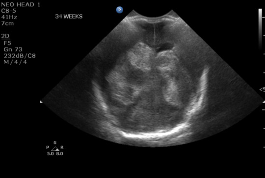
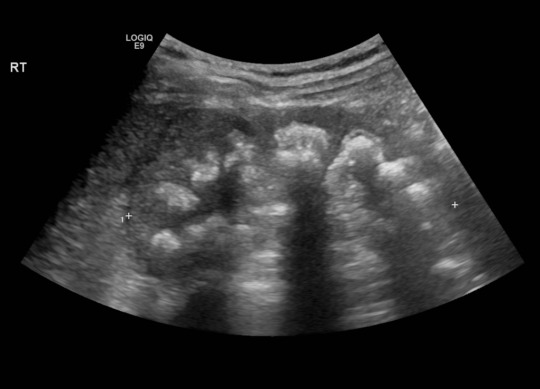
Today's case is an intracranial ultrasound of a 2-week-old neonate born at 24 weeks. A single coronal image depicts the ventricles which should be hypoechoic but are instead nearly completely filled with echogenic material, compatible with intraventricular hemorrhage (IVH). There is likely extension into periventricular white matter on the right.
IVH is a common complication seen in preterm neonates and is often described in four grades, as outlined below. This case is a grade 4 IVH.
Grade 1. Bleeding occurs just in a small area of the ventricles.
Grade 2. Bleeding also occurs inside the ventricles.
Grade 3. Ventricles are enlarged by the blood.
Grade 4. Bleeding into the brain tissues around the ventricles.
(Credit Children's Hospital of Philadelphia, https://www.chop.edu/conditio.../intraventricular-hemorrhage)
Case courtesy of Ryan Thibodeau, Radiopaedia.org, rID: 166948
12 notes
·
View notes
Text
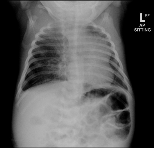

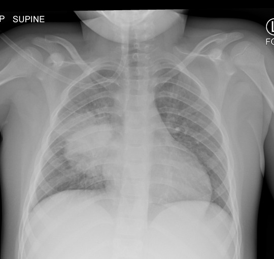
Here's a companion case to yesterday's. This is a neonate with respiratory distress.
...
...
...
...
The right mid to lower lung is hyperinflated. There is leftward medisastinal shift, including prominent medisatinal fat (that's not a mass in the left lung). CT shows cystic expansion of the right lung (2nd image). This is a congenital pulmonary airway malformation (CPAM) formerly known as congenital cystic airway malformation (CCAM). CPAM is a multicystic mass with abnormal bronchial proliferation. Differential diagnosis includes pulmonary sequestration, congenital lobar overinflation, and bronchogenic cyst (usually not aerated).
Case courtesy of Jeremy Jones, Radiopaedia.org, rID: 22542
#FOAMrad#TeachingRounds#FOAMed#Radiology#RadEd#RadCases#radres#PedsRad#Pediatrics#xray#CTRad#CT#Neonatology
22 notes
·
View notes
Text

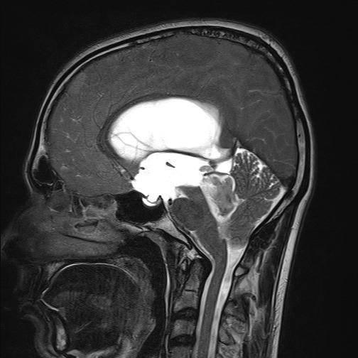
A posterior fossa tumor arising from the fourth ventricle in a child is likely an ependymoma (image 1, Case courtesy of Jini P Abraham, Radiopaedia.org, rID: 98653).
A tumor arising from the brainstem, particularly the pons, is likely a diffuse midline glioma, although pilocytic astrocytomas and gangliogliomas can also arise here (image 2, Case courtesy of Jeremy Jones, Radiopaedia.org, rID: 68486).
#TeachingRounds#FOAMed#FOAMRad#pediatrics#neurology#pedsneuro#neurosurgery#radiology#pedsrad#neuroradiology#peds
6 notes
·
View notes
Text
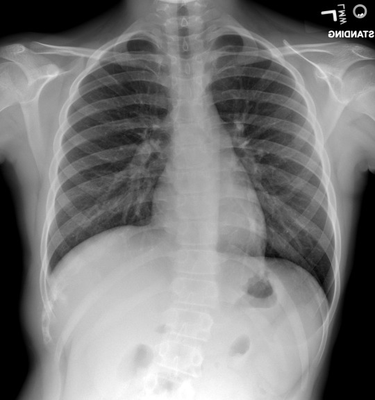
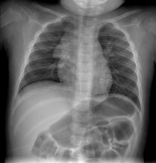
This is an 11-year-old girl with several months of right chest pain.
...
...
...
...
This case is your reminder that not all important chest x-ray findings are in the heart and lungs. There is a mixed lytic/sclerotic lesion in the right lateral 10th rib. At CT this was shown to be an expansile mass with a large soft tissue component. In a patient of this age, you should be thinking sarcoma, and this was indeed Ewing sarcoma.
Case courtesy of Brian Gilcrease-Garcia, Radiopaedia.org, rID: 65212
#FOAMrad#TeachingRounds#FOAMed#Radiology#RadEd#RadCases#radres#PedsRad#Pediatrics#xray#oncology#pedsonc#pediatriconcology
15 notes
·
View notes
Text

Today's case is a 25-year-old woman with bilateral echogenic, shadowing renal medullas, compatible with medullary nephrocalcinosis. Radiograph also shows the bilateral calcium deposits. In this condition, calcium salts deposit in the renal medulla and predispose the patient to infection and obstructing renal stones. Differential diagnosis includes hypercalcemia (esp in hyperparathyroidism), hypervitaminosis D, renal tubular acidosis type I, papillary necrosis, medullary sponge kidney, and other rarer entities. This patient renal tubular acidosis.
Case courtesy of Vikas Shah, Radiopaedia.org, rID: 164910
3 notes
·
View notes
Text
This is a case of a 7 year-old boy who presented with vomiting, diarrhea, and shortness of breath.
...
...
...
...
There is a right lung mass or consolidation with irregular margins, and an adjacent airspace opacity. History and location of the finding are strongly suggestive of round pneumonia. Imaging follow-up is not required but this can be done 30 days after treatment if needed. In this patient, follow-up chest radiograph after 6 weeks was clear.
Case courtesy of Yusra Sheikh, Radiopaedia.org, rID: 68757
4 notes
·
View notes
Text
This is a 2 year-old boy, history withheld.
...
...
...
...
Left lung is hyperinflated. Right lung is clear. This is a classic appearance of the lungs due to inhaled foreign body. Due to a "check valve" effect, inhaled air can pass into the lung but not out upon expiration. Less common causes of unilateral hyperinflated lung include bronchial compression by a hilar mass or bronchogenic cyst, pulmonary sling, and post-infectious obliterative bronchiolitis (Swyer-James syndrome).
Case courtesy of Angela Byrne, Radiopaedia.org, rID: 8128
2 notes
·
View notes
Text
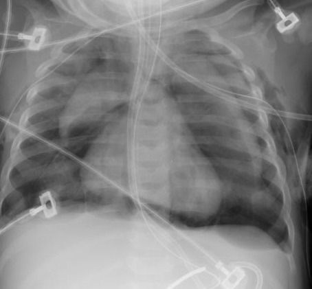
This is a companion case to yesterday's normal chest radiograph. This is an intubated neonate.
...
...
...
...
...
Lucencly outlining the thymus ("spinnaker sign") and heart is compatible with pneumomediastinum. Lucency around the heart creates the so-called "continuous diaphragm sign" of pneumomediastinum. CT revealed tiny bilateral pneumothoraces. The likely cause in this patient is barotrauma.
Case courtesy of The Radswiki, Radiopaedia.org, rID: 11791
2 notes
·
View notes
Text
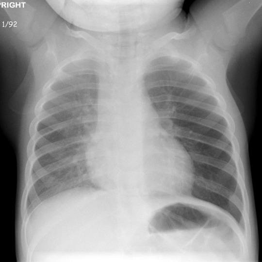
This week we will present a series of pediatric chest radiographs.
Today's case is a 3-year-old with cough and fever for 3 days. Fine wheezing was heard in both lungs.
...
...
...
...
This is a normal chest radiograph. The lungs are clear. The "thymic sail" sign is seen, which is simply normal thymus in the anterior mediastinum.
2 notes
·
View notes
Text
This 1-year-old patient has abnormal metaphyses, and you might be tempted to say this is hypovitaminosis D, but the sclerotic ring at the epiphyseal margins (Wimberger ring) and the lucent metaphyseal band (Trummelfeld zone) adjacent the sclerotic provisional zone of calcification at the growth plate are more suggestive of scurvy/hypovitaminosis C.
Case courtesy of Laughlin Dawes, Radiopaedia.org, rID: 29571
2 notes
·
View notes
Text
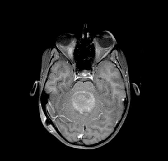
Next, we will consider supratentorial pediatric brain tumors, starting with the most common low-grade tumors: low-grade astrocytoma (LGA), pleomorphic xanthoastrocytoma (PXA), dysembryoplastic neuroepithelial tumor (DNET), and ganglioglioma. LGAs have variable appearance at MRI, but do tend to have mild enhancement. PXAs tend to have a large cyst with an enhancing solid component, but can have multiple cystic areas, and as such the imaging features overlap with gangliogliomas which tend to have multiple cystic areas. DNETs have cystic and solid components without robust enhancement. PXAs and ganglioglioma have a predilection for the temporal lobe, DNETs for frontal or temporal, and LGAs can occur anywhere (but favor the supratentorial brain). Today’s case is a 10-year-old boy who presented with 4-month history of headache and seizure. MRI reveals a cystic lesion with an enhancing nodule in the right parietal lobe. This was a PXA. See our post from July 8, 2023 for an example of a ganglioglioma.
Case courtesy of Luis Felipe Camarillo, Radiopaedia.org, rID: 94934
#TeachingRounds#FOAMed#FOAMRad#pediatrics#neurology#pedsrad#pedsneuro#neurosurgery#radiology#neuroradiology#peds
1 note
·
View note
Text
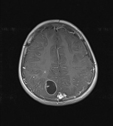
The posterior fossa is the most common location for intracranial tumors in children. They may arise from the cerebellum, brainstem, or the fourth ventricle.
Today we will focus on the cerebellum. Medulloblastoma is the most common tumor, accounting for ~25% of all pediatric brain tumors and 40% of posterior fossa tumors. Medulloblastomas are densely cellular and rarely contain cysts, calcifications, or hemorrhage. At MRI, they are seen as T1 iso- or hypointense, heterogeneously T2 hyperintense, enhancing masses with restricted diffusion (due to high cellularity). If more aggressive features are present, atypical teratoid/rhabdoid tumor (ATRT) is a consideration, especially in an infant or toddler. If the tumor contains cystic areas (“cyst with enhancing nodule”), juvenile pilocytic astrocytoma is the more likely diagnosis.
Medulloblastomas metastasize via the subarachnoid space so the spine must also be imaged, and the supra-tentorial brain should be scrutinized for additional lesions.
Today’s case is a 10-year-old patient with an enhancing cerebellar mass, which demonstrated restricted diffusion (not shown). After resection, pathology confirmed the diagnosis of medulloblastoma.
Case courtesy of Antonio Rodrigues de Aguiar Neto, Radiopaedia.org, rID: 70432
#TeachingRounds#FOAMed#FOAMRad#pediatrics#neurology#pedsneuro#neurosurgery#radiology#pedsrad#neuroradiology#peds
0 notes
Text
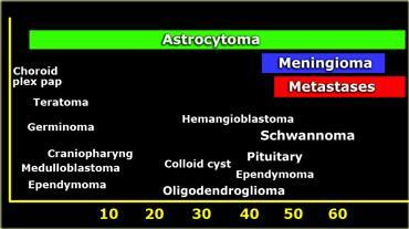
This week we will discuss intracranial tumors that occur in the pediatric population (as a follow-up to our week of posts on adult brain tumors).
A patient's age is a major consideration when considering the differential diagnosis for an intracranial tumor. Today's image depicts the age ranges at which common tumors tend to occur. Astrocytomas can occur at any age but for other types of tumors, there is a divide between those that commonly occur in adults and those that commonly occur in children.
Image credit: radiologyassistant.nl
#TeachingRounds#FOAMed#FOAMRad#pediatrics#neurology#pedsneuro#neurosurgery#radiology#pedsrad#neuroradiology#peds
0 notes
Text
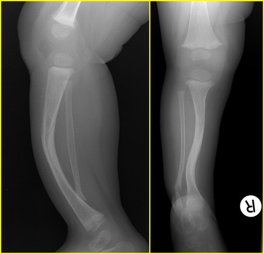
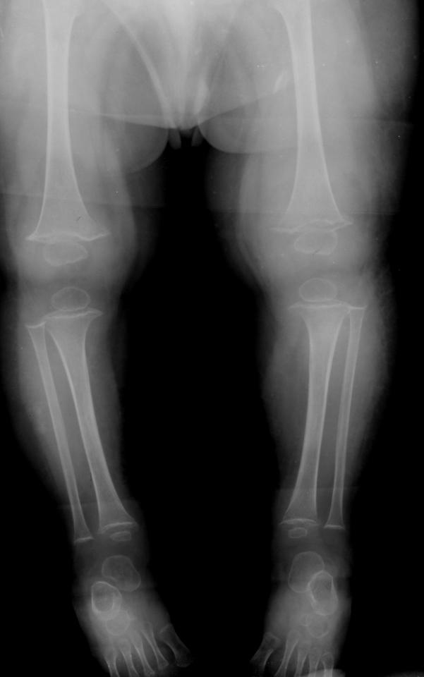
This is 1-year-old male with difficulty walking. Leg radiograph shows anterior and lateral bowing of the tibia, with a fibular pseudoarthrosis. Findings are diagnostic of type 1 neurofibromatosis (NF-1).
NF-1 is a genetic disorder with autosomal dominant inheritance pattern and multi-system involvement. Musculoskeletal manifestations are often overlooked. The spine, ribs, and lower extremities are the most commonly affected. Multiple non-ossifying fibromas are often seen at the metaphyses of long bones. Read more here: https://radiopaedia.org/.../neurofibromatosis-type-1...
Case courtesy of Henry Knipe, Radiopaedia.org, rID: 41320
1 note
·
View note
Text
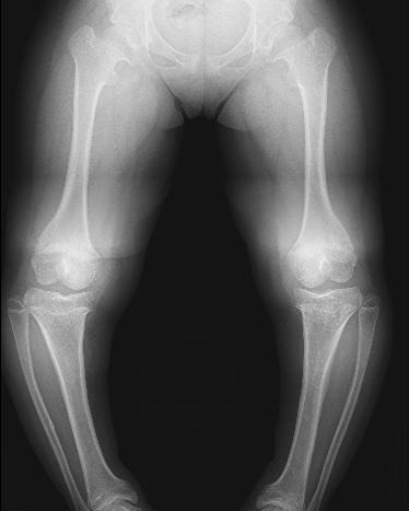
Today's case is a lower extremity radiograph of a 14-year-old patient showing proximal shortening of the lower extremities (femurs are shorter than they should be relative to the tibias), bilateral varus deformity (aka genu varum), and metaphyseal flaring. Findings are diagnostic of achondroplasia.
Case courtesy of Dr Muhammad Essam, Radiopaedia.org, rID: 29184
1 note
·
View note
Text
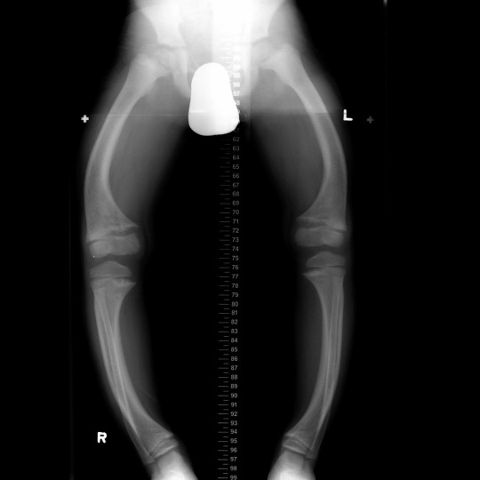
Here is a case of a 6 year-old with leg bowing. Radiograph shows bowing of long bones of the lower extremities with metaphyseal fraying and cupping. Findings are diagnostic of Rickets disease (hypovitaminosis D).
Case courtesy of Dr Angela Byrne, Radiopaedia.org, rID: 8116
1 note
·
View note
Last Seen Blogs

oiforfoxsake
Kel

crystalice09
Crystal Ice

mahitoastley
Mahito

ghethugiandep
Sans titre

dichtung-und-wahrheit
Dichtung und Wahrheit