
supermandreaming
Superman Dreaming
Keep on, Keepin' on!
3889 posts
Don't wanna be here? Send us removal request.
Last Seen Blogs

granpuccino
All About Granblue Fantasy
blueisgood
Blue is Good

percysgreekfire
Internet Vibes

fluff269
SG

coughloop
the bird and the flower had fun for an hour
Text
Peeking into electronics prototyping with the Arduino Uno
With so many passionate engineers here at Tumblr, meetings can get a little, well, heated. When staff turns up the fire in the kitchen, it’s time to crack a window.
Open windows can have their disadvantages. Wasted heat, wasted cooling, and unexpected visitors.

Don’t let them in.
However, it’s easy to forget to close the windows when leaving the room. To that end, I decided to build a reminder system during our most recent Hack Day.

The system, as shown above, uses an Arduino Uno to which a flexiforce pressure sensor has been wired. When the window is opened, a “start up” tune is played, and a timer begins. Once a certain amount of time has passed (say, 30 minutes,) a warning sound plays. Then, a reminder sound will play at another customizable interval. There’s also a button that activates a snooze feature to mute the reminder sound, during which an LED will blink. The logic for the main loop is below. Since this is Hack Day code, keep in mind it may be a bit wonky.
void loop() { timer.run(); int sensorValue = analogRead(sensorPin); int buttonState = digitalRead(buttonPin); if (started && buttonState == HIGH) snooze(); int difference = sensorValue - lastVoltage; if (difference < 0) difference = difference * -1; if ((difference < 150) && (lastVoltage > -1)) return; if (sensorValue <= threshold) { //window is open if(! started) { playTune(); started = true; } if(! triggered) { timer.enable(open_start_id); timer.enable(reminder_id); } lastVoltage = sensorValue; } else { //window is closed if(started) { //reset everything open_start_id = timer.setTimeout(warnAfter, play_warn); triggered = false; started = false; } timer.disable(open_start_id); timer.disable(reminder_id); lastVoltage = sensorValue; } }
And since we’d be remiss in not showing you this baby in action:
https://aloria.tumblr.com/post/143120428399
Apologies to Lipps Inc.
A list of components can be found here: https://aloria.tumblr.com/post/143121827147/arduino-project-components. Also, https://www.tumblr.com/jobs
- @aloria
79 notes
·
View notes

Text
Think dark and act bright
Wear black and drink white
Be cold and look hot
See the full but expect the half
Fear no one and scare everyone
Respect all but love one
- Hedonist Poet
7K notes
·
View notes
Photo

Recognising the basic structure of language is not unique to humans
Publishing in Nature Communications, the team led by Dr Ben Wilson and Professor Chris Petkov explain how using an imaging technique to explore the brain activity in humans and monkeys has identified the evolutionary origins of cognitive functions in the brain that underpin language and allow us to evaluate orderliness in sequences of sounds.
This new knowledge will help our understanding of how we learn – and lose – language such as in aphasia after a stroke or in dementia.
Brain activity of humans and monkeys
Scanning the brains of humans and macaque monkeys, the research team has identified the area at the front of the brain which in both humans and monkeys recognises when sequences of sounds occur in a legal order or in an unexpected, illegal order.
Professor Petkov said: “Young children learn the rules of language as they develop, even before they are able to produce language. So, we used a ‘made up’ language first developed to study infants, which our lab has shown the monkeys can also learn. We then determined how the human and monkey brain evaluates the sequences of sounds from this made up language.”
The team first had the humans and monkeys listen to example sequences from the made up language, allowing them to hear what were correct orderings in the sequence of sounds. They then scanned the brain activity of both species as they listened to new sequences that either had a correct order or could not have been generated by the made up language.
Functional magnetic resonance imaging (fMRI) revealed that in both groups a corresponding region of the brain – the ventral frontal and opercular cortex – responded to the order that both species had learned to expect.
Evidence some human and monkey brain functions are similar
These results suggest that the function of this frontal region, which is one of the areas involved in processing the order of words in a sentence in human language, is shared in both humans and primates, revealing its evolutionary origins. This brain region seems to monitor the orderliness, or organisation, of what is heard, which is an important cognitive function that provides a foundation for the more complex language abilities of humans.
These results provide first evidence that some of the functions of this brain area, which include understanding language in humans, are shared by other animals.
Professor Petkov adds: “This will help us answer questions on how we learn language and on what goes wrong when we lose language, for example after a brain injury, stroke or dementia.”
Building on these developments, the Newcastle University team, with their neurology collaborators in Cambridge and Reading Universities have begun a project to study the function of this brain region and its role in language impairment in aphasic patients with stroke, which might lead to better diagnosis and prognosis of language impairment.
Professor Petkov explains: “Identifying this similarity between the monkey and human brain is also key to understanding the brain regions that support language but are not unique to us and can be studied in animal models using state-of-the-art neuroscientific technologies.”
239 notes
·
View notes
Photo


Hannibal Ibarra - https://www.facebook.com/hannibal.ibarra
4K notes
·
View notes
Photo


Space sleep study to understand ageing
Tomorrow the first official British astronaut Tim Peake will blast into space for 6 months.
He’ll be doing lots of experiments and tests that aren’t possible on Earth because of the unique conditions on the ISS 400km above Earth. He’ll even be running the London Marathon from space.
But living in microgravity for so long will take a toll on his body; astronauts experience bone and muscle loss, diminished immune systems and increased inflammation, complaints that also are common in the elderly.
To help find out why this happens an experiment at the University of Surrey with the European Space Agency is mimicking the effects of microgravity. Young healthy men will spend two weeks living in the lab ‘normally’ before spending 60 days constantly in beds that are slightly tilted to simulate the microgravity on board the ISS.
By investigating how these conditions disrupt sleep and body clocks, the study will help figure out the genetic processes that contribute to the health problems experienced by both the elderly and astronauts in space.
Read more
Photos: NASA and Victor Zelentsov
619 notes
·
View notes
Photo


Mystery Solved: How Young Stars Appear In Old Clusters
“But surprisingly, bluer, brighter, hotter stars often appear in these older clusters anyway! Even in cases where there’s no evidence of a recent star-forming event, these blue straggler stars can frequently be found. For a long time, we thought the occasional merger of two low-mass stars gave rise to a heavier, blue star. But there’s an alternative.”
When stars are born, it normally happens in groups of thousands or more, all at once. They range from just 8% the mass of our Sun, which are dim, red, and long-lived, to dozens or even hundreds of times as massive: the brightest, bluest and shortest-lived stars of all. Because they burn through their fuel the fastest, these most massive stars cease to exist the most quickly. Yet even in the oldest star clusters available for study, stars that are blue and bright still appear! These blue stragglers were once thought to form from the merger of smaller, redder stars, but two recent studies now suggest an alternative: these stars are formed when one star siphons material off of a binary companion, increasing its own mass and creating a dead white dwarf out of its neighbor.
2K notes
·
View notes
Photo
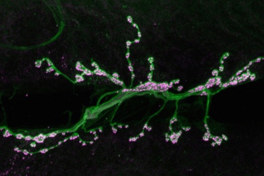
(Image caption: Motor neurons (green) form synapses (highlighted in magenta) on muscle fibers in a fruit fly. MIT neuroscientists have discovered a pathway that contributes to strengthening these synapses. Credit: Troy Littleton)
Neuroscientists reveal how the brain can enhance connections
When the brain forms memories or learns a new task, it encodes the new information by tuning connections between neurons. MIT neuroscientists have discovered a novel mechanism that contributes to the strengthening of these connections, also called synapses.
At each synapse, a presynaptic neuron sends chemical signals to one or more postsynaptic receiving cells. In most previous studies of how these connections evolve, scientists have focused on the role of the postsynaptic neurons. However, the MIT team has found that presynaptic neurons also influence connection strength.
“This mechanism that we’ve uncovered on the presynaptic side adds to a toolkit that we have for understanding how synapses can change,” says Troy Littleton, a professor in the departments of Biology and Brain and Cognitive Sciences at MIT, a member of MIT’s Picower Institute for Learning and Memory, and the senior author of the study, which appears in the Nov. 18 issue of Neuron.
Learning more about how synapses change their connections could help scientists better understand neurodevelopmental disorders such as autism, since many of the genetic alterations linked to autism are found in genes that code for synaptic proteins.
Richard Cho, a research scientist at the Picower Institute, is the paper’s lead author.
Rewiring the brain
One of the biggest questions in the field of neuroscience is how the brain rewires itself in response to changing behavioral conditions — an ability known as plasticity. This is particularly important during early development but continues throughout life as the brain learns and forms new memories.
Over the past 30 years, scientists have found that strong input to a postsynaptic cell causes it to traffic more receptors for neurotransmitters to its surface, amplifying the signal it receives from the presynaptic cell. This phenomenon, known as long-term potentiation (LTP), occurs following persistent, high-frequency stimulation of the synapse. Long-term depression (LTD), a weakening of the postsynaptic response caused by very low-frequency stimulation, can occur when these receptors are removed.
Scientists have focused less on the presynaptic neuron’s role in plasticity, in part because it is more difficult to study, Littleton says.
His lab has spent several years working out the mechanism for how presynaptic cells release neurotransmitter in response to spikes of electrical activity known as action potentials. When the presynaptic neuron registers an influx of calcium ions, carrying the electrical surge of the action potential, vesicles that store neurotransmitters fuse to the cell’s membrane and spill their contents outside the cell, where they bind to receptors on the postsynaptic neuron.
The presynaptic neuron also releases neurotransmitter in the absence of action potentials, in a process called spontaneous release. These “minis” have previously been thought to represent noise occurring in the brain. However, Littleton and Cho found that minis could be regulated to drive synaptic structural plasticity.
To investigate how synapses are strengthened, Littleton and Cho studied a type of synapse known as neuromuscular junctions, in fruit flies. The researchers stimulated the presynaptic neurons with a rapid series of action potentials over a short period of time. As expected, these cells released neurotransmitter synchronously with action potentials. However, to their surprise, the researchers found that mini events were greatly enhanced well after the electrical stimulation had ended.
“Every synapse in the brain is releasing these mini events, but people have largely ignored them because they only induce a very small amount of activity in the postsynaptic cell,” Littleton says. “When we gave a strong activity pulse to these neurons, these mini events, which are normally very low-frequency, suddenly ramped up and they stayed elevated for several minutes before going down.”
Synaptic growth
The enhancement of minis appears to provoke the postsynaptic neuron to release a signaling factor, still unidentified, that goes back to the presynaptic cell and activates an enzyme called PKA. This enzyme interacts with a vesicle protein called complexin, which normally acts as a brake, clamping vesicles to prevent release neurotransmitter until it’s needed. Stimulation by PKA modifies complexin so that it releases its grip on the neurotransmitter vesicles, producing mini events.
When these small packets of neurotransmitter are released at elevated rates, they help stimulate growth of new connections, known as boutons, between the presynaptic and postsynaptic neurons. This makes the postsynaptic neuron even more responsive to any future communication from the presynaptic neuron.
“Typically you have 70 or so of these boutons per cell, but if you stimulate the presynaptic cell you can grow new boutons very acutely. It will double the number of synapses that are formed,” Littleton says.
The researchers observed this process throughout the flies’ larval development, which lasts three to five days. However, Littleton and Cho demonstrated that acute changes in synaptic function could also lead to synaptic structural plasticity during development.
“Machinery in the presynaptic terminal can be modified in a very acute manner to drive certain forms of plasticity, which could be really important not only in development, but also in more mature states where synaptic changes can occur during behavioral processes like learning and memory,” Cho says.
The study is significant because it is among the first to reveal how presynaptic neurons contribute to plasticity, says Maria Bykhovskaia, a professor of neurology at Wayne State University School of Medicine who was not involved in the research.
“It was known that the growth of neural connections was determined by activity, but specifically what was going on was not very clear,” Bykhovskaia says. “They beautifully used Drosophila to determine the molecular pathway.”
Littleton’s lab is now trying to figure out more of the mechanistic details of how complexin controls vesicle release.
224 notes
·
View notes
Photo


Baking tip #1: Booze.
Have you ever added vodka to your apple pie? There’s a scientific reason for boozing up a pie.
We’ve gathered a few scientific baking tips from @scienceandfood that will help you make a perfect apple pie this Thanksgiving…
Baking a better pie with science →
1K notes
·
View notes
Photo

Mental Maps: Route-Learning Changes Brain Tissue
Fifteen years ago, a study showed that the brains of London cab drivers had an enlargement in the hippocampus, a brain area associated with navigation. But questions remained: Did the experience of navigating London’s complex system of streets change their brains, or did only the people with larger hippocampi succeed in becoming cab drivers?
Now, Carnegie Mellon University scientists have determined that learning detailed navigation information causes the hippocampal brain changes. Published in NeuroImage, Tim Keller and Marcel Just show for the first time that brief navigation training changes a person’s brain tissue and improves how that changed tissue communicates with other brain areas involved with navigation. The findings establish a critical link between structural and functional brain alterations that happen during spatial learning. They also illustrate that the changes are related to how neural activity synchronizes – or communicates – between the hippocampus and other regions that are important for navigation understanding and learning.
“The hippocampus has long been known to be involved in spatial learning, but only recently has it been possible to measure changes in human brain tissues as synapses become modified during learning,” said Keller, a senior research scientist in CMU’s Department of Psychology and Center for Cognitive Brain Imaging (CCBI). “Our findings provide a better understanding of what causes the hippocampal changes and how they are related to communication across a network of areas involved in learning and representing cognitive maps of the world around us.”
To examine how the hippocampus changes, Keller and Just recruited 28 young adults with little experience playing action video games. For 45 minutes, the participants played a driving simulation game. One group practiced maneuvering along the same route 20 times. The control group drove for the same amount of time, but along 20 different routes. Before and after each training session, each participant’s brain was scanned using diffusion-weighted imaging (DWI),which measures water molecule movement in the brain,and functional magnetic resonance imaging (fMRI), which analyzes brain activity.
The researchers found that the group that practiced the same route over and over — the spatial learning group — increased their speed at completing the driving task more than the group practicing on different routes, indicating that they learned something specific about the spatial layout of the virtual environment. The spatial learning group also improved their ability to order a sequence of random pictures taken along the route and to draw a 2-D map representing the route.
Importantly, only the spatial learning group showed brain structural changes in a key spatial learning part of the hippocampus, the left posterior dentate gyrus. There also were increases in the synchronization of activity – or functional connectivity – between this region and other cortical areas in the network of brain regions responsible for spatial cognition. And, the amount of the structural change was directly related to the amount of behavioral improvement each person showed on the task.
“The new discovery is that microscopic changes in the hippocampus are accompanied by rapid changes in the way the structure communicates with the rest of the brain,” said Just, the D.O. Hebb University Professor of Psychology in the Dietrich College of Humanities and Social Sciences and director of the CCBI. “We’re excited that these results show what re-wiring as a result of learning might refer to. We now know, at least for this type of spatial learning, which area changes its structure and how it changes its communication with the rest of the brain.”
102 notes
·
View notes
Photo


How Beetles Breathe Underwater
Air-breathing aquatic bugs and beetles don’t hold their breath the way sea mammals do, nor do they have gills like fish.
The answer lies in their small size. Insect scuba strategies hinge on a property of water that relative giants like us usually overlook: surface tension.
67 notes
·
View notes
Quote
I’m not denying the power of beauty but it is absolutely nothing compared to the power of words.
Hedonist Poet (via hedonistpoet)
7K notes
·
View notes
Photo

New study maps the progression of Parkinson’s disease within the brain
Scientists at the Montreal Neurological Institute and Hospital -The Neuro, at McGill University and the McGill University Health Centre, have made advances in understanding the process involved in the progression and spread of Parkinson’s disease (PD) within the brain.
The study, published in September issue of eLIFE Journal, focused on understanding the process that drives the disease’s progression by mapping the distribution and degree of atrophy, characteristic of the disease, in certain brain regions and identify the paths leading the spread from affected to healthy tissue.
“Past studies have failed to consistently demonstrate regional brain atrophy in earlier stages of the disease due to samples of subjects that were too small and to methods that were less sensitive in detecting all aspects of the disease’s impact on the brain. We now have the means to map the disease with greater sensitivity than previously possible,” says Dr. Alain Dagher, senior author of the study.
The researchers had access to an unprecedented number of MRI scans and clinical data through the open source Parkinson’s Progression Markers Initiative (PPMI) database. Thanks to this wealth of data, researchers were able to analyse MRI scans which show the structure of the brains of 230 people in the early stages of Parkinson’s disease and compare them to those from age-matched healthy individuals. This allowed them to identify the set of brain regions that show atrophy in the early stages of the disease.
“The atrophy pattern on MRI is compatible with a disease process that spreads via brain networks – something that had never been shown in human patients before, and would support the hypothesis that PD is caused by a “toxic agent” that spreads trans-neuronally,” says Dr. Alain Dagher.
The findings add new evidence to the hypothesis that brain cells in Parkinson’s patients might deteriorate according to a prion-like mode of disease propagation, in which a toxic agent spreads from brain cell to brain cell utilizing the normal connections of the brain. Similar mechanisms have been proposed for diseases ranging from Alzheimer’s Disease to Bovine Spongiform Encephalopathy. The process would involve the spread of alpha-synuclein, a toxic misfolded protein with the ability to make copies of itself and infect neighbouring cells while traveling through the brain’s neuronal highways.
The patients who were enrolled in this study will continue to be evaluated on a yearly basis providing researchers with more data to continue mapping how the disease progresses throughout the brain and furthering our understanding of its causes.
Current treatment options help control or minimize symptoms including tremors, slowness of movement, stiffness or rigidity, and loss of balance. The findings of this study hold exciting therapeutic implications. In the longer term, it will help researchers develop new techniques to assess the efficacy of drugs that could target the culprit protein and might eventually lead to treatments that will prevent, slow, halt or even reverse the progression of PD.
121 notes
·
View notes
Photo


This Breathtaking Scene That Happens When Sunlight Hits This Mosque
The Nasir al-Molk Mosque in Iran, often referred to as the ‘Pink Mosque’, is without a doubt as one of the most colorful mosque in the world.
The whole building is flooded with color when sunlight hits in. Using stained glass, which is not common, creates this color spectacle. However, these images seem like they’re from a fairytale story. Source:theonlinecentral
5K notes
·
View notes
Quote
When things go wrong, take a moment to be thankful for the things going right.
(via staypozitive)
2K notes
·
View notes


Photo

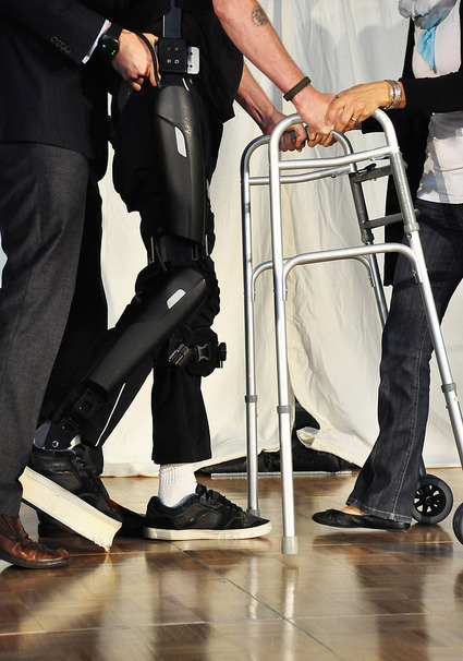
Captain Trevor Greene partners with SFU to walk again
Former Canadian soldier Trevor Greene, who survived a debilitating brain injury while on duty in Afghanistan in 2006, has recovered his ability to walk again with the help of a customized exoskeleton, his personal determination and support of researchers at Simon Fraser University.
Greene demonstrated his progress at SFU’s Surrey campus.
Told he would likely never walk again after a vicious axe attack, Greene began working with Dr. Ryan D’Arcy, a neuroscientist and SFU professor, in 2009. D’Arcy became involved with Greene’s recovery after watching a documentary about him.
He asked Greene to partner with him in a research project to explore how brain plasticity affects motor functions. Plasticity refers to the brain’s ability to reorganize its neural pathways and synapses in response to different behaviours, thoughts or emotions.
The two have since met regularly for D’Arcy to collect functional magnetic resonance imaging (fMRI) scans of Greene’s brain, which D’Arcy uses to track how the brain rewires itself.
In an article published in The Journal of Head Trauma Rehabilitation this month, D’Arcy and his research team challenge the current assumptions that after a traumatic brain injury, any further recovery ceases to happen over the long-term. His team discovered physical functions can be recovered through rehabilitation even six years after an injury.
In 2014, D’Arcy called on Carolyn Sparrey, an assistant professor in the School of Mechatronic Systems Engineering (MSE) who has extensive experience in biomechanics, to see if she could customize an exoskeleton that would suit the unique requirements of the 6’4” Greene.
Exoskeletons are typically designed for those with spinal cord injuries as an assistive technology providing lower leg movement. Sparrey notes that this is the first time exoskeleton technology has been used for a person with a brain injury.
Today, Greene is able to walk upright with assistance, outfitted with a custom-made exoskeleton from Israel-based company, ReWalk. In the future he plans to walk unassisted. Ultimately, he says his goal is to make it to Everest base camp.
A ReWalk company trainer has supported Greene by customizing the motorized exoskeleton so that Greene can wear the battery pack as a backpack.
“Trevor has been extremely committed to his rehabilitation program,” says D’Arcy, who is also co-chair of Innovation Boulevard.
Greene’s positive attitude was never more poignantly demonstrated than when he stood, using parallel bars, at his 2010 wedding to wife Deborah.
“This newest dimension in his rehabilitation, wearing exoskeletons to walk again, enables SFU faculty members to track research milestones in a real-life scenario while making a positive impact on his life,” says D’Arcy.
Dr. Joy Johnson, SFU’s vice-president, research, says: “This is such a heartwarming story of courage and determination, and of the power of collaboration to push beyond seemingly impenetrable boundaries. This is why SFU places such value on interdisciplinary research and open innovation—they help turn ideas into action, so that people may benefit.”
The Royal Canadian Legion raised funds for the ReWalk device. SFU investigators are donating their time, expertise and specialized equipment to assist with the project.
223 notes
·
View notes