mymedicalblog
My Medical Blog
Resident Doctor in Hepatology. Brazil.
703 posts
Don't wanna be here? Send us removal request.
Last Seen Blogs

s1yfox14
Look it's gay!

toptenwave
Untitled

entrancinghoneypie
soft and sweet

arafilez
𝅄ㅤARAㅤ ꒱

coronel-things
Watcher

Photo
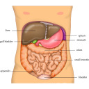
Mesentery is now classified as an organ.
Researchers have classified a brand-new organ inside our bodies, one that’s been hiding in plain sight in our digestive system this whole time. Although we now know about the structure of this new organ, its function is still poorly understood, and studying it could be the key to better understanding and treatment of abdominal and digestive disease.
Known as the mesentery, the new organ is found in our digestive systems, and was long thought to be made up of fragmented, separate structures. But recent research has shown that it’s actually one, continuous organ.
The evidence for the organ’s reclassification is now published in The Lancet Gastroenterology & Hepatology. “In the paper, which has been peer reviewed and assessed, we are now saying we have an organ in the body which hasn’t been acknowledged as such to date,” said J Calvin Coffey, a researcher from the University Hospital Limerick in Ireland, who first discovered that the mesentery was an organ.
So what is the mesentery? It’s a double fold of peritoneum - the lining of the abdominal cavity - that attaches our intestine to the wall of our abdomen, and keeps everything locked in place. One of the earliest descriptions of the mesentery was made by Leonardo da Vinci, and for centuries it was generally ignored as a type of insignificant attachment. Over the past century, doctors who studied the mesentery assumed it was a fragmented structure made of separate sections, which made it pretty unimportant.
But in 2012, Coffey and his colleagues showed through detailed microscopic examinations that the mesentery is actually a continuous structure. And while that doesn’t change the structure that’s been inside our bodies all along, with the reclassification comes a whole new field of medical science that could improve our health outcomes.
“When we approach it like every other organ… we can categorise abdominal disease in terms of this organ,” said Coffey. “Now we have established anatomy and the structure. The next step is the function. If you understand the function you can identify abnormal function, and then you have disease. Put them all together and you have the field of mesenteric science … the basis for a whole new area of science.”
“This is relevant universally as it affects all of us.”
(Source).
901 notes
·
View notes
Text
Liver: disseminated histoplasmosis

This is a high power view of a section of liver from an adult with histoplasmosis and HIV infection.
It shows a cluster of macrophages containing many yeasts. The dark nucleus and pale/white cytoplasm of the fungus are evident. H&E stain.
101 notes
·
View notes
Photo


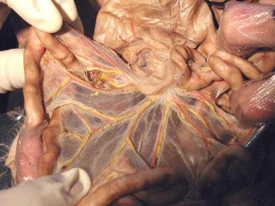
3D printed Liver
I fee like my blog is becoming a 3-D printing blog.. but I can’t stop! How amazing is this? Honestly, looking at a liver, it just seems like a big hard block of tissue. It’s very hard to visualize the vessels going through the liver, and when you are removing a tumor or cancerous tissue, it can be very difficult.
Dr. Zein’s team from Cleveland Clinic has used this method in over 30 cases so far with very high success rates. It has allowed his team to decrease the amount of time in operation, and prepare more thoroughly!
I love these transparent livers.. I really need one for my room.
219 notes
·
View notes
Text
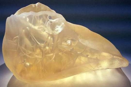
♥️ You can find LOVE in the strangest of places ♥️
Have a happy and histological Valentine’s Day!
Which ♥️ will you send?
Repost and comments with the number heart you give them!
i♡histo
Key (to my hearts)
1 ♥️ in a pancreas
2 ♥️ in a Pap smear
3 ♥️ in a thyroid
4 ♥️ in a breast
5 ♥️ in a sweat gland
6 ♥️ in an inflamed lesion
7 ♥️ in a bronchiole
8 ♥️ in an alveolus
9 ♥️ in an anal canal
10 ♥️ in a liver
11 ♥️ in an artery
12 ♥️ in a cervix
13 ♥️ in a colonic crypt
14 ♥️ in a parotid gland
15 ♥️ in a lymph node
16 ♥️ in an ovary
17 ♥️ in a vaginal smear
18 ♥️ in a lung
19 ♥️ in a breast
20 ♥️ in a vagina
21 ♥️ in a salivary gland
22 ♥️ in a gallbladder
23 ♥️ in a scalp
24 ♥️ in a vein
25 ♥️ in a cervix
26 ♥️ in a pineal gland
27 ♥️ in a uterus
28 ♥️ in an epididymis
29 ♥️ in a vagina
30 ♥️ in a bone
31 ♥️ in a hair
32 ♥️ in a venule
33 ♥️ in a thyroid smear
34 ♥️ in a testicle
35 ♥️ in a heart
36 ♥️ in a nipple
167 notes
·
View notes


Photo

Twins interact with each other in the womb
The University of Padova recently discovered that twins are as close in the womb as they are outside of it. At 14 weeks of gestation, twins were seen reaching for each other. Four weeks later, twins in the womb touched each other more often than they touched their own bodies. The twins were gentle to each other and made distinct gestures toward each other. (Source)
33K notes
·
View notes

Photo
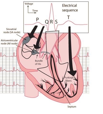
Electrical sequence of the heart for the visual learner
636 notes
·
View notes
Photo
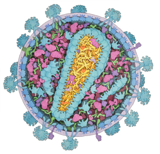
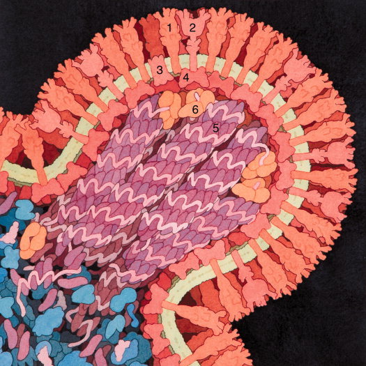

HIV virus particle, budding influenza virus and HIV in blood serum as illustrated by David S. Goodsell.
Goodsell is a professor at the Scripps Research Institute and is widely known for his scientific illustrations of life at a molecular scale. The illustrations are usually based on electron microscopy images and available protein structure data, which makes them more or less accurate. Each month a new illustrated protein structure can be found in Protein Data Bank molecule of the month section and you can read more on how his art is made here.
1K notes
·
View notes
Note
What makes one a medblr?
Medblr: (n) 1. One who blogs on tumblr about medicine 2. A person working in a healthcare field who is also on tumblr and blogs about whatever the heck he or she wants.
247 notes
·
View notes
Photo
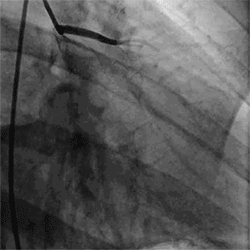
Cardiac catheterization is a medical procedure used to diagnose and treat some heart conditions. A long, thin, flexible tube called a catheter is put into a blood vessel in your arm, groin, or neck and threaded to your heart. Through the catheter, your doctor can do diagnostic tests and treatments on your heart.
For example, your doctor may put a special type of dye in the catheter. The dye will flow through your bloodstream to your heart. Then, your doctor will take x-ray pictures of your heart. The dye will make your coronary arteries visible on the pictures. This test is called coronary angiography.
The dye can show whether a waxy substance called plaque has built up inside your coronary arteries. Plaque can narrow or block the arteries and restrict blood flow to your heart. The buildup of plaque in the coronary arteries is called coronary heart disease or coronary artery disease.
Doctors also can use ultrasound during cardiac catheterization to see blockages in the coronary arteries. Ultrasound uses sound waves to create detailed pictures of the heart’s blood vessels. Doctors may take samples of blood and heart muscle during cardiac catheterization or do minor heart surgery.
Cardiologists usually do cardiac catheterization in a hospital. You’re awake during the procedure, and it causes little or no pain. However, you may feel some soreness in the blood vessel where the catheter was inserted. Cardiac catheterization rarely causes serious complications.
(x)
936 notes
·
View notes


Photo
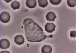

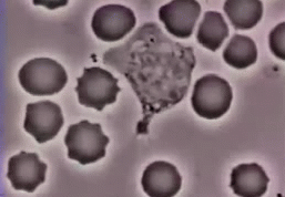
The following is a white blood cell chasing a bacterium. It eventually ends up swallowing it. The following white blood cell is specifically a neutrophil. They end up ingesting the microbe a process known as phagocytosis.
VIDEO
17K notes
·
View notes