#radiography
Text


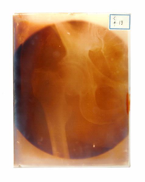
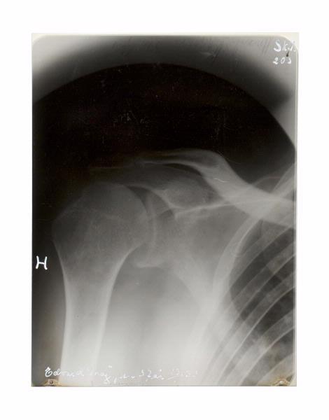
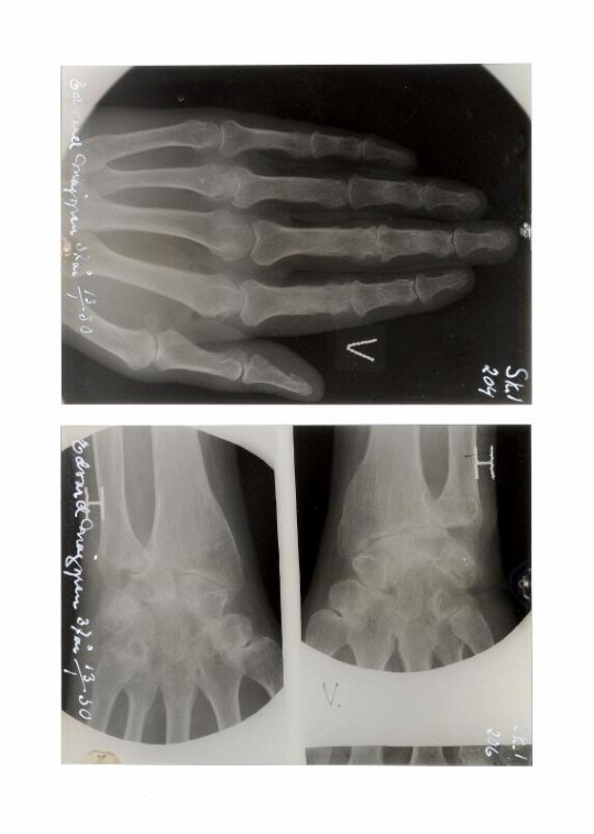
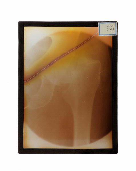
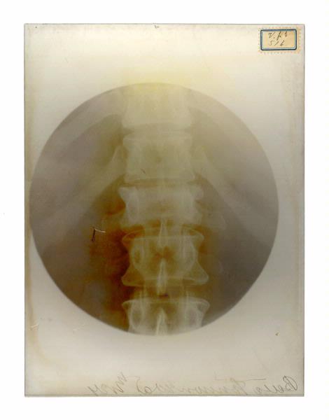

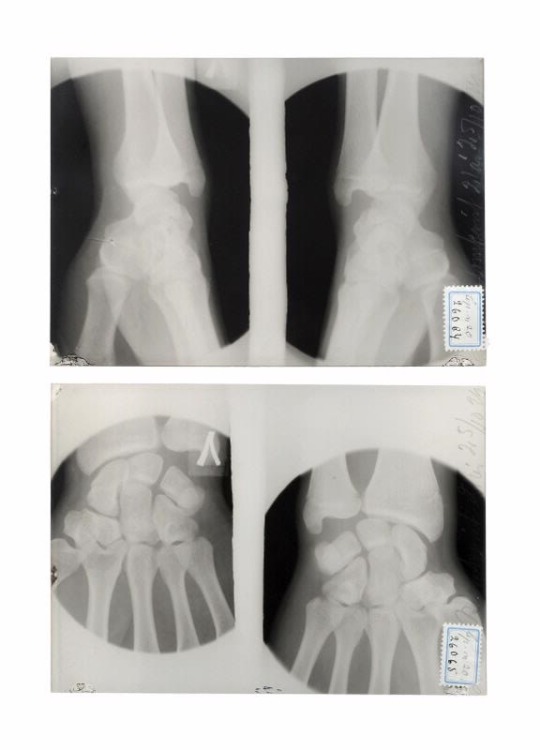

x-ray images, 1916-1931
#anatomy#x-ray image#x-ray images#vintage#medical history#x-ray#radiology#radiography#1910's#20's#30's#*
3K notes
·
View notes
Text
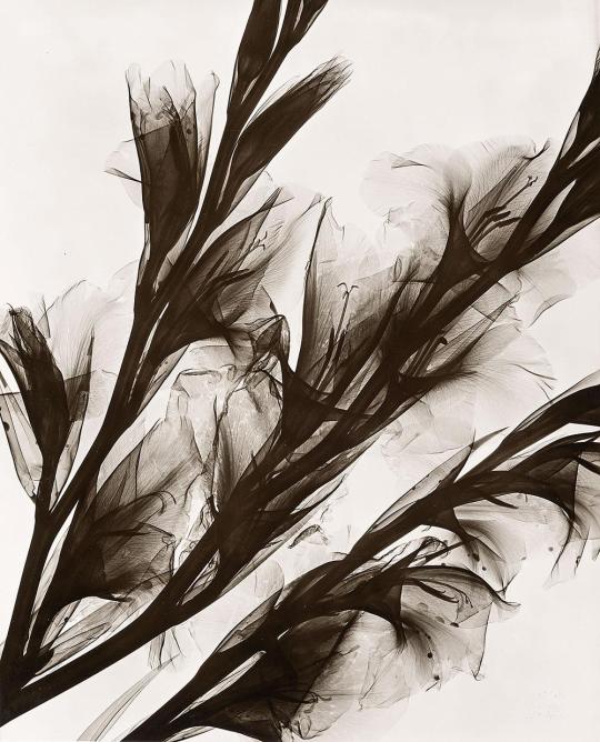
Herbert W. Franke (1927-2022) ~ Radiographische Gladiolen, Kontakt, 1956
Aus Experimentalserie (Experimental series) Alternative Sichtweisen | src Schneider-Henn (link to pdf)
#Herbert W. Franke#Herbert Franke#X-ray photography#x-ray#flowers#blumen#gladiola#1950s#fiori#fleurs#gladiolus#radiography
51 notes
·
View notes
Text
Radley's sneaky spicy cheatsheet for imaging modalities!
X-ray
Super quick zappyzap! One-to-four images per body part in the UK, unless you're doing fancy orthopaedic projections. Fast, cheap, and very good at looking at bones / basic abdominal or lung pathology / size of your heart. You hold still for like, one second. Then - ZOOP! You're done.
CT
X-ray BUT MORE X-RATED! Much, much higher dose, because it's basically taking a ridiculous amount of x-ray images while spinning the gantry (the 'camera', so to speak) around you at very high speed, and then algorithmically compiling those images to create a 3d digital construction of your innards! Very useful for looking at internal organs, especially with contrast media that makes pathology all shiny and pretty. Sometimes slower & always more expensive than X-ray, but much faster & cheaper than MRI. Patients can't move for the duration (unless you're doing cardiac stuff that takes a picture only in the lull between heartbeats - very cool!).
MRI
CT BUT WITH MAGNETS (okay it's not really much like CT except that it creates slices of the body in all three planes). There's no ionising radiation! It produces really, really clear, gorgeous pictures that show off soft tissue beautifully! We like that!! But... it's also really slow, really expensive, and really claustrophobic. A lot of patients don't enjoy it, and who can blame 'em? Who wants to be stuck in a small tube for a half hour while it makes horrid boomy noises at you? And you're not allowed to move at all? (we don't even like you to breathe too fast or too slow!)
Ultrasound
SOUNDWAVES GO BOUNCYBOUNCE. Though it produces a relatively unclear image that you need a lotta extra training to decipher, the tech is super cheap and available, and it's very quick to use! There's no ionising radiation involved! AND you can use it for realtime imaging - so, you can see a foetus move as it happens, rather than this movement messing up your entire image, or having to be carefully planned around!
Fluoroscopy
X RAY BUT VIDEO. This is a constant (and therefore high dose) real-time 'video' taken with X-ray, which can visualise movements within the body. You can watch contrast media (shiny juice) shift around to ensure that systems are functioning correctly! You can watch surgeons push their guide wires/stents/etc. into place, to be sure they hit the right spot! Or you can inject Shiny Juice into the blood vessels and, with angiography, watch it flow around the heart/brain to find blockages!
Nuclear med
WE STICK THE RADIOACTIVE STUFF IN YOU. For instance, we give you a radioactive tracer in a solution with stuff that binds well to bony metastases.... and BOOM we can see all your bony metastases on a PET-CT/MRI because they're glowing red-hot! Or we IV a nuclear tracer into your heart and make you exercise/give you meds that raise your heart rate and BOOM we can see whether your heart has an adequate blood supply during exertion!
41 notes
·
View notes
Text
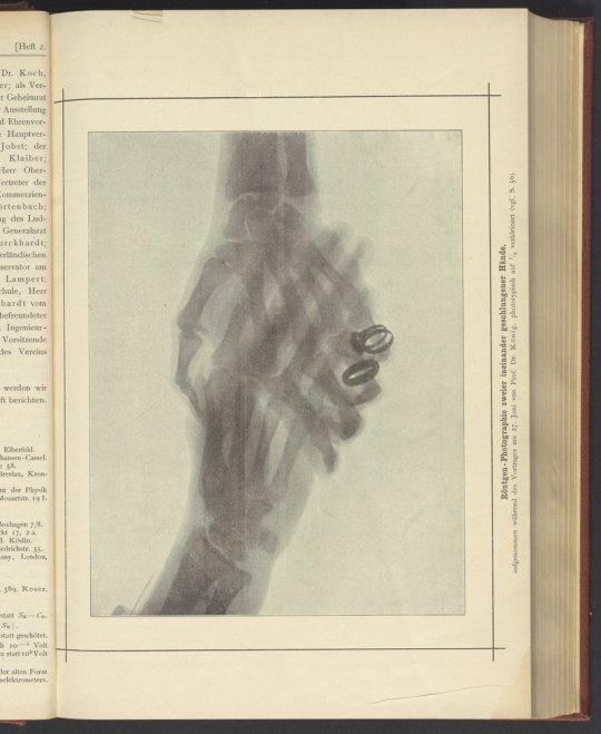
Morbid or romantic? You decide.
This is: Röntgen-Photographie zweier ineinander geschlungener Hände
#x-ray#rontgen#morbid#romantic#halloween#skeleton#hands#hand holding#radiography#electrochemistry#othmer library#history of science#spooky#old books#othmeralia
182 notes
·
View notes
Text
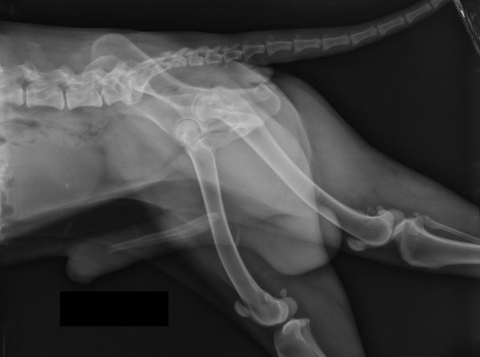
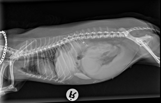
a geriatric dog presented with a little bit (?) of poopin troubles. what do you see in this radiograph?

a huge prostate pressing onto his rectum.
This is Benign Prostatic Hypertrophic (BPH). The recommended treatment is castration. And while we wait for the prostate to shrink, some laxative could help him be a little more comfortable.
18 notes
·
View notes
Text
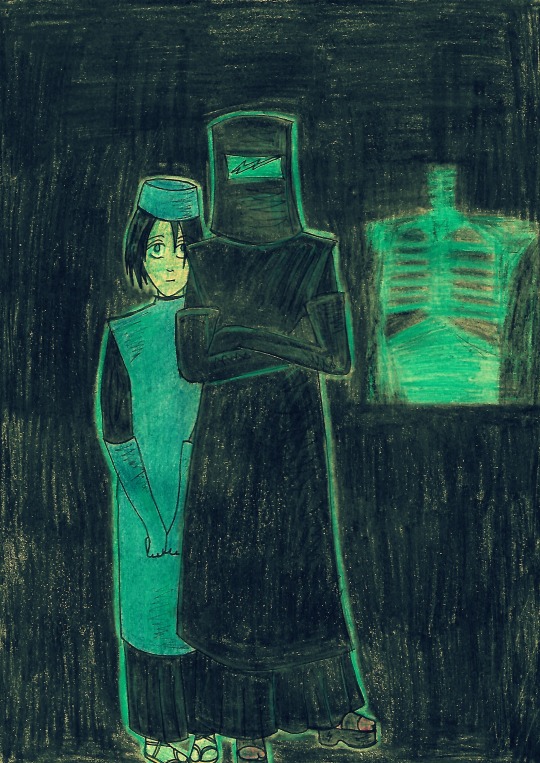
Happy B-day Hana-chan! I will protect you from all bullies, but also give you dangerous work <3
pencils, marker & filters
5 notes
·
View notes
Text
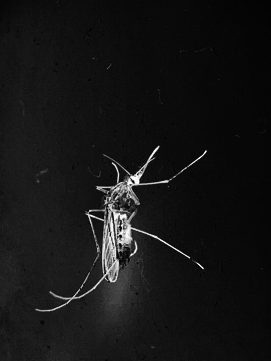
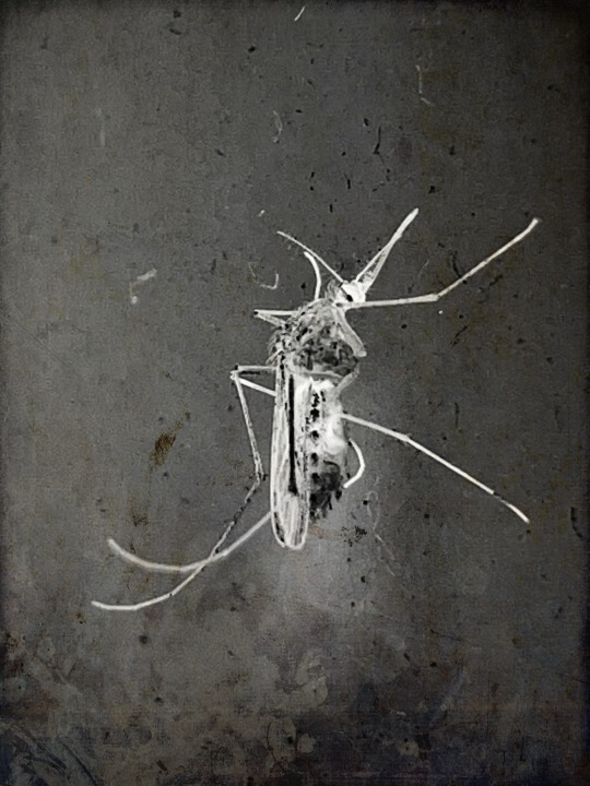
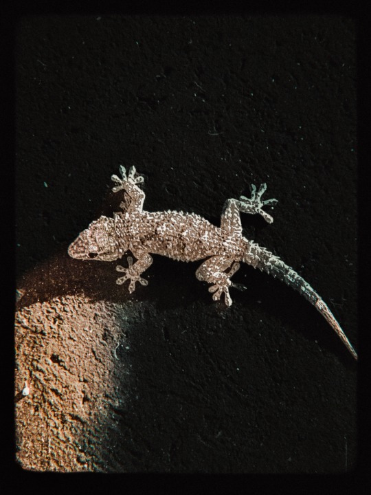
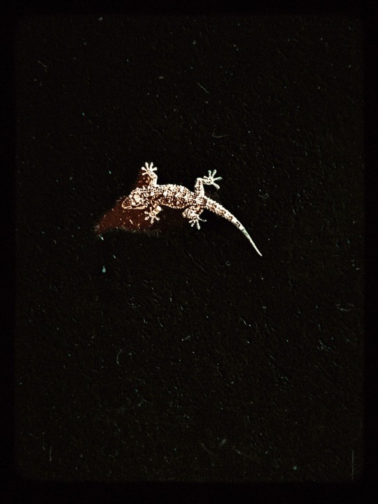
Ninnananna amore mio___Lullaby my love
#alchimilla#katia celestini#artists on tumblr#photographers on tumblr#magic photography#enchanted photography#gothic photography#life and death#gothic art#aesthetic mood#radiography#creatures#little animals#geko#mosquito
7 notes
·
View notes
Text
He is so pretty, he is so well behaved
#animals#medical imaging#wrapping him#he is so pretty#he is so well behaved#rat#rats#vet#under the weather#radiography#photos of animals having medical imaging or procedures done that look silly are far and few between here#at the vet
11 notes
·
View notes
Text
Underground Radio (No Money Remix) is on the 13 (ALBUM) LYS 030
#radio#radioshow#radiojavan#radiohead#radiohost#radiostation#radiopersonality#radiology#RadioDJ#radiolife#Radioactive#radiofrequency#radiocontrol#radiorecord#RadioPlay#radiofrequencia#radio1#radiologia#RadioStations#radiointerview#radiocity#radioshows#radiocitymusichall#radiodeejay#radiography#radioonline#radioone#radiologist#Radiofrecuencia#radiomir
4 notes
·
View notes
Photo

From the series : “The Wedding of the Earth ” R.Tanaka
http://rafamonzo.tumblr.com / http://tanaka-clan.tumblr
#photography#black&white#leaves#radiography#experimental landscape#experimental photography#altered landscape#altered photography#film photography#experimental print#altered perception#the wedding of the Earth
19 notes
·
View notes
Text

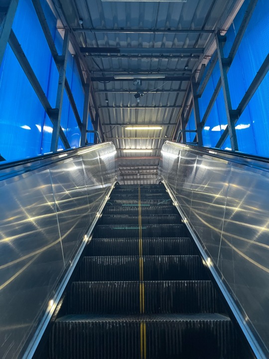


3 May 2023
Loads of train travel this summer, and already fell sick from heat exhaustion 🥵 Had my first presentation 👩💻this week on Allergy and Anaphylaxis from the Canine and Feline Medicine course that I’m taking this semester (that i spent the entire weekend on), which got postponed to next week ⏭️
I’ve been exploring the new metro line 🚈🚉 that directly connects my house 🏠 to my bf’s which is super great cause the traffic’s 🚥 shit
#vet student#vetmed#vet studyblr#veterinary student#vet school#vetblr#veterinary#studyblr#vet stuff#veterinary medicine#study blog#studyspo#study aesthetic#train travel#metro station#radiography#xray#commuter train#daily commute#heat exhaustion#summer
16 notes
·
View notes
Text
It’s SPOOKY SCARY SKELETONS MONTH
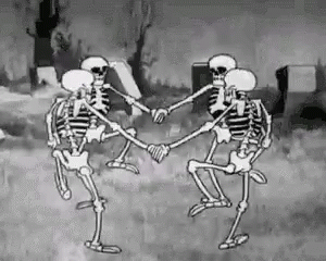
So let’s talk about yer bones! Yeah, that’s right, Captian Holt. I said –

An adult has (roughly) 206 bones (I say ‘roughly’! You can have non-pathological anatomical variation, such as lumbarised sacral vertebrae (an extra bone in your back) or accessory sesamoids like the flabella (a little bone at the back of the knee!))
A newborn has (roughly) 300 bones
That's a big difference! Almost 100 bones of difference! Where do they go?
Well, you see - as you get older, every time you come into the hospital we steal more of your bones...
Just kidding.
...Or am I
As an embryo, your skeleton is completely composed of cartilage. This gradually ossifies as you age, until, as an adult, you have a full skeleton, with only the interactive portions of joints being capped with hyaline cartilage.
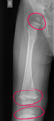
[Paediatric normal whole leg radiograph, showing epiphyseal plates around the head of the femur, the femoral condyles, the proximal tibia, the distal tibia and the lateral malleoli that can mimic fractures. Courtesy of radiopaedia]
See all those weird blobs? Those are bones in the process of fusing together! The transverse lines that could be mistaken for fractures are actually epiphyseal plates – hyaline cartilage bridges between the shaft of a bone and what will become its tip, which don't attenuate x-rays, and thus appear black on our radiographs! This is where bone growth occurs - the cartilage forms a sort of template matrix that then ossifies into bone.
Compare our paediatric radiograph to the AP knee radiographs of an adult with no visible pathology:
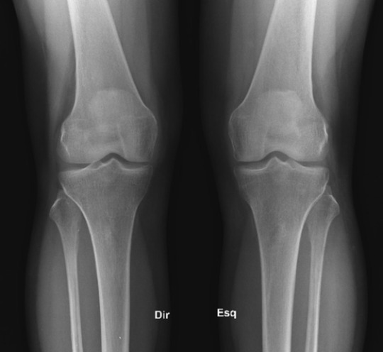
[Adult normal AP knee radiographs, showing fully fused bones. Courtesy of radiopaedia]
See how all those ragged pieces have joined up? That’s endochondral ossification, BABY!
This is how we can figure out the age of a paediatric service user from their bones! Certain bones form at different times.
Let’s check out the carpal bones – all those fiddly little bones in your wrist! Anyone who’s binged Hatecrimes MD – sorry, House MD as often as I have will know the classic acronym for remembering the names of these bones. Moving thumb side to pinkie side, we have…
Scared (Scaphoid - red)
Lovers (Lunate - dark blue)
Hate (Hamate - green)
To (Triquetrum - yellow)
Try (Trapezium - orange)
The (Trapezoid - light blue)
Coolest (Capitate - purple)
Positions (Pisiform - pink)
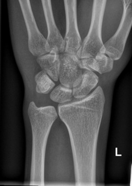
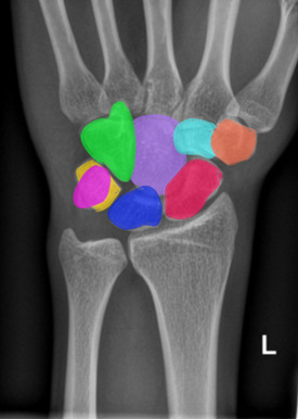
[Normal adult wrist radiograph, shown with and without coloured carpal bones. Courtesy of radiopaedia.]
But did you know that these bones form at different times?
The Capitate ossifies at 1-3 months
The Hamate ossifies at 2-4 months
The Triquetrum ossifies at 2-3 years
The Lunate ossifies at 2-4 years
The Scaphoid, Trapezium and Trapezoid ossify at 4-6 years
And the Pisiform ossifies at 8-12 years
So, I can look at this picture, and tell you that this child is approximately 3 years old, because their Capitate and Hamate have ossified, and their Triquetrum is just visible, starting to ossify below the thumb (circled!)
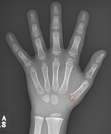
[Normal wrist radiograph of a paediatric patient, triquetral ossification centre circled. Courtesy of Radiopaedia.]
Your bones continue to form and fuse until you reach about 25! Your olecranon (the bump of your elbow) starts to ossify at 6-11 years, and fuses at 13-16 years! We can look at the base of your fifth metatarsal to age afab folks who are approx. 10 and amab folks who are approx. 12, as this bone fuses in the 2-4 years following these ages! The medial end of the clavicle can be used to assess your age from approximately 18-22, and your facial bones continue to ossify into adulthood! How cool is that?
If you’re over 25, fret not – there’s still plenty of funky stuff happening to your bones. But we’ll get into all of that next time, when we take a look at the function of osteoblasts and osteoclasts and explore all the cool little jobs that your bones perform within your body - it's more than you might think! So, tune in next time for more Bone facts...

And thank you for reading!
#medblr#halloween#spooky#skeleton#science side of tumblr#radiography#radiology#medicine#human anatomy#x rays#radley irradiates people#spoopy
26 notes
·
View notes
Text
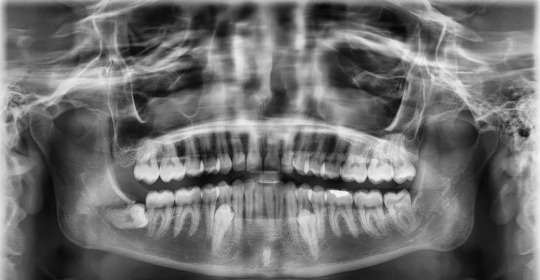
#dientes#anatomy#Supernumerarios#Supernumeraryteeth#Tooth#anatomia#radiography#Radiografia#horroredit#horror#scary aesthetic
9 notes
·
View notes
Text
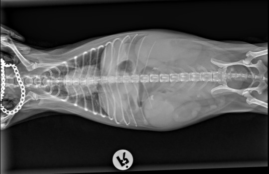
a geriatric intact female dog hasn’t eaten anything for three days.
dog did not look too depressed, but at abdominal palpation she cramped and expressed pain.
Here are the x ray above, what is your diagnosis?
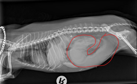
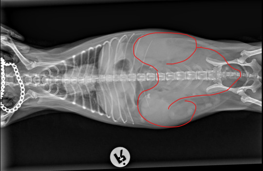
Closed pyometra.
The dog was sent to emergency surgery, and treated for sepsis (as blood result showed super high wbc) for a few days. The dog was healthy at the day of suture removal.
15 notes
·
View notes
Text
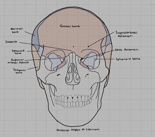
Anterior cranium bones
11 notes
·
View notes
Text
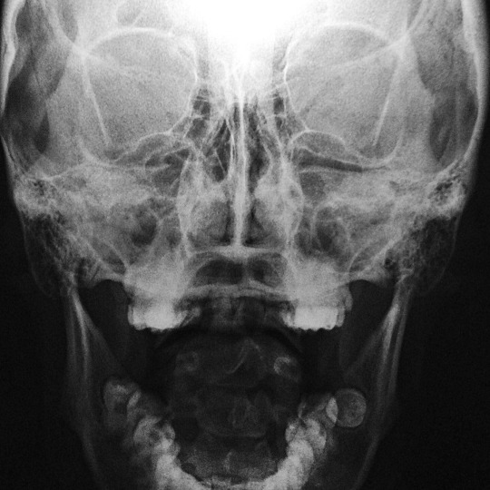
¿Y que sigue despues de la muerte?
ESTO
#skull#skull aesthetic#black and white#blackandwithephotography#radiography#myself#afterdeath#vscophoto#vscocam
4 notes
·
View notes
Last Seen Blogs
design-coalition-photo-journal
J&Co Design Coalition Photo Journal
charizardstolemynickname
i aint a nerd i promise

sillykomaeda
Full Of AUTISM

justbelikemakaras
Where the rage Bard and Prince hide

no-quenos-blog-pn
Sin título