#plant microscopy
Text
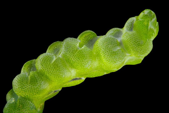
Gametophyte of Pleurocladula albescens.
114 notes
·
View notes
Text
That post that called the relationship btwn plants and mycorrhizal fungi “yuri stuff” lives rent free in my head
62 notes
·
View notes
Text

Plant cells under the microscope.
8 notes
·
View notes
Photo

Special microscope shows different anti-icing strategies of plant leaves
When environmental temperatures go below zero, ice crystals are formed on many leaves of evergreen plants. Nevertheless, they usually survive frost phases unharmed. Using a special cryo-scanning electron microscope, researchers from the Zoological Institute of Christian-Albrechts-Universität zu Kiel (CAU) were able to take high-resolution images of icing processes on surfaces of plants native to Germany and Antarctica at the micro- and nanoscales for the first time. In the process, they discovered various tiny structures on the leaf surfaces with which the plants protect themselves against low temperatures.
A better understanding of these protective strategies could also be interesting for the protection of crops or artificial surfaces such as airplanes. The results were published in the journal The Science of Nature.
Inspiration for artificial anti-icing surfaces
Airplanes are treated with special liquids or are built with heatable surfaces to protect them from icing. Science and industry worldwide are investigating suitable coatings for aviation. "However, many of our wild plants have developed their own natural protection against icing over the course of evolution," explains Professor Stanislav Gorb, head of the research group Functional Morphology and Biomechanics. For more than 20 years, the zoologist has been studying the surfaces of plants at CAU together with his wife Dr. Elena Gorb, a botanist by training.
Read more.
#Materials Science#Science#Icing#Plants#Materials Characterization#Electron microscopy#Biomimicry#Surfaces#Cryogenics#Kiel University
28 notes
·
View notes
Text

I captured a leaf stomata under the microscope yesterday at work!
#was testing removing a stomata with nail polish and tape:')#biology#my posts#microscopy#microscope#plants#botany#plant science#studyblr
2 notes
·
View notes
Text
An example of induced aerenchyma occurs in maize (corn; Zea mays) (Figure 24.19).
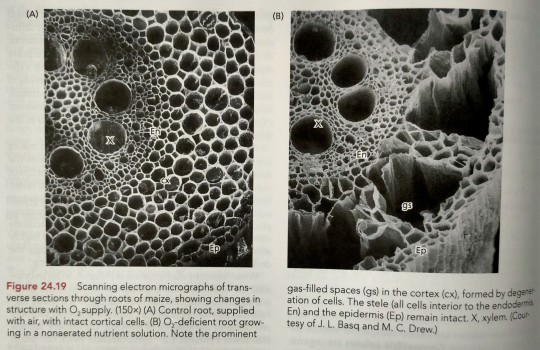
"Plant Physiology and Development" int'l 6e - Taiz, L., Zeiger, E., Møller, I.M., Murphy, A.
#book quotes#plant physiology and development#nonfiction#textbook#corn#maize#zea mays#trypophobia#scanning electron micrograph#microscopy#aerenchyma#plant cells
8 notes
·
View notes
Text
Other specialized epidermal cells, such as lithocysts, bulliform cells, silica cells, and cork cells (Figure 19.9), are found only in certain groups of plants and are not studied as well.
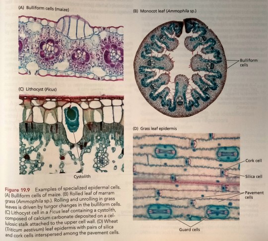
"Plant Physiology and Development" int'l 6e - Taiz, L., Zeiger, E., Møller, I.M., Murphy, A.
#book quote#plant physiology and development#nonfiction#textbook#plant cells#epidermis#epidermal cells#lithocysts#bulliform cells#silica cells#cork cells#maize#corn#zea mays#ammophila#ficus#grass#pavement cells#guard cells#microscopy
7 notes
·
View notes
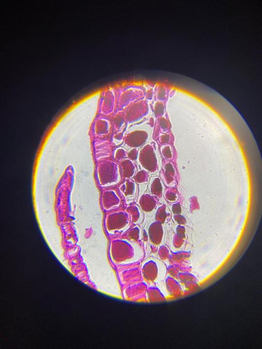
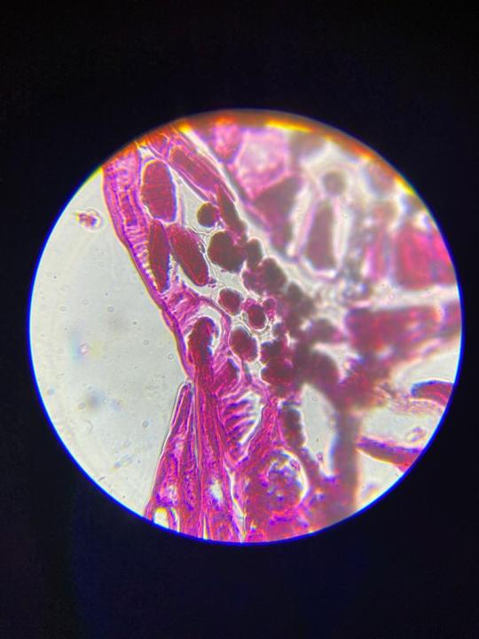
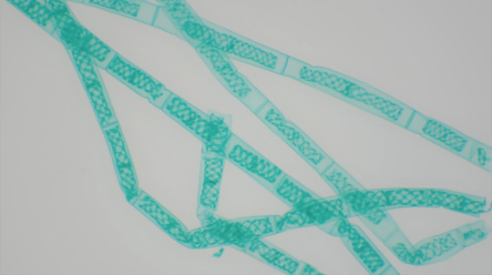
Text

the beauty of microscopy II
juncus effusus
🔬instagram🔬
#mine#plant aesthetic#botanical#botanic academia#botanicart#microscope#microscopy#botanical microscopy#aesthetic#nature#stem academia#stem#women in stem#green aesthetic#laboratory aesthetic#laboratory#Lab aesthetic#botany#botanical illustration#botanical garden#stem student#cell aesthetic#cells at work#university#scienceblr#science tumblr#science
2 notes
·
View notes
Text
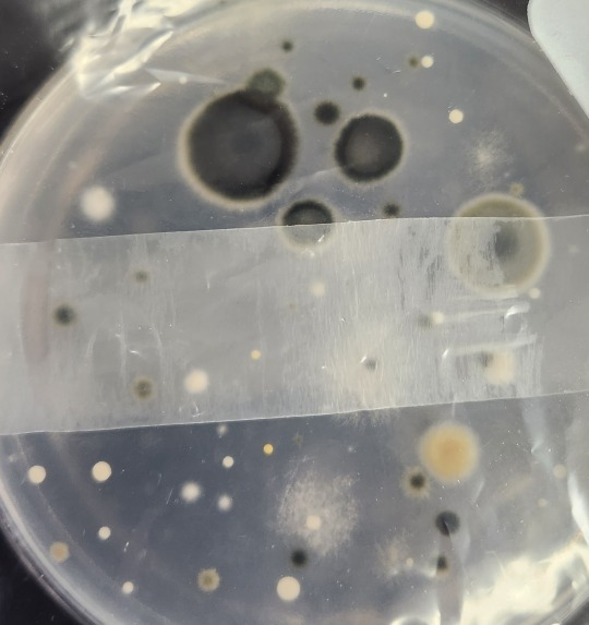
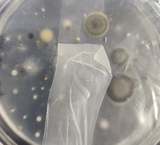
Fungal spore growth on agar after being left by potted plants
4 notes
·
View notes
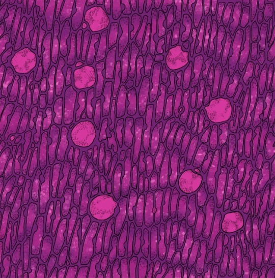
Photo


These tiny “mouths” in the picture are stomata, the pores on plants’ leaves that allow plants to “breathe”. Plants need to take in carbon dioxide and release oxygen. Stomata facilitate gas exchange in photosynthesis by letting these gasses in and out of the plant tissue. Plants are able to open and close their stomata, which is important since water vapour also escapes via the stomata. Closed stomata help prevent the plant from drying out. These images are from my undergraduate research project which focused on the molecular signals controlling stomata opening and closing. These picture are the epidermis of Arabidopsis thaliana peeled off and seen under a light microscope.
#katia plant scientist#plants#plant#plant biology#plant science#Microscopy#light microscope#stomata#gas exchange#science facts#science#biology#plant anatomy#leaves
14 notes
·
View notes
Text
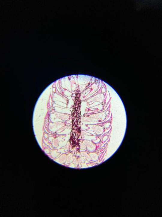

Different Sections of a Pine Young Staminate Cone
One of the prettiest prepped mounts that came with the microscope.
3 notes
·
View notes
Text

Hazelnut (male flower), overlay of 7 channel autofluorescence microscopy. Imaged with ZEISS Axio Observer, Axiocam, Colibri 7.
49 notes
·
View notes
Text

*has a silly time*
#After having microscopy for like half a year i finally know what im doing and its fun#Cuz when i got the gear at first i had no fucking clue what im looking at and it was boring as fuck tbh#Plants are rly cool cooler than animals i think#Photo#Also i need to go to the shop and get glue for making specimen cuz since the time i got the last tiny bottle it all dried up to stone (augh#I even have tiny microtome with changable razzor thatssocool i think
4 notes
·
View notes
Photo
Pond water plants under the microscope.
25 notes
·
View notes
Text


More of my microscope drawing, the leaf cells and digestive glands of a Venus flytrap, now with vivid, unrealistic, and completely unnecessary recolors. As people who followed me from Twitter could tell you, step one of my creative process is to draw a thing, step two is to recolor it pink. So, I did the thing and it was fun, and now I'm going to throw it into my folder of things to put in my store, but I only put up an item a week (trying to appease the algorithm gods), so if you want one of these now, let me know and I'm (broke) totally willing to put it up early. But also, these are all small for use as computer wallpaper, if you feel so inclined.
#venus flytrap#carnivorous plants#small artist#artists on tumblr#art#microscope#microscopy#botany#science#groovy
4 notes
·
View notes
Photo

Celery Leaf Popular prints, shirts, & stickers available on my Etsy. Línk in bio #microscope #microscopy #sciart #science #nature #optics #imaging #plant #plants #plantcell #photomicrograph #biology #microbiology #cell #etsy #Infinitesimal #Prints #shopping #etsyshop #etsyseller #crosssection #celery #leaf #vegetable #vegetables (at Cal Poly Humboldt) https://www.instagram.com/p/Ci7_m18pkDU/?igshid=NGJjMDIxMWI=
#microscope#microscopy#sciart#science#nature#optics#imaging#plant#plants#plantcell#photomicrograph#biology#microbiology#cell#etsy#infinitesimal#prints#shopping#etsyshop#etsyseller#crosssection#celery#leaf#vegetable#vegetables
3 notes
·
View notes
Last Seen Blogs


deardreamweaver
Dear Dream Weaver,

lanterne
les aristocrats à la

xvi-lewisknight
Lewis Knight
chdarling-tle
The Last Enemy Project